. 2012 Sep;32(17):3392-402.
doi: 10.1128/MCB.00070-12. Epub 2012 Jun 25.

GATA6 is required for proliferation, migration, secretory cell maturation, and gene expression in the mature mouse colon

Affiliations

GATA6 is required for proliferation, migration, secretory cell maturation, and gene expression in the mature mouse colon

Eva Beuling et al. Mol Cell Biol. 2012 Sep.

Abstract

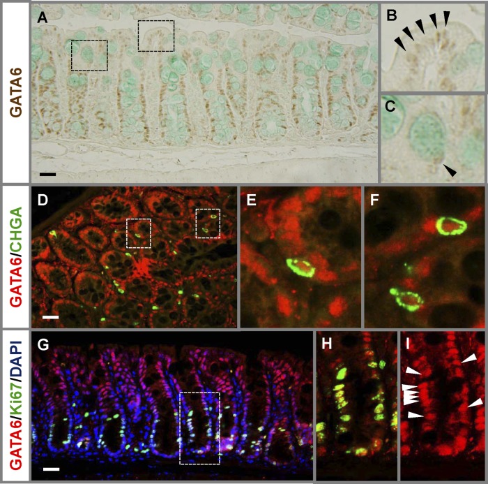
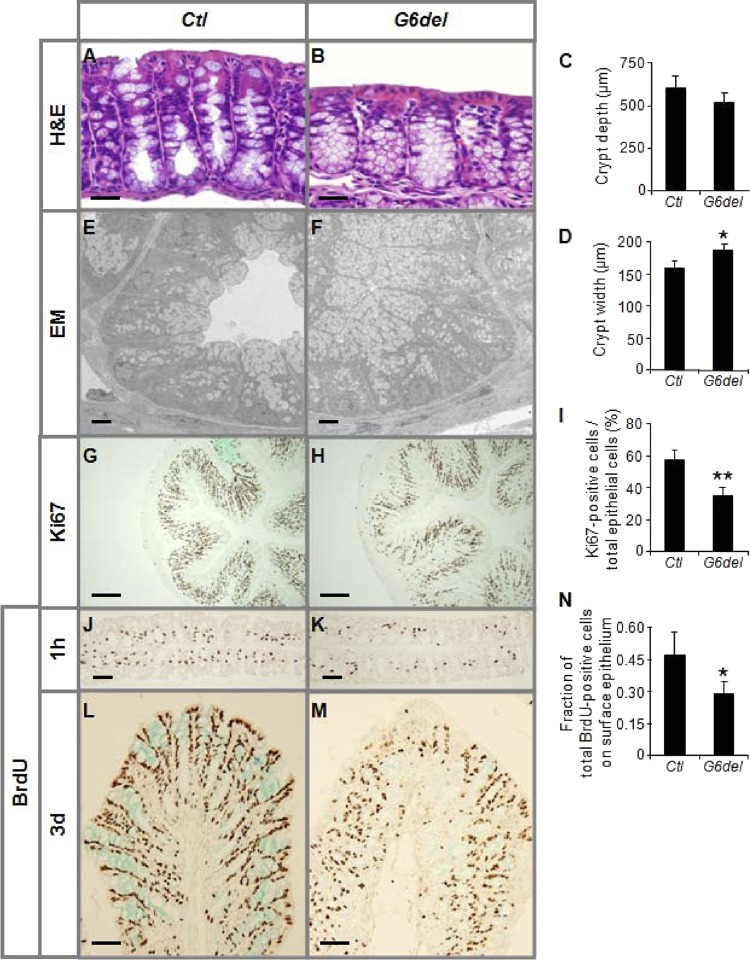
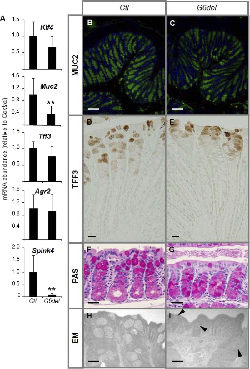
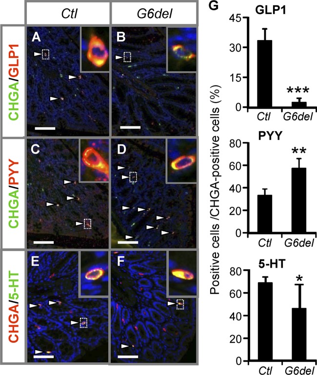
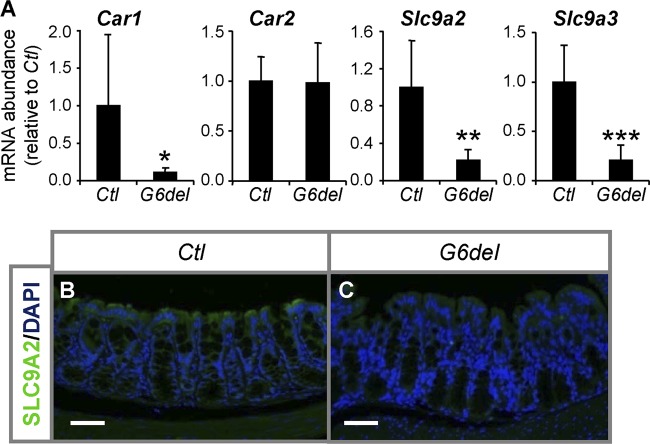
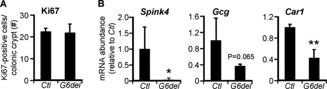
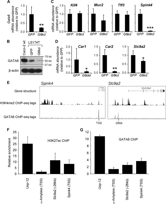
Controlled renewal of the epithelium with precise cell distribution and gene expression patterns is essential for colonic function. GATA6 is expressed in the colonic epithelium, but its function in the colon is currently unknown. To define GATA6 function in the colon, we conditionally deleted Gata6 throughout the epithelium of small and large intestines of adult mice. In the colon, Gata6 deletion resulted in shorter, wider crypts, a decrease in proliferation, and a delayed crypt-to-surface epithelial migration rate. Staining techniques and electron microscopy indicated deficient maturation of goblet cells, and coimmunofluorescence demonstrated alterations in specific hormones produced by the endocrine L cells and serotonin-producing cells. Specific colonocyte genes were significantly downregulated. In LS174T, the colonic adenocarcinoma cell line, Gata6 knockdown resulted in a significant downregulation of a similar subset of goblet cell and colonocyte genes, and GATA6 was found to occupy active loci in enhancers and promoters of some of these genes, suggesting that they are direct targets of GATA6. These data demonstrate that GATA6 is necessary for proliferation, migration, lineage maturation, and gene expression in the mature colonic epithelium.


Figures

Fig 1
Fig 1
GATA6 is expressed in all differentiated and proliferating cells in the mature mouse colonic epithelium. (A to C) Immunostaining reveals that GATA6 is expressed in nuclei of surface colonocytes (B) and goblet cells (C) (arrowheads). (D to I) Coimmunofluorescence with GATA6 and CHGA (D to F) or GATA6 and Ki67 (G to I) reveals that GATA6 is also expressed in CHGA-positive enteroendocrine cells and Ki67-positive cells in the proliferative compartment. Arrowheads indicate examples of GATA6-positive cells. Bars: 100 μm.
Fig 2
Fig 2
Intestinal Gata6 deletion results in a reduction in the proliferation and migration rates in the mature mouse colonic epithelium. (A to D) H&E staining reveals an increase in goblet-like structures in crypts of G6del colon (A and B), a modest (nonsignificant) reduction in crypt depth (C), and significant increase in crypt width (D). (E and F) Transmission EM confirmed an accumulation of goblet-like cells in crypts. (G to K) The number of Ki67-positive cells as a percentage of total epithelial cells is decreased in G6del colon compared to Ctl colon (G to I) and is supported by a decrease in the number of BrdU-positive cells in G6del colon harvested 1 h after BrdU injection (J and K). (L to N) BrdU immunostaining 3 days after BrdU injection revealed a significantly lower percentage of cells at the surface epithelium (n = 4 in each group). *, P < 0.05; **, P < 0.01. Bars: A and B, 100 μm; E and F, 10 μm; G and H, 400 μm; J, K, L, and M, 200 μm.
Fig 3
Fig 3
Intestinal Gata6 deletion results in impaired terminal differentiation of goblet cells in the mature mouse colon. (A) Quantitative RT-PCR shows a decrease in Muc2 and Spink4 mRNA abundance in G6del colon compared to Ctl colon (n = 7 in each group). (B and C) Immunostaining reveals a reduction in MUC2-positive goblet cells, mainly in the lower half of the crypts, in G6del colon compared to Ctl colon. (D and E) Immunostraining reveals that TFF3-positive goblet cells localize higher in the crypts in G6del compared to Ctl colon. (F and G) PAS staining demonstrates an absence of intense staining in the upper half of the crypt of G6del colon. (H and I) Transmission EM reveals disorganized Golgi structures (arrowheads) in crypt goblet cells of G6del colon. **, P < 0.01. Bars: B and C, 400 μm; D and E, 50 μm; F and G, 100 μm; H and I, 40 μm.
Fig 4
Fig 4
Intestinal Gata6 deletion results in alterations in enteroendocrine subpopulations in the colon. (A to G) Cell counts on sections stained with GLP1 and CHGA (A and B), PYY and CHGA (C and D), or 5-HT and CHGA (E and F) reveal decreases in the numbers of GLP1-positive and 5-HT-positive and an increase in the number of PYY-positive cells per total number of CHGA-positive cells in G6del compared to Ctl colon (G) (n = 4 in each group). Arrowheads indicate positive cells. Bars: 200 μm.
Fig 5
Fig 5
GATA6 is required for the expression of specific colonocyte genes in the mature mouse colon. (A) Quantitative RT-PCR shows decreases in Car1, Slc9a2, and Slc9a3 mRNA abundance in G6del colon compared to Ctl colon (n = 7 in each group). *, P < 0.05; **, P < 0.01; ***, P < 0.001. (B and C) Immunofluorescence analysis performed with anti-SLC9A2 antibody shows a reduction in intensity in the fluorescence signal (green) for SLC9A2 protein in surface colonocytes of G6del colon compared to Ctl colon. Bars: 100 μm.
Fig 6
Fig 6
Short-term Gata6 deletion. (A) The number of Ki67-positive cells as a percentage of total epithelial cells is unchanged in G6del colon compared to Ctl colon. (B) Quantitative RT-PCR shows decreases in Car1, Spink4, and Gcg mRNA abundance in G6del colon compared to Ctl colon (n = 3 or 4 in each group). *, P < 0.05; **, P < 0.01.
Fig 7
Fig 7
GATA6 occupies active chromatin sites in the Spink4 and Slc9a2 genes. (A and B) LS174T cells infected with a lentivirus expressing an shRNA for Gata6 (G6kd) demonstrate a decrease in Gata6 mRNA abundance (qRT-PCR; n = 4 independent infections) (A) and GATA6 protein abundance (Western blotting) (B) compared to control LS174T cells infected with a lentivirus expressing an shRNA for GFP. (C and D) Among the goblet (C) and colonocyte (D) markers, Spink4, Car1, Car2, and Slc9a2 were downregulated in the Gata6 knockdown LS174T cells. (E) Using publicly available ChIP-seq databases of the human intestinal cell line Caco-2 (37), GATA6 enrichment was found in enhancers, identified by H3K4me2 analysis (6, 15, 16), that were assigned to Spink4 and Slc9a2. (F) ChIP analysis using LS174T cells and an antibody for the H3K27ac enhancer mark, demonstrating enrichment at these sites. (G) ChIP analysis using LS174T cells and a GATA6 antibody, showing GATA6 enrichment at these sites.

Similar articles

Cited by

References

    1. Al-azzeh ED, Fegert P, Blin N, Gott P. 2000. Transcription factor GATA-6 activates expression of gastroprotective trefoil genes TFF1 and TFF2. Biochim. Biophys. Acta 1490: 324–332 - PubMed
    1. Ayanbule F, Belaguli NS, Berger DH. 2011. GATA factors in gastrointestinal malignancy. World J. Surg. 35: 1757–1765 - PubMed
    1. Bachmann O, et al. 2004. The Na+/H+ exchanger isoform 2 is the predominant NHE isoform in murine colonic crypts and its lack causes NHE3 upregulation. Am. J. Physiol. Gastrointest. Liver Physiol. 287: G125–G133 - PubMed
    1. Barker N, et al. 2007. Identification of stem cells in small intestine and colon by marker gene Lgr5. Nature 449: 1003–1007 - PubMed
    1. Belaguli NS, et al. 2010. GATA6 promotes colon cancer cell invasion by regulating urokinase plasminogen activator gene expression. Neoplasia 12: 856–865 - PMC - PubMed

Publication types

MeSH terms

Substances

LinkOut - more resources