. 2012 Sep;32(17):3414-27.
doi: 10.1128/MCB.06678-11. Epub 2012 Jun 25.

Relocalization of junctional adhesion molecule A during inflammatory stimulation of brain endothelial cells

Affiliations

Relocalization of junctional adhesion molecule A during inflammatory stimulation of brain endothelial cells

Svetlana M Stamatovic et al. Mol Cell Biol. 2012 Sep.

Abstract

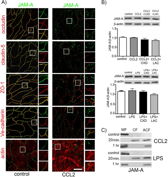
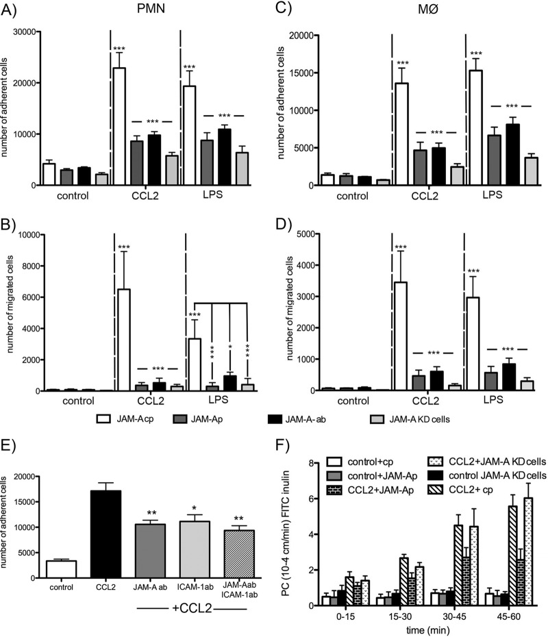
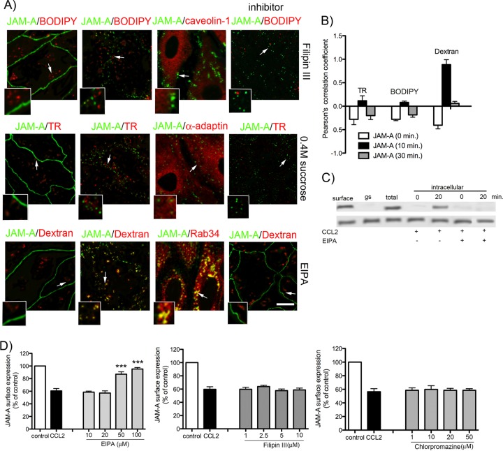
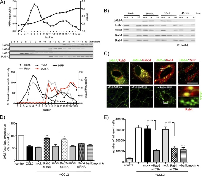
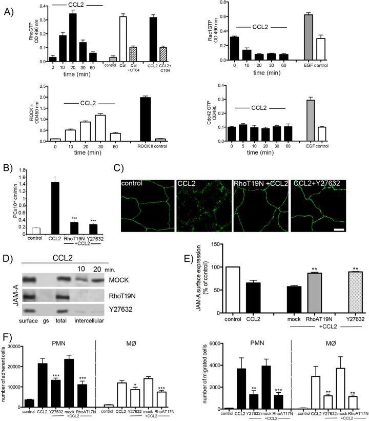
Junctional adhesion molecule A (JAM-A) is a unique tight junction (TJ) transmembrane protein that under basal conditions maintains endothelial cell-cell interactions but under inflammatory conditions acts as a leukocyte adhesion molecule. This study investigates the fate of JAM-A during inflammatory TJ complex remodeling and paracellular route formation in brain endothelial cells. The chemokine (C-C motif) ligand 2 (CCL2) induced JAM-A redistribution from the interendothelial cell area to the apical surface, where JAM-A played a role as a leukocyte adhesion molecule participating in transendothelial cell migration of neutrophils and monocytes. JAM-A redistribution was associated with internalization via macropinocytosis during paracellular route opening. A tracer study with dextran-Texas Red indicated that internalization occurred within a short time period (~10 min) by dextran-positive vesicles and then became sorted to dextran-positive/Rab34-positive/Rab5-positive vesicles and then Rab4-positive endosomes. By ~20 min, most internalized JAM-A moved to the brain endothelial cell apical membrane. Treatment with a macropinocytosis inhibitor, 5-(N-ethyl-N-isopropyl)amiloride, or Rab5/Rab4 depletion with small interfering RNA oligonucleotides prevented JAM-A relocalization, suggesting that macropinocytosis and recycling to the membrane surface occur during JAM-A redistribution. Analysis of the signaling pathways indicated involvement of RhoA and Rho kinase in JAM-A relocalization. These data provide new insights into the molecular and cellular mechanisms involved in blood-brain barrier remodeling during inflammation.


Figures

Fig 1
Fig 1
(A) Double immunostaining for JAM-1 and TJ proteins (occludin, claudin-5, and ZO-1), adherens junction protein (Ve-cadherin), and phalloidin-Alexa 596 staining for actin in mBMECs under resting conditions (control, nontreated cells) or with treatment with CCL2 (100 ng/ml) for 30 min. All samples were examined using confocal microscopy. Notice the close association of JAM-A with occludin, claudin-5, and ZO-1 under control conditions, while CCL2 induced relocalization of JAM-A away from the lateral cell border. Boxes indicate locations of high magnification of JAM-A immunostaining with TJ and AdJ proteins as well as the actin cytoskeleton. Bar, 50 μm. (B) During CCL2 or LPS exposure, there were no changes in total JAM-A protein levels. Treatment with an inhibitor of protein synthesis, cycloheximide (CXD; 5 μg/ml), or an inhibitor of protein degradation, lactacystin (LAC; 1 μM), did not affect the total level of protein expression determined by Western blot analysis. Blots represent one of the three independent experiments. Data represent means of 3 independent experiments. (C) mBMECs were exposed to either CCL2 (100 ng/ml) or LPS (5 μg/ml) for 20 or 60 min and then underwent cell fractionation and Western blotting. Under control conditions, JAM-A was found in the membrane fraction (MF; Triton X-100-insoluble fraction). CCL2 and LPS treatment for 20 min resulted in movement of JAM-A to the cytosolic fraction (CF; Triton X-100-soluble fraction) and actin cytoskeletal fraction (ACF; Triton X-100-insoluble fraction). By 60 min, most JAM-A returned to the membrane fraction, while some remained in the actin cytoskeleton fraction. Blots represent one of three successful experiments.
Fig 2
Fig 2
(A) (Left) Time-lapse confocal microscopy. bEnd.3 monolayers expressing GFP–JAM-A were exposed to CCL2 (100 ng/ml). Images were obtained every 5 min from 0 to 120 min. Representative images are from some critical time points during CCL2-induced alterations in brain endothelial cell barrier permeability. There is a fragmented pattern of JAM-A staining at the cell-cell border of endothelial cells during CCL2 exposure. (Right) TEER after exposure (0 to 120 min) to CCL2 or LPS. Data represent averages ± SDs for 5 independent experiments. (B) Triple-label immunostaining with CellLight membrane-CFP Bacmam 2.0 (blue), anti-JAM-A antibody (green), and anti-ZO-1 antibody (red) supported by optical z-stack section and three-dimensional analysis clarified that after exposure to CCL2 for 30 min, JAM-A redistributed onto the brain endothelial cell apical surface, where it may play a role as a leukocyte adhesion molecule. Bar, 50 μm. (C) Cell-based ELISA of JAM-A surface expression upon exposure to CCL2 (100 ng/ml) or LPS (5 μg/ml). Cells were treated for different time points (0 to 60 min), and every sample was then incubated with anti-JAM-A antibody, followed by HRP-conjugated secondary antibody and fixation. Notice that over a short time (10 to 20 min) JAM-A disappears from the cell surface, with return and expression on the apical membrane from 30 to 60 min. Data represent averages ± SDs for 3 independent experiments. *, P < 0.05 compared with control nontreated cells; **, P < 0.01 compared with control nontreated cells; ***, P < 0.001 compared with control nontreated cells. ODS 450, optical density at 450 nm.
Fig 3
Fig 3
Freshly prepared neutrophils (polymorphonuclear leukocytes [PMNs]) (A and B) and macrophages (MØ) (C and D) were labeled with calcein-AM and layered on top of mBMEC monolayers previously treated with CCL2, LPS, or vehicle in the presence of JAM-A-inhibitory peptide (JAM-Ap; 1 μg/ml), control JAM-A peptide (JAM-A-cp; 1 μg/ml), or neutralizing anti-JAM-A antibody (JAM-A-Ab; 10 μg/ml). As a control, we also used JAM-A-KD cells, generated by stable transfection of bEnd.3 cells with JAM-A shRNA. Cells were incubated for 2 h, and then the medium with nonadherent cells was removed and the sample was washed and fixed. The fluorescence was read on a fluorescent reader. Both CCL2 and LPS increased the number of adherent (A and C) and migrated (B and D) neutrophils and macrophages, and that was blocked by treatment with JAM-A-inhibitory peptide or JAM-A antibody or if JAM-A-KD cells were used. Data represent averages ± SDs for 3 independent experiments. *, P < 0.05; ***, P < 0.001. (E) Adhesion assay for neutrophils (polymorphonuclear leukocytes) under conditions of exposure of brain endothelial cell monolayer to CCL2. The adhesion was blocked by adding either neutralizing anti-JAM-A (10 μg/ml; R&D Systems), neutralizing anti-ICAM-1 (10 μg/ml; R&D Systems), or a cocktail of anti-JAM-A and anti-ICAM-A antibodies (both at a concentration of 10 μg/ml) (pretreated and treated for 1 h). Notice the significant reduction in polymorphonuclear leukocyte adhesion if JAM-A or ICAM-1 is blocked. However, there was no amplifying effect if two neutralizing antibodies were applied. Data represent averages ± SDs for 3 independent experiments. *, P < 0.05 compared with cells treated with CCL2 only; **, P < 0.01 compared with cells treated with CCL2 only. ab, antibody. (F) Recombinant murine CCL2 was applied at the top and bottom of a Transwell system. The permeability coefficient (PC) for FITC-inulin was evaluated from 0 to 60 min. CCL2 induced a time-dependent increase in permeability. Adding JAM-A-neutralizing peptide showed a partial protection of this opening of the brain endothelial cell barrier, but CCL2 still increased permeability in JAM-A-KD cells. Data represent averages ± SDs for 5 independent experiments.
Fig 4
Fig 4
(A) Internalization of surface-biotinylated JAM-A protein. Confluent mBMECs treated with and without cycloheximide (CXD) were surface biotinylated at 0°C and then exposed to CCL2 for 60 min at 37°C to allow internalization. Any membrane-bound biotin was removed by glutathione solution (glutathione stripping [gs]). Lanes 1 and 2, biotinylated JAM-A at the cell surface; lane 3, glutathione stripping of surface biotin; lanes 4 and 5, total cell lysate; lane 6 and 7, portion of biotinylated-internalized proteins. Adjusted blot showing the time course (0 to 60 min) of internalized biotinylated JAM-A during mBMEC exposure to CCL2. (B) Quantification of internalized JAM-A during the exposure to CCL2 and opening of the brain endothelial cell barrier. The percent internalized protein was estimated as the percentage of the total biotinylated JAM-A. GADPH (glyceraldehyde-3-phosphate dehydrogenase) represents an internal loading control. Data represent averages ± SDs of 5 independent experiments.
Fig 5
Fig 5
(A) Morphological analysis of JAM-A–GFP internalization. Briefly, bEnd.3 cell monolayers were first exposed to the indicated concentration of the tracer BODIPY-TR-ceramide (BODIPY; 5 μM; caveola pathway), Texas Red-transferrin conjugate (TR; 10 μg/ml; clathrin pathway), and dextran (10 kDa)-Texas Red conjugate (dextran; 0.5 mg/ml; macropinocytosis) for 30 min at 4°C, and then CCL2 was added at a concentration of 100 ng/ml and samples were placed in an incubator at 37°C for 10 min. Cells were then fixed. In separate experiments, bEnd.3 cells stably transfected with GFP–JAM-A were exposed to CCL2 for 0 to 60 min, fixed, and processed for immunocytochemistry using mouse anti-caveolin-1, anti-α-adaptin, or anti-Rab34 antibodies. Both the tracer study and immunocytochemistry indicated a close association of JAM-A with dextran and Rab34 immunostaining (small boxes). Arrows, magnified colocalization of JAM-A with tracers and antibody-labeled internalization pathways. In the inhibition study (indicated in column inhibitors), cells were preincubated with a certain inhibitor, either filipin III (caveola-dependent internalization), 0.4 M sucrose (clathrin-dependent pathway), or EIPA (100 mM; macropinocytotic uptake), followed by incubation with CCL2 for 10 min. Only the inhibition of macropinocytosis prevented JAM-A movement from the cell border. Bar, 50 μm. (B) Quantification of colocalization of JAM-A with internalization vesicles based on Pearson's correlation coefficient of GFP–JAM-A/BODIPY-TR ceramide, JAM-A–GFP/transferrin-Texas Red, and JAM-A–GFP/dextran-Texas Red. There was a high degree of correlation between total JAM-A and dextran. Other tracers did not show any colocalization pattern. Error bars indicate means ± SDs. (C) The internalization pathway via macropinocytosis was also confirmed in a biotin internalization assay. In confluent mBMECs, JAM-A was biotinylated at 0°C and then exposed to either CCL2 for 20 min at 37°C or CCL2 and the inhibitor of macropinocytosis, EIPA. Surface, biotinylated JAM-A at the cell surface; gs, glutathione stripping (amount of membrane-bound biotin which was removed by glutathione solution); total, total cell lysate; intracellular, the amount of JAM-A internalized during the period from 0 to 20 min in the presence of CCL2 or CCL2 and EIPA. The macropinocytosis inhibitor prevented JAM-A internalization during CCL2 exposure. The blot represents one of three experiments performed. (D) Dose-dependent inhibition of CCL2-induced internalization of JAM-A after exposure to CCL2 for 10 min and in the presence of selected inhibitors: EIPA (10 to 100 μM), filipin III (1 to 10 μM), and chlorpromazine (1 to 50 μM, clathrin-dependent internalization). JAM-A surface expression under these conditions was evaluated by cell-based ELISA, and it is presented as the percentage of the JAM-A under control conditions. Again, only EIPA at a dose of 50 to 100 mM was able to block internalization of JAM-A. Data represent averages ± SDs for 3 independent experiments. ***, P < 0.001 compared with control nontreated cells.
Fig 6
Fig 6
(A) After exposure of mBMEC to CCL2 (100 ng/ml) for 20 min, endosome-rich fractions were prepared using a continuous sucrose gradient (15 to 40%). Twenty fractions were collected and analyzed by Western blotting. Fraction 1 is the top of the gradient and has the lowest density of sucrose. The fraction and its endosomal content were also labeled with an HRP (1 mg/ml) pulse, incubated for 5 min, and chased for 40 min. Fractions 14 to 20 mostly contain Rab5+ vesicles (early endosomes) and fractions 11 to 16 contain Rab4+ vesicles (recycling endosomes), while fractions 5 to 8 contain Rab7+ vesicles (late endosomes). The Western blot represents one of three successful experiments. Densitometric analysis of Rab5, Rab4, Rab7, and JAM-A expression in collected fractions indicated the correlation of JAM-A presence with Rab5+ and Rab4+ endosomes. The graph is a summary of the three independent experiments. (B) Isolated endosomes were pulled down using JAM-A antibody-coated beads. Western blot assays for Rab5, Rab34, Rab4, and Rab7 were performed to assess the JAM-A presence in certain Rab+ vesicles. Total, whole collected fraction; B, endosomal fraction bound to JAM-A antibody-coated magnetic Dynabeads; UB, unbound fraction not absorbed to JAM-A antibody-coated Dynabeads. The blot represents one of three successful experiments. IP, immunoprecipitation. (C) Immunocytochemistry analysis of JAM-A–GFP localization with Rab5+, Rab34+, Rab4+, and Rab7+ vesicles during CCL2 exposure (20 min). Rab5 or Rab34 siRNA prevented JAM-A internalization. Rab4 siRNA did not prevent internalization, but it did prevent JAM-A from appearing on the apical membrane (zoom confocal images). Bar, 20 μm. (D and E) Preventing JAM-A internalization via macropinocytosis (Rab5 siRNA and Rab34 siRNA), preventing JAM-A recycling by depleting Rab4 vesicles (Rab4 siRNA), or treatment with an inhibitor of emptying of the recycling endosomes (bafilomycin A1, 50 nM) reduced the internalization of JAM-A (D) after 30 min of treatment as well as neutrophil adhesion (P < 0.001). Notice that Rab4 inhibition did not affect the internalization of JAM-A but via inhibition of JAM-A recycling affected the adhesion of neutrophils on the brain endothelial cell surface. All data represent averages ± SDs for 3 independent experiments. *, P < 0.05 compared with control or mock-transfected cells; **, P < 0.01 compared with control or mock-transfected cells; ***, P < 0.001 compared with control or mock-transfected cells.
Fig 7
Fig 7
Role of RhoA and Rho kinase in JAM-A internalization. (A) Time course of RhoA Rac1, Cdc42, and ROCK II activation during exposure to CCL2 in brain endothelial cells. Rho GTP, Rac1, and Cdc42 levels and ROCK II activity were measured with RhoA–, Rac1–, or Cdc42–small G-protein activation assay (G-lisa) or ROCK activation assay using MYPT1 as a substrate. The positive controls for RhoA GTP levels and for Rac1 GTP and Cdc42 GTP levels were Swiss 3T3 cells treated with calpeptin (Cal; 0.1 mg/ml) and epidermal growth factor (EGF; 10 ng/ml), respectively. The specificity of the RhoA activation was determined under conditions of blocking the RhoA activation in Swiss 3T3 cells (calpeptin induced) and brain endothelial cells (CCL2 induced) by CT04 inhibitor (1 μg/ml; cytoskeleton). Data represent averages ± SDs for 3 independent experiments. OD 490 nm, optical density at 490 nm. (B) Permeability coefficient for FITC-inulin of brain endothelial cell monolayers exposed to CCL2 for 30 min. The brain endothelial cell monolayer was transfected with pCMVRhoT19N or pretreated with Y27632 to block RhoA or Rho kinase and treated with CCL2 for 30 min. The absence of RhoA or Rho kinase activation significantly diminished the increased permeation for FITC-inulin at the brain endothelial cell monolayer. Data represent averages ± SDs for 3 independent experiments. ***, P < 0.001. (C) mBMEC monolayers were mock transfected or transfected with pCMVRhoT19N or pretreated with Rho kinase inhibitor Y27632. Cells were then treated with CCL2 (100 ng/ml). Inhibiting either Rho or Rho kinase diminished CCL2-induced relocalization of JAM-A (absence of the punctate pattern of immunostaining). Bar, 10 μm. (D) Inhibition of RhoA or Rho kinase activity in mBMECs also prevented CCL2-induced internalization of biotin-labeled JAM-A, also evaluated by cell-based ELISA for JAM-A (E). Data represent averages ± SDs for 3 independent experiments. **, P < 0.01 compared with cells treated only with CCL2. (F) Adhesion and migration assay for neutrophils (polymorphonuclear leukocytes [PMN]) and macrophages (MØ) under conditions of exposure of brain endothelial cell monolayer to CCL2, transient depletion of RhoA activity via RhoT19N dominant inactive mutant, or deprivation of Rho kinase activity via Rho kinase inhibitor Y27632 (10 μM). Notice the significant reduction in polymorphonuclear leukocyte and MØ adhesion and migration if RhoA and Rho kinase activity is blocked. Data represent averages ± SDs for 3 independent experiments. *, P < 0.05 compared with cells treated only with CCL2; **, P < 0.01 compared with cells treated only with CCL2; ***, P < 0.001 compared with cells treated only with CCL2.

References

    1. Abbott NJ. 2000. Inflammatory mediators and modulation of blood-brain barrier permeability. Cell. Mol. Neurobiol. 20: 131– 147 - PMC - PubMed
    1. Babinska A, et al. 2007. The F11 receptor (F11R/JAM-A) in atherothrombosis: overexpression of F11R in atherosclerotic plaques. Thromb. Haemost. 97: 272– 281 - PubMed
    1. Bazzoni G. 2011. Pathobiology of junctional adhesion molecules. Antioxid. Redox Signal. 15: 1221– 1234 - PubMed
    1. Bazzoni G, Dejana E. 2004. Endothelial cell-to-cell junctions: molecular organization and role in vascular homeostasis. Physiol. Rev. 84: 869– 901 - PubMed
    1. Bazzoni G, et al. 2000. Homophilic interaction of junctional adhesion molecule. J. Biol. Chem. 275: 30970– 30976 - PubMed

Publication types

MeSH terms

LinkOut - more resources