. 2012 Sep;32(17):3438-51.
doi: 10.1128/MCB.00564-12. Epub 2012 Jun 25.

Coordinate regulation of TPL-2 and NF-κB signaling in macrophages by NF-κB1 p105

Affiliations

Coordinate regulation of TPL-2 and NF-κB signaling in macrophages by NF-κB1 p105

Huei-Ting Yang et al. Mol Cell Biol. 2012 Sep.

Abstract

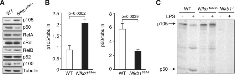
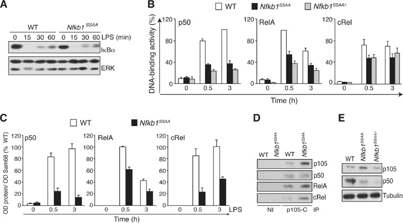
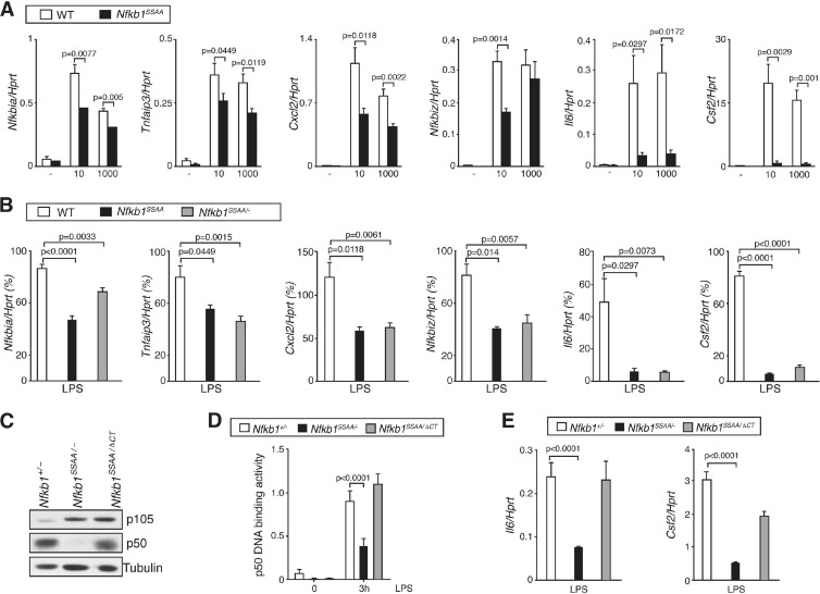
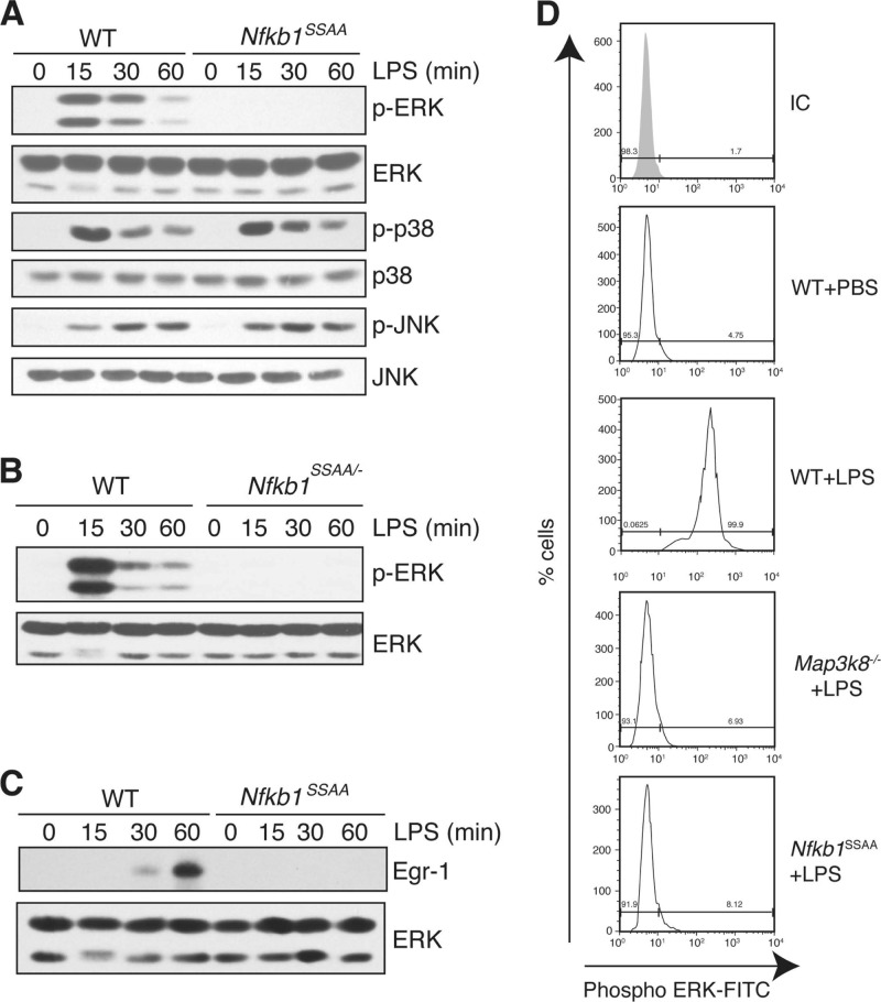
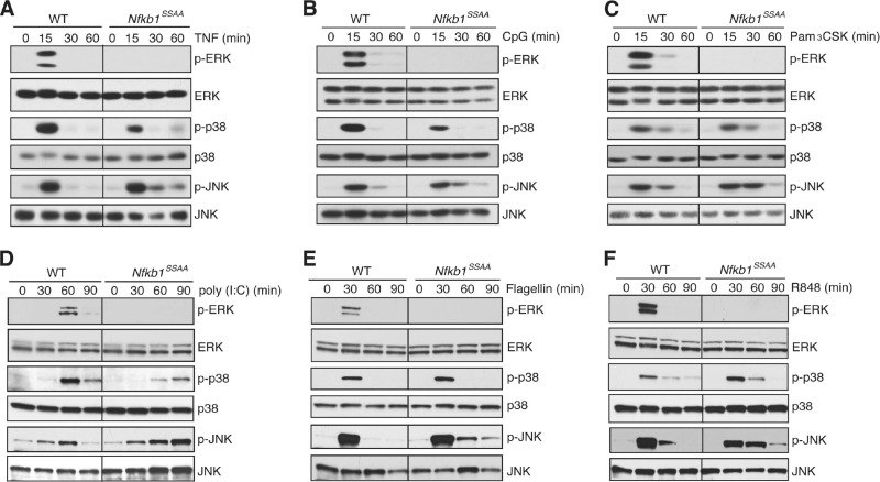
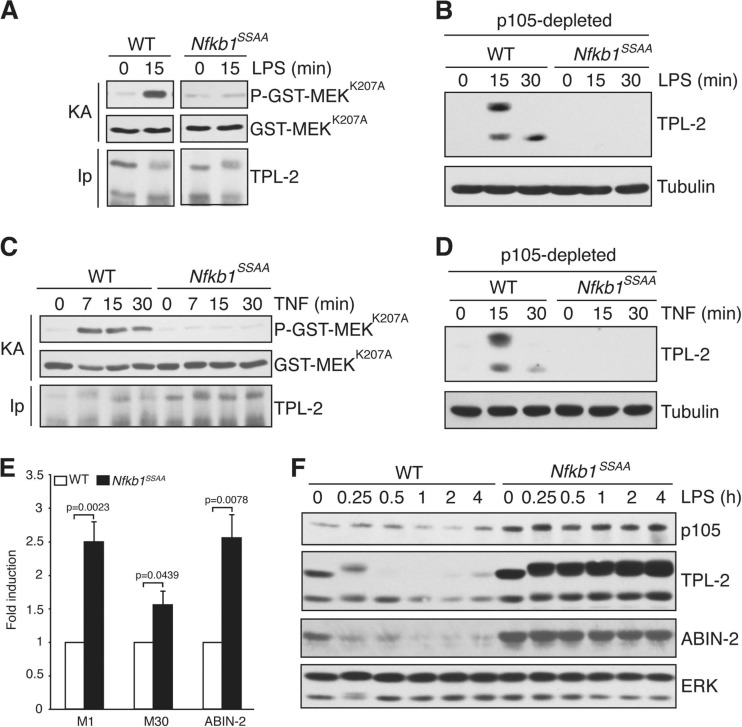
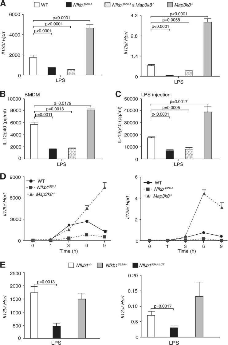
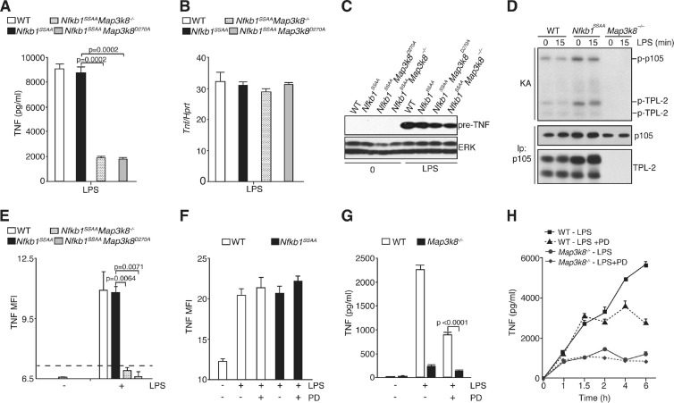
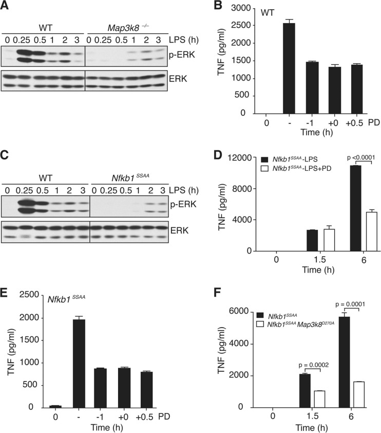
The role of IκB kinase (IKK)-induced proteolysis of NF-κB1 p105 in innate immune signaling was investigated using macrophages from Nfkb1(SSAA/SSAA) mice, in which the IKK target serines on p105 are mutated to alanines. We found that the IKK/p105 signaling pathway was essential for TPL-2 kinase activation of extracellular signal-regulated kinase (ERK) mitogen-activate protein (MAP) kinase and modulated the activation of NF-κB. The Nfkb1(SSAA) mutation prevented the agonist-induced release of TPL-2 from its inhibitor p105, which blocked activation of ERK by lipopolysaccharide (LPS), tumor necrosis factor (TNF), CpG, tripalmitoyl-Cys-Ser-Lys (Pam(3)CSK), poly(I · C), flagellin, and R848. The Nfkb1(SSAA) mutation also prevented LPS-induced processing of p105 to p50 and reduced p50 levels, in addition to decreasing the nuclear translocation of RelA and cRel. Reduced p50 in Nfkb1(SSAA/SSAA) macrophages significantly decreased LPS induction of the IκBζ-regulated Il6 and Csf2 genes. LPS upregulation of Il12a and Il12b mRNAs was also impaired although specific blockade of TPL-2 signaling increased expression of these genes at late time points. Activation of TPL-2/ERK signaling by IKK-induced p105 proteolysis, therefore, induced a negative feedback loop to downregulate NF-κB-dependent expression of the proinflammatory cytokine interleukin-12 (IL-12). Unexpectedly, TPL-2 promoted soluble TNF production independently of IKK-induced p105 phosphorylation and its ability to activate ERK, which has important implications for the development of anti-inflammatory drugs targeting TPL-2.


Figures

Fig 1
Fig 1
Nfkb1SSAA mutation reduces LPS-induced p105 processing to p50. (A) Lysates from BMDM were immunoblotted. The Nfkb1SSAA/SSAA genotype is shown in this figure and in subsequent figures as Nfkb1SSAA. (B) p105 and p50 levels in BMDM were assayed by immunoblotting. Bands were quantified by laser densitometry and are expressed relative to tubulin loading control (error bars, SEM). (C) BMDM were metabolically pulse-labeled for 1 h with [35S]methionine-cysteine (−) and then chased for 1 h with nonradioactive medium containing 100 ng/ml LPS (+). p105/p50 were immunoprecipitated from cell lysates with p105N antibody and resolved by SDS-PAGE, and labeled bands were detected by autoradiography. In panels A and C, results are representative of three independent experiments. In panel B, data were pooled from five independent experiments.
Fig 2
Fig 2
Nfkb1SSAA mutation impairs LPS activation of NF-κB. (A) BMDM were stimulated for the indicated times with LPS. Cell lysates were immunoblotted. (B) BMDM, cultured in triplicate, were stimulated with LPS (1 μg/ml) for 0.5 and 3 h. Binding of p50, RelA, and cRel to an NF-κB oligonucleotide was determined by ELISA (error bars, SEM). (C) BMDM were cultured in triplicate and stimulated with LPS (1 μg/ml). The amounts of the indicated Rel subunits in nuclear fractions were determined by immunoblotting and densitometry (error bars, SEM). (D) BMDM lysates were immunoprecipitated (IP) with p105C antiserum or nonimmune (NI) serum. Isolated proteins were immunoblotted. (E) Lysates from BMDM were immunoblotted. In panels A, D, and E, results are representative of at least three independent experiments. In panels B and C, results were pooled from five independent experiments.
Fig 3
Fig 3
Nfkb1SSAA mutation reduces LPS-induced expression of NF-κB-dependent genes. (A) Triplicate cultures of BMDM were stimulated for 1 h with LPS at 10 or 1000 ng/ml. Expression of the mRNAs shown was determined by quantitative RT-PCR (error bars, SEM). (B) BMDM were stimulated for 1 h with LPS at 10 ng/ml and analyzed as described in panel A. (C) Lysates of BMDM were immunoblotted. (D) BMDM were stimulated with LPS (1 μg/ml) for 3 h. Binding of p50 to an NF-κB oligonucleotide was determined by ELISA (error bars, SEM). (E) Expression of the mRNAs shown was determined as described in panel A, following stimulation of BMDM with 1 μg/ml LPS for 1 h. All results are representative of at least three independent experiments.
Fig 4
Fig 4
Nfkb1SSAA mutation blocks LPS activation of ERK. (A to C) BMDM were stimulated for different times with LPS. Lysates were immunoblotted. (D) WT, Map3k8−/−, and Nfkb1SSAA/SSAA mice were injected intraperitoneally with LPS (200 μg) or PBS. Peritoneal cells were aspirated after 10 min, and intracellular phospho-ERK (p-ERK) in F4/80+ cells was monitored by flow cytometry. All results are representative of at least three independent experiments.
Fig 5
Fig 5
Nfkb1SSAA mutation blocks activation of ERK by TNFR1 and multiple TLRs. Lysates from BMDM, stimulated for the indicated times with TNF (A), CpG (B), Pam3CSK (C), poly(I · C) (D), flagellin (E), or R848 (F) or left unstimulated (0 min), were immunoblotted. All results are representative of at least two independent experiments.
Fig 6
Fig 6
Nfkb1SSAA mutation prevents activation of TPL-2 MEK kinase activity. (A and C) TPL-2 was immunoprecipitated (IP) from lysates of BMDM with or without LPS (A) or TNF (C) stimulation and assayed for its ability to phosphorylate GST-MEKK207A (KA) in vitro. To ensure that equivalent amounts of TPL-2 were assayed for each condition, immunoprecipitations were carried out with cell lysates in excess. (B and D) Lysates of BMDM, stimulated with LPS (B) or TNF (D), were depleted of p105 by immunoprecipitation and then immunoblotted. (E and F) Lysates of BMDM, unstimulated (E) or stimulated for the indicated times with LPS (F), were immunoblotted. Due to alternative translational initiation on a second methionine (at residue 30), TPL-2 is expressed as two isoforms, M1-TPL-2 and M30-TPL-2 (1). In panels A, B, C, D, and F, results are representative of three independent experiments. In panel E, data were pooled from six independent experiments.
Fig 7
Fig 7
Nfkb1SSAA mutation reduces LPS induction of IL-12. (A and B) BMDM were stimulated for 6 h with LPS (10 ng/ml). In panel A, expression of Il12a and Il12b mRNAs was determined by quantitative RT-PCR (error bars, SEM). In panel B, IL-12 p40 in culture supernatants was measured by ELISA (error bars, SEM). (C) WT, Nfkb1SSAA/SSAA, Map3k8−/−, and Nfkb1SSAA/SSAA Map3k8−/− mice were injected intraperitoneally with LPS. After 6 h, IL-12 p40 in serum was measured by ELISA (error bars, SEM of 6 mice). (D) Triplicate cultures of BMDM were stimulated with LPS for the times shown. Expression of Il12a and Il12b mRNAs was determined by quantitative RT-PCR (error bars, SEM). (E) Levels of Il12a and Il12b mRNAs in LPS-stimulated BMDM were measured as described in panel A (error bars, SEM). All results are representative of at least three independent experiments.
Fig 8
Fig 8
Nfkb1SSAA mutation does not inhibit LPS induction of sTNF production. (A and B) BMDM were cultured in triplicates and stimulated with LPS for 6 h (A) or 1 h (B). The Tpl2D270A/D270A genotype is shown here and subsequently as Tpl2D270A. In panel A, TNF in culture supernatants was assayed by ELISA (error bars, SEM). In panel B, Tnf mRNA was quantified as described in the legend of Fig. 3A (error bars, SEM). (C) Lysates were prepared from unstimulated or LPS-stimulated BMDM of the indicated genotypes and immunoblotted. (D) p105 was quantitatively immunoprecipitated from BMDM lysates of the indicated genotypes with or without LPS stimulation (100 ng/ml) and subjected to an in vitro kinase assay (KA). 32P-labeled bands were revealed by autoradiography after SDS-PAGE. The amounts of TPL-2 and p105 isolated were determined by immunoblotting. (E) BMDM were stimulated for 1 h with LPS (1 μg/ml) and fixed, and surface pre-TNF expression was assayed by flow cytometry. The dotted line shows the mean fluorescence intensity (MFI) for negative-control antibody staining. (F) WT or Nfkb1SSAA/SSAA BMDM were stimulated with LPS (1 μg/ml) with or without PD184352 (PD) for 1 h, and surface pre-TNF expression was determined by flow cytometry. (G) BMDM were cultured for 6 h with LPS with or without PD184352 or with control medium (0). Culture supernatants were assayed for TNF (triplicate cultures; error bars, SEM). (H) BMDM were stimulated for the indicated times with LPS with or without PD or with control medium (0). PD was added 1 h before LPS stimulation. TNF in culture supernatants was assayed (error bars, SEM). All results are representative of at least three independent experiments.
Fig 9
Fig 9
Delayed regulation of sTNF production by ERK following LPS stimulation. (A and C) BMDM were stimulated with LPS. Lysates were subjected to immunoblotting. (B and E) WT or Nfkb1SSAA/SSAA BMDM were treated with PD184352 (PD) 1 h prior to LPS addition (−1), coincident with LPS addition (0), or 0.5 h after LPS addition. Vehicle control (−) was added 1 h prior to LPS stimulation. After 6 h of culture, TNF in supernatants was assayed by ELISA (error bars, SEM). (D) Nfkb1SSAA/SSAA BMDM were stimulated with LPS with or without PD184352. Supernatants were assayed for TNF (error bars, SEM). (F) Nfkb1SSAA and Nfkb1SSAA/SSAA Tpl2D270A/D270A (labeled as Nfkb1SSAA Tpl2D270A) BMDM were stimulated with LPS, and supernatants were assayed for TNF (error bars, SEM). All results are representative of at least three independent experiments.

References

    1. Aoki M, et al. 1993. The human cot proto-oncogene encodes two protein serine/threonine kinases with different transforming activities by alternative initiation of translation. J. Biol. Chem. 268: 22723– 22732 - PubMed
    1. Babu GR, et al. 2006. Phosphorylation of NF-κB1/p105 by oncoprotein kinase Tpl2: implications for a novel mechanism of Tpl2 regulation. Biochim. Biophys. Acta 1763: 174– 181 - PubMed
    1. Banerjee A, et al. 2008. NF-kB1 and c-Rel cooperate to promote the survival of TLR4-activated B cells by neutralizing Bim via distinct mechanisms. Blood 112: 5063– 5073 - PMC - PubMed
    1. Beinke S, et al. 2003. NF-κB p105 negatively regulates TPL-2 MEK kinase activity. Mol. Cell. Biol. 23: 4739– 4752 - PMC - PubMed
    1. Beinke S, Ley SC. 2004. Functions of NF-κB1 and NF-κB2 in immune cell biology. Biochem. J. 382: 393– 409 - PMC - PubMed

Publication types

MeSH terms

LinkOut - more resources