. 2012 Jul 2;209(7):1273-87.
doi: 10.1084/jem.20120033. Epub 2012 Jun 25.

Identification of broadly protective human antibodies to Pseudomonas aeruginosa exopolysaccharide Psl by phenotypic screening

Affiliations

Identification of broadly protective human antibodies to Pseudomonas aeruginosa exopolysaccharide Psl by phenotypic screening

Antonio DiGiandomenico et al. J Exp Med. .

Abstract

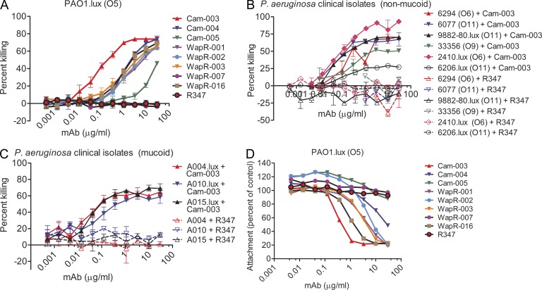
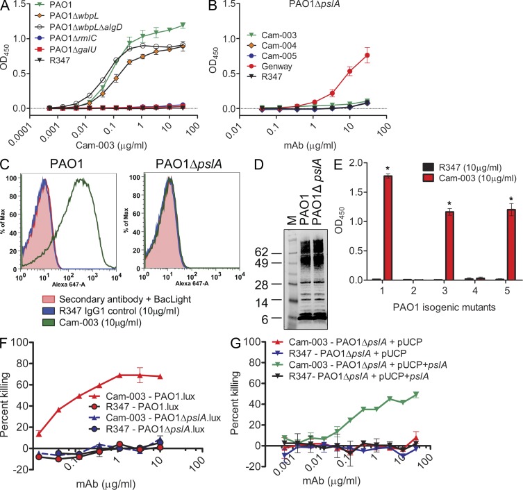
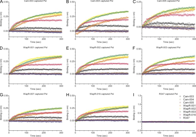
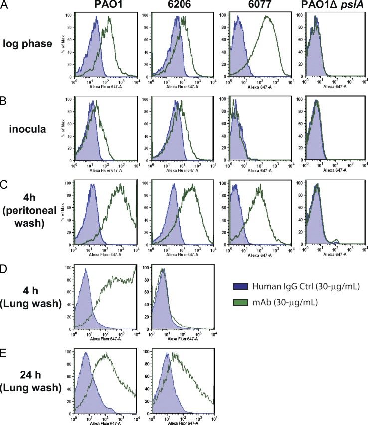
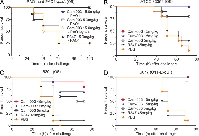
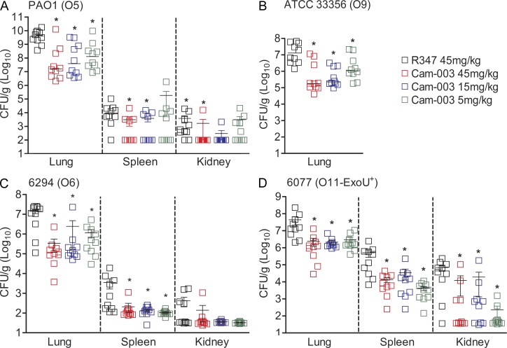
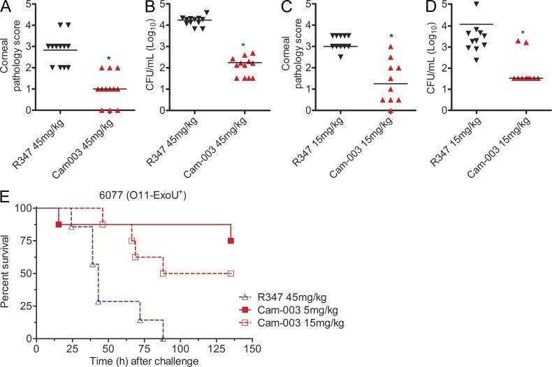
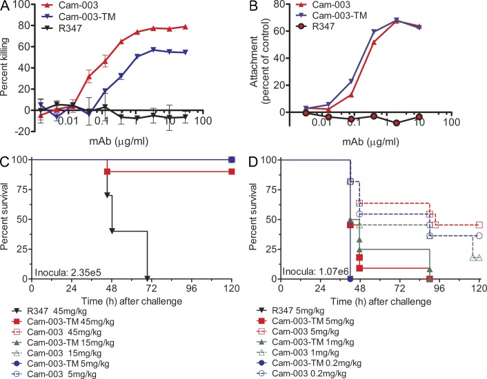
Pseudomonas aeruginosa is a leading cause of hospital-associated infections in the seriously ill, and the primary agent of chronic lung infections in cystic fibrosis patients. A major obstacle to effective control of P. aeruginosa infections is its intrinsic resistance to most antibiotic classes, which results from chromosomally encoded drug-efflux systems and multiple acquired resistance mechanisms selected by years of aggressive antibiotic therapy. These factors demand new strategies and drugs to prevent and treat P. aeruginosa infections. Herein, we describe a monoclonal antibody (mAb) selection strategy on whole P. aeruginosa cells using single-chain variable fragment phage libraries derived from healthy individuals and patients convalescing from P. aeruginosa infections. This approach enabled identification of mAbs that bind three distinct epitopes on the product of the Psl. This exopolysaccharide is important for P. aeruginosa attachment to mammalian cells, and for the formation and maintenance of biofilms produced by nonmucoid and mucoid P. aeruginosa isolates. Functional screens revealed that mAbs to one epitope exhibit superior activity in opsonophagocytic killing and cell attachment assays, and confer significant protection in multiple animal models. Our results indicate that Psl is an accessible serotype-independent surface feature and promising novel protective antigen for preventing P. aeruginosa infections. Furthermore, our mAb discovery strategy holds promise for application to other bacterial pathogens.


Figures

Figure 1.
Figure 1.
Functional activity screening of antibodies derived from phage scFv patient libraries. In vitro functional screens included OPK assays and cell attachment assays using the lung epithelial cell line A549. R347, an isotype-matched human mAb that does not bind P. aeruginosa antigens, was used as a negative control. (A) Opsonophagocytosis assay with P. aeruginosa serogroup O5 strain PAO1, which was engineered to be luminescent (PAO1.lux), with dilutions of purified mAbs derived from phage panning. (B) Opsonophagocytosis assay with Cam-003 and heterologous serotype P. aeruginosa nonmucoid clinical isolates. Strains 9882–80, 2410, and 6206 were engineered to be luminescent (lux). (C) Opsonophagocytosis assay with mucoid P. aeruginosa clinical isolates that were engineered to be luminescent (lux). (A–C) The lines represent the mean percentage of killing and error bars represent the standard deviation. Percent killing was calculated relative to results obtained in assays run in the absence of antibody. (D) Log-phase PAO1.lux were added to a confluent monolayer of human cell line A549 cells after the addition of antibody at a MOI of 10 followed by analysis of RLUs after repeated washing to remove unbound P. aeruginosa. Wells lacking antibody were used as the comparative control. (A–D) Results are representative data from three independent experiments performed for each antibody and P. aeruginosa isolate.
Figure 2.
Figure 2.
Identification of the P. aeruginosa Psl exopolysaccharide as the target of antibodies derived from phenotypic screening. (A and B) Reactivity of antibodies was determined by whole-cell ELISA on plates coated with indicated P. aeruginosa strains: (A) wild-type PAO1, PAO1ΔwbpL, PAO1ΔwbpLΔalgD, PAO1ΔrmlC, and PAO1ΔgalU, and (B) PAO1ΔpslA. In B, mAb (Genway Biosciences) specific to a P. aeruginosa outer membrane protein was used as a positive control. (C) FACS binding analysis of Cam-003 to PAO1 and PAO1ΔpslA. Cam-003 is indicated by a green line; an isotype matched non–P. aeruginosa–specific human IgG1 antibody was used as a negative control and is indicated by the blue line. Washed cells were stained with BacLight to differentiate live from dead cells. Staining with the secondary antibody alone plus BacLight was used as an additional control. (D) LPS purified from PAO1 and PAO1ΔpslA was resolved by SDS-PAGE and immunoblotted with antisera derived from mice vaccinated with PAO1ΔwapRΔalgD, a double mutant strain deficient in O-antigen transport to the outer membrane and alginate production. (E) Cam-003 ELISA binding data with isogenic mutants of PAO1. Mutant 1, PAO1ΔwbpLΔalgD; mutant 2, PAO1ΔwbpLΔalgDΔpslA; mutant 3, PAO1ΔwbpLΔalgDΔpelA; mutant 4, PAO1ΔwbpLΔalgDΔpslA + pUCP; mutant 5, PAO1ΔwbpLΔalgDΔpslA + pUCP+pslA. * indicates P < 0.005 using the Mann-Whitney U test when comparing Cam-003 versus R347 binding. (F and G) Opsonophagocytosis assay using Cam-003 and negative control R347 with indicated luminescent (lux) strains of P. aeruginosa (F) or strains complemented with pUCP+pslA (G). R347 was used as a negative control in all experiments. (A–C, F, and G). (A–G) Results are representative data from three independent experiments.
Figure 3.
Figure 3.
Anti-Psl antibody capture of enriched Psl isolated from whole P. aeruginosa cells. To confirm that all of the antibodies bound the same antigen, we developed a capture binding assay using an Octet platform with total carbohydrate extracts prepared from P. aeruginosa, which was deficient in O-antigen, alginate, and Pel polysaccharide production. In addition, the enriched carbohydrate extract was exhaustively digested with proteinase K. Antibodies were bound to aminopropylsilane sensors, followed by the addition of enriched carbohydrate. After washing, binding of the other indicated mAbs to mAb-captured carbohydrate (Psl exopolysaccharide) was assessed. (A–I) Data are representative results from three independent experiments.
Figure 4.
Figure 4.
Anti-Psl binding to P. aeruginosa passaged in vivo. To test if Psl expression is maintained in vivo, BALB/c mice were injected intraperitoneally with P. aeruginosa isolates, followed by harvesting of bacteria by peritoneal lavage 4 h after infection. The presence of Psl was analyzed with a control antibody and Cam-003 by FACS. (A) For the positive control, Cam-003 was assayed for binding to strains grown in vitro to log-phase from an overnight culture (∼5 × 10⁸/ml). (B) The inocula for each strain were prepared to 5 × 10⁸ CFU/ml, which were suspended from an overnight TSA plate grown to lawn and tested for reactivity to Cam-003. (C) 4 h after intraperitoneal challenge, bacteria was harvested from mice by peritoneal lavage and assayed for Cam-003 binding. (D) 4 h after intranasal challenge, bacteria was harvested from mice by BAL and assayed for Cam-003 binding. (E) 24 h after intranasal challenge, bacteria was harvested from mice by BAL and assayed for Cam-003 binding. Three animals were used in each group for the peritoneal lavage, and eight mice were used for BAL at each time point. (A–E) Results are representative data from five independent experiments.
Figure 5.
Figure 5.
Psl mAb Cam-003 protects mice against lethal challenge in a P. aeruginosa acute pneumonia model. In each experiment, BALB/c mice were treated with PBS (B–D) or antibody (Cam-003 or negative control R347; A–D) 24 h before intranasal infection with PAO1 (4.4e7 CFU) and PAO1ΔpslA (3e7 CFU; A), 33356 (3e7 CFU; B), 6294 (7e6 CFU; C), or 6077 (1e6 CFU; D). Animals were monitored for survival between 72 and 120 h after infection. In all experiments, PBS and R347 served as negative controls. (A) A PBS control was not tested in this experiment because previous results indicated no difference in survival versus mice treated with R347. In its place, we used challenge with PAO1ΔpslA as an additional control. Results are represented as Kaplan-Meier survival curves; differences in survival were calculated by the log-rank test for Cam-003 versus R347. (A) Cam-003 (15 mg/kg, P = 0.0028; 5 mg/kg, P = 0.0028); (B) Cam-003 (45 mg/kg, P = 0.0012; 15 mg/kg, P = 0.0012; 5 mg/kg, P = 0.0373); (C) Cam-003 (45 mg/kg, P = 0.0007; 15 mg/kg, P = 0.0019; 5 mg/kg, P = 0.0212); (D) Cam-003 (45 mg/kg, P < 0.0001; 15 mg/kg, P < 0.0001; 5 mg/kg, P = 0.0001). 10 animals were used in each group. Results are representative data from three (A and D) and two independent experiments (B and C).
Figure 6.
Figure 6.
Anti-Psl mAb, Cam-003, reduces organ burden after induction of acute pneumonia. BALB/c mice were treated with antibody 24 h before infection with PAO1 (1.1e7 CFU; A), 33356 (1e7 CFU; B), 6294 (6.25e6; C), and 6077 (1e6 CFU; D). 24 h after infection, animals were euthanized, followed by harvesting of organs for identification of viable CFUs. Differences in viable CFU were determined by the Mann-Whitney U test for Cam-003 versus R347. (A) Lung, Cam-003 (45 mg/kg, P = 0.0015; 15 mg/kg, P = 0.0021; 5 mg/kg, P = 0.0015); spleen, Cam-003 (45 mg/kg, P = 0.0120; 15 mg/kg, P = 0.0367); kidneys, Cam-003 (45 mg/kg, P = 0.0092; 15 mg/kg, P = 0.0056; (B) lung, Cam-003 (45 mg/kg, P = 0.0010; 15 mg/kg, P < 0.0001; 5 mg/kg, P = 0.0045); (C) lung, Cam-003 (45 mg/kg, P = 0.0003; 15 mg/kg, P = 0.0039; 5 mg/kg, P = 0.0068); spleen, Cam-003 (45 mg/kg, P = 0.0057; 15 mg/kg, P = 0.0230; 5 mg/kg, P = 0.0012); (D) lung, Cam-003 (45 mg/kg, P = 0.0005; 15 mg/kg, P = 0.0003; 5 mg/kg, P = 0.0007); spleen, Cam-003 (45 mg/kg, P = 0.0015; 15 mg/kg, P = 0.0089; 5 mg/kg, P = 0.0089); kidneys, Cam-003 (45 mg/kg, P = 0.0191; 15 mg/kg, P = 0.0355; 5 mg/kg, P = 0.0021). (A–D) The lungs, spleen, and kidneys from 10 animals (for each antibody-treated group) were used. Results are representative data from two independent experiments for each P. aeruginosa strain.
Figure 7.
Figure 7.
Anti-Psl mAb Cam-003 is active in P. aeruginosa keratitis and thermal injury models. C3H/HeN mice were treated with control R347 antibody or Cam-003 at 45 mg/kg (A and B) or 15 mg/kg (C and D) 24 h before infection with 6077 (O11-cytotoxic, 2e6 CFU). The corneal infection model was performed as previously described (DiGiandomenico et al., 2007). Differences in pathology scores and viable CFU were determined by the Mann-Whitney U test. P = 0.0001 (A); P < 0.0001 (B); P = 0.0003 (C); P = 0.0015 (D). (E) The thermal injury model was performed using CD-1 mice as previously described (DiGiandomenico et al., 2007). Survival analysis from Cam-003– and R347-treated CF-1 mice in a P. aeruginosa thermal injury model after 6077 infection (2 × 10⁵ CFU; log-rank: R347 vs. Cam-003 15 mg/kg, P = 0.0094; R347 vs. Cam-003 5 mg/kg, P = 0.0017). (A–D) 12 animals were used in each group. (E) Number of animals in each treatment group (R347, n = 7; Cam-003 5 mg/kg, n = 8; Cam-003 15 mg/kg, n = 8). Results are representative data from five (A–D) and from three independent experiments (E).
Figure 8.
Figure 8.
A Cam-003 Fc mutant antibody, Cam-003-TM, has diminished OPK and in vivo efficacy but maintains anti-cell attachment activity. (A) PAO1.lux OPK assay with Cam-003 and Cam-003-TM, which harbors mutations in the Fc domain that reduces Fc interactions with Fcγ receptors (Oganesyan et al., 2008). R347 was used as a negative control. (B) PAO1 cell attachment assay with Cam-003 and Cam-003-TM. Wells lacking antibody was used as the comparative control. (C, D) P. aeruginosa strain 6077 acute pneumonia model using BALB/c mice inoculated with (C) 2.35e5 or (D) 1.07e6 comparing efficacy of Cam-003 versus Cam-003-TM. Mice were treated with antibody 24 h before challenge. (C-D) Ten animals were used in each group. Results are representative data from (A) three independent experiments. (B-D) two independent experiments.

Similar articles

Cited by

References

    1. Beerli R.R., Bauer M., Buser R.B., Gwerder M., Muntwiler S., Maurer P., Saudan P., Bachmann M.F. 2008. Isolation of human monoclonal antibodies by mammalian cell display. Proc. Natl. Acad. Sci. USA. 105:14336–14341 10.1073/pnas.0805942105 - DOI - PMC - PubMed
    1. Borlee B.R., Goldman A.D., Murakami K., Samudrala R., Wozniak D.J., Parsek M.R. 2010. Pseudomonas aeruginosa uses a cyclic-di-GMP-regulated adhesin to reinforce the biofilm extracellular matrix. Mol. Microbiol. 75:827–842 10.1111/j.1365-2958.2009.06991.x - DOI - PMC - PubMed
    1. Buchwald U.K., Pirofski L. 2003. Immune therapy for infectious diseases at the dawn of the 21st century: the past, present and future role of antibody therapy, therapeutic vaccination and biological response modifiers. Curr. Pharm. Des. 9:945–968 10.2174/1381612033455189 - DOI - PubMed
    1. Burns J.L., Gibson R.L., McNamara S., Yim D., Emerson J., Rosenfeld M., Hiatt P., McCoy K., Castile R., Smith A.L., Ramsey B.W. 2001. Longitudinal assessment of Pseudomonas aeruginosa in young children with cystic fibrosis. J. Infect. Dis. 183:444–452 10.1086/318075 - DOI - PubMed
    1. Byrd M.S., Sadovskaya I., Vinogradov E., Lu H., Sprinkle A.B., Richardson S.H., Ma L., Ralston B., Parsek M.R., Anderson E.M., et al. 2009. Genetic and biochemical analyses of the Pseudomonas aeruginosa Psl exopolysaccharide reveal overlapping roles for polysaccharide synthesis enzymes in Psl and LPS production. Mol. Microbiol. 73:622–638 10.1111/j.1365-2958.2009.06795.x - DOI - PMC - PubMed

Publication types

MeSH terms