. 2012 Jun 26:8:91.
doi: 10.1186/1746-6148-8-91.

The complement system of the goat: haemolytic assays and isolation of major proteins

Affiliations

The complement system of the goat: haemolytic assays and isolation of major proteins

Isabel Moreno-Indias et al. BMC Vet Res. .

Abstract

Background: The aim of the present study was to develop a haemolytic assay for the study of the complement system in dairy goats (Capra aegagrus hircus) and to characterize the major goat complement system proteins.

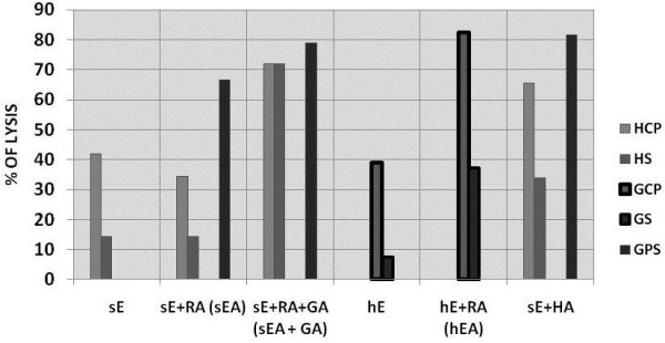
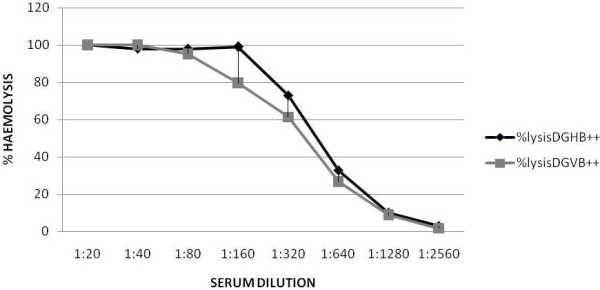
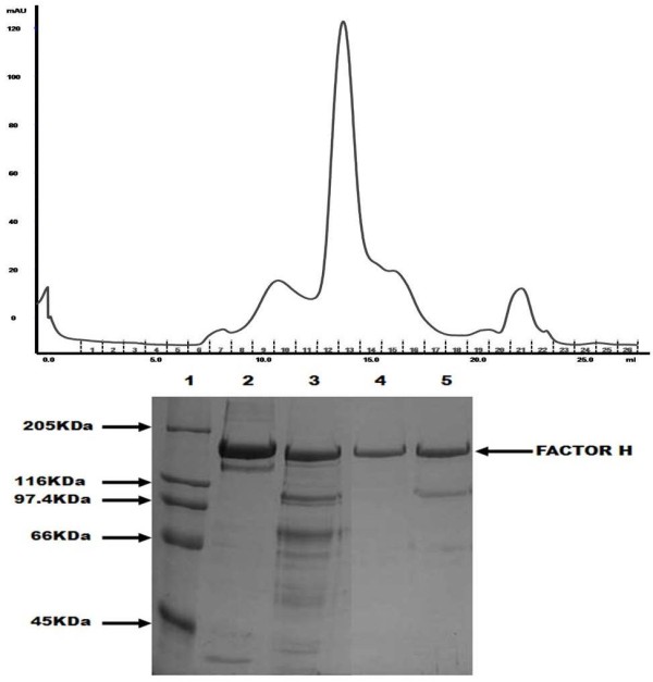
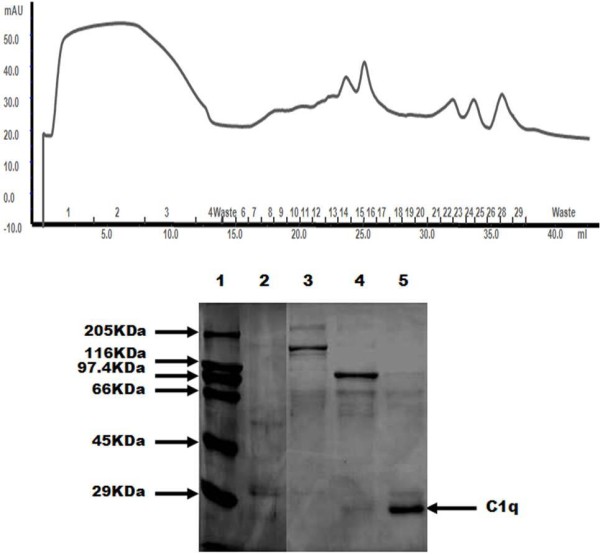
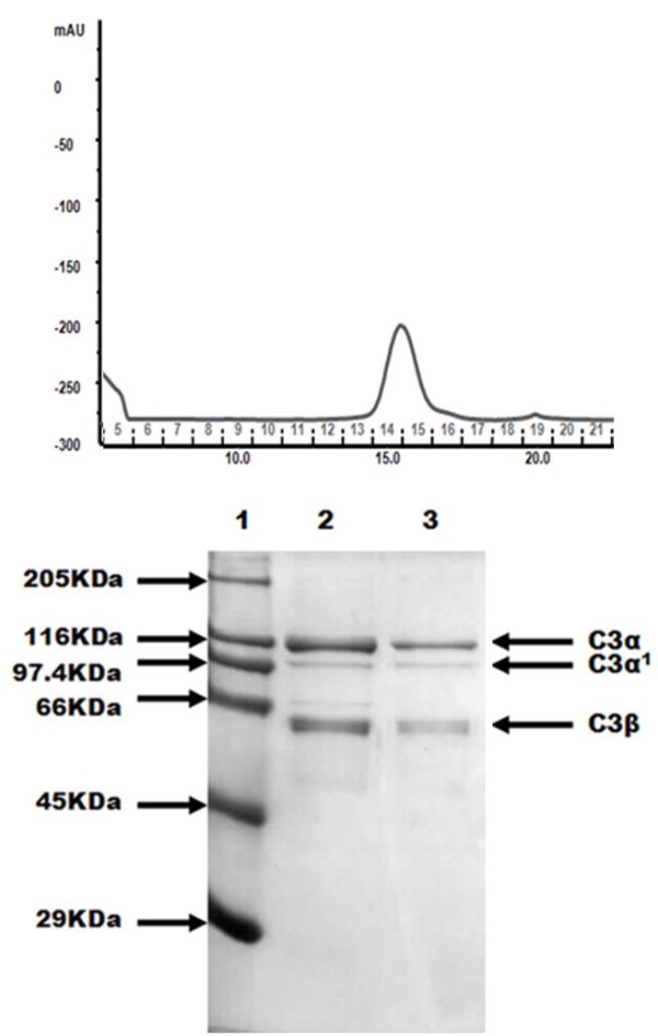
Results: The commonly used sheep erythrocyte sensitized with rabbit antibodies were not sensitive to lysis by goat serum, but the combination of human red blood cells (RBC) plus rabbit antibodies was the best option found for goat complement assay. A buffer based on HEPES instead of the classical veronal (barbitone) was developed. Three proteins were isolated: factor H, C1q and C3 and these were compared with the corresponding human proteins. A novel affinity chromatography technique was developed for isolation of factor H.

Conclusions: Human RBC plus rabbit antibodies were a suitable option for haemolytic assays. The isolated proteins are similar to the human counterparts.


Figures

Figure 1
Figure 1
ca="yes"Lysis of different cells, with and without antibody by human, goat and guinea-pig plasma/serum (HCP and HS, GCP and GS, and GPS, respectively). Sheep (sE) and human (hE) erythrocytes were pre-coated or not with rabbit or rabbit + goat antibodies. Erythrocytes (50 μl of 109/ml) in DGVB++ were incubated with 100 μl of 1/5 dilution of plasma/serum for 1 hour at 37 °C. Percentage of cell lysis was calculated as indicated in methods section.sE, hE: sheep and human erythrocytes without pre-coated; sEA, hEA = sheep and human pre-coated with rabbit antibodies; RA, GA, HA = rabbit, goat and human antibodies, respectively.
Figure 2
Figure 2
Comparison of HEPES (DGHB++) and Veronal (DGVB++) buffers. Serial 2-fold dilutions of human serum were prepared in either buffer, starting at 1/20 (5%). Dilutions (100 μl) were mixed with 100 μl of 108/ml sheep EA cells, in the same buffer, and incubated for 1hour, at 37 °C. Percentage lysis was calculated as in methods.
Figure 3
Figure 3
Titration of sensitising antibody and effects of “classical pathway”versus“alternative pathway” condition. Human RBC (E) were used without sensitization or sensitised with 10 μl anti(human RBC) antiserum per ml of 109 cells/ml (EA1) or with 100 μl antiserum per ml of 109 cells/ml (EA2). Two-fold serial dilutions of goat serum were made in either DGHB++ or DGHB-Mg-EGTA, starting at 1:1 (50%). Dilutions (100 μl) were incubated with E, EA1 or EA2 cells (100 μl of 108/ml) in the same buffer for 1hour at 37 °C. Percentage lysis was calculated, and the serum concentration (% serum) required for 50% lysis calculated. CH50 (the number of complement lysis units in 100% serum).
Figure 4
Figure 4
Comparison of multiple samples. Ten goat serum samples were assayed (as described in the legend for fig. 3) using DGHB++ and DGHB-Mg-EGTA buffers, and highly sensitised EA (equivalent to EA2 in fig. 3). The titre of each sample CH50 units in the assay is shown for both buffer conditions.
Figure 5
Figure 5
Isolation of Goat Factor H. a) Gel flitration of goat FH on Superose 6. b) SDS-PAGE analysis of reduced goat FH fractions. lane 1: Molecular weight standard, lane 2: human Factor H, lane 3: Factor H before the gel filtration, lane 4: fraction 12 from the gel filtration, lane 5: fraction 13 from the gel filtration. Gel was stained with Coomassie blue.
Figure 6
Figure 6
Purification of goat C1q. a) Ion Exchange (monoS) chromatogram of C1q. b) SDS-PAGE analysis of reduced goat C1q fractions. Lane 1: Molecular weight standard, lane 2: human C1q, (with a contaminant ~50 kDa), lane 3: fraction 16 from the ion exchange, lane 4: fraction 22, lane 5: fraction 24. Fractions were concentrated with Strataclean beads. Gel was stained with Coomassie blue.
Figure 7
Figure 7
Isolation of C3. a) Gel filtration of goat C3 on Superose 6. b) SDS-PAGE of the final step gel filtration on Superose 6. Lane 1: Molecular weight standard, lane 2 and 3: fractions of the gel filtration (14, 15). Fractions were concentrated with Strataclean beads. Gel was stained with Coomassie blue.

Similar articles

Cited by

References

    1. Mayilyan KR, Kang YH, Dodds AW, Sim RB. In: Innate Immunity of Plants, Animals and Humans. Heine H, editor. Berlín: Springer-Verlag Berlin Heidelberg; 2008. The complement system in innate immunity; pp. 219–236. 21.
    1. Walport MJ. Complement: first of two parts. N Engl J Med. 2001;344:1058–1066. doi: 10.1056/NEJM200104053441406. - DOI - PubMed
    1. Rainard P. The complement in milk and defense of the bovine mammary gland against infections. Vet Res. 2003;34:647–670. doi: 10.1051/vetres:2003025. - DOI - PubMed
    1. Seelen MA, Roos A, Wieslander J, Mollnes TE, Sjoholm AG, Wurzner R, Loos M, Tedesco F, Sim RB, Garred P, Alexopoulos E, Turner MW, Daha MR. Functional analysis of the classical, alternative, and MBL pathways of the complement system: standardization and validation of a simple ELISA. J Immunol Methods. 2005;296:187–198. doi: 10.1016/j.jim.2004.11.016. - DOI - PubMed
    1. Matsushita M, Endo Y, Fujita T. Cutting edge: complement-activating complex of ficolin and mannose-binding lectin-associated serine protease. J Immunol. 2000;164:2281–2284. - PubMed

Publication types

LinkOut - more resources