

Investigating ligand-receptor interactions at bilayer surface using electronic absorption spectroscopy and fluorescence resonance energy transfer
- PMID: 22734511
- PMCID: PMC3439585
- DOI: 10.1021/la300724z
Investigating ligand-receptor interactions at bilayer surface using electronic absorption spectroscopy and fluorescence resonance energy transfer
Abstract
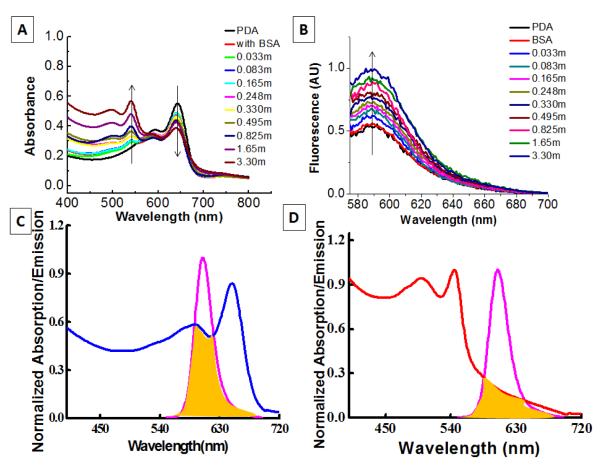
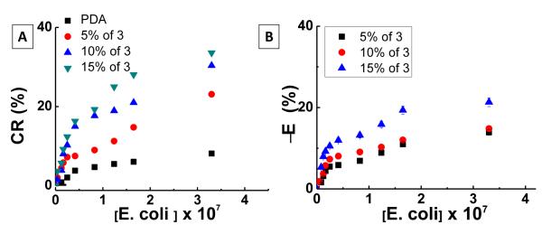
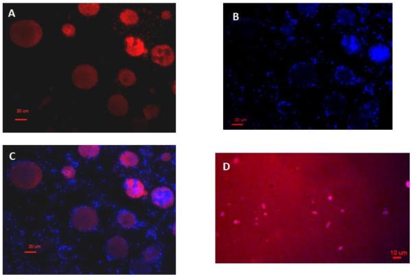
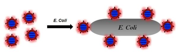
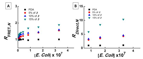
We investigate interactions between receptors and ligands at bilayer surface of polydiacetylene (PDA) liposomal nanoparticles using changes in electronic absorption spectroscopy and fluorescence resonance energy transfer (FRET). We study the effect of mode of linkage (covalent versus noncovalent) between the receptor and liposome bilayer. We also examine the effect of size-dependent interactions between liposome and analyte through electronic absorption and FRET responses. Glucose (receptor) molecules were either covalently or noncovalently attached at the bilayer of nanoparticles, and they provided selectivity for molecular interactions between glucose and glycoprotein ligands of E. coli. These interactions induced stress on conjugated PDA chain which resulted in changes (blue to red) in the absorption spectrum of PDA. The changes in electronic absorbance also led to changes in FRET efficiency between conjugated PDA chains (acceptor) and fluorophores (Sulphorhodamine-101) (donor) attached to the bilayer surface. Interestingly, we did not find significant differences in UV-vis and FRET responses for covalently and noncovalently bound glucose to liposomes following their interactions with E. coli. We attributed these results to close proximity of glucose receptor molecules to the liposome bilayer surface such that induced stress were similar in both the cases. We also found that PDA emission from direct excitation mechanism was ~2-10 times larger than that of the FRET-based response. These differences in emission signals were attributed to three major reasons: nonspecific interactions between E. coli and liposomes, size differences between analyte and liposomes, and a much higher PDA concentration with respect to sulforhodamine (SR-101). We have proposed a model to explain our experimental observations. Our fundamental studies reported here will help in enhancing our knowledge regarding interactions involved between soft particles at molecular levels.
Figures
References
-
- Karlsson KA. Microbial recognition of target-cell glycoconjugates Curr. Opin. Struct. Biol. 1995;5:622. - PubMed
-
- Guo CX, Boullanger P, Liu T, Jiang L. Size effect of polydiacetylene vesicles functionalized with glycolipids on their colorimetric detection ability. J. Phys. Chem. B. 2005;109:18765–18771. - PubMed
-
- Sharon N, Lis H. Lectins as cell recognition molecules. Science. 1989;246:227. - PubMed
-
- Spevak W, Nagy JO, Charych DH. Molecular assemblies of functionalized polydiacetylenes. Adv. Mater. 1995;7:85–89.
-
- Kim TH, Swager TM. A fluorescent self-amplifying wavelength-responsive sensory polymer for fluoride ions. Angew. Chem. Int. Ed. 2003;42:4803. - PubMed
Publication types
MeSH terms
Substances
Grants and funding
LinkOut - more resources
Full Text Sources
Other Literature Sources
Research Materials
