. 2012 Jun 26:7:44.
doi: 10.1186/1750-1172-7-44.

Molecular genetic studies and delineation of the oculocutaneous albinism phenotype in the Pakistani population

Affiliations

Molecular genetic studies and delineation of the oculocutaneous albinism phenotype in the Pakistani population

Thomas J Jaworek et al. Orphanet J Rare Dis. .

Abstract

Background: Oculocutaneous albinism (OCA) is caused by a group of genetically heterogeneous inherited defects that result in the loss of pigmentation in the eyes, skin and hair. Mutations in the TYR, OCA2, TYRP1 and SLC45A2 genes have been shown to cause isolated OCA. No comprehensive analysis has been conducted to study the spectrum of OCA alleles prevailing in Pakistani albino populations.

Methods: We enrolled 40 large Pakistani families and screened them for OCA genes and a candidate gene, SLC24A5. Protein function effects were evaluated using in silico prediction algorithms and ex vivo studies in human melanocytes. The effects of splice-site mutations were determined using an exon-trapping assay.

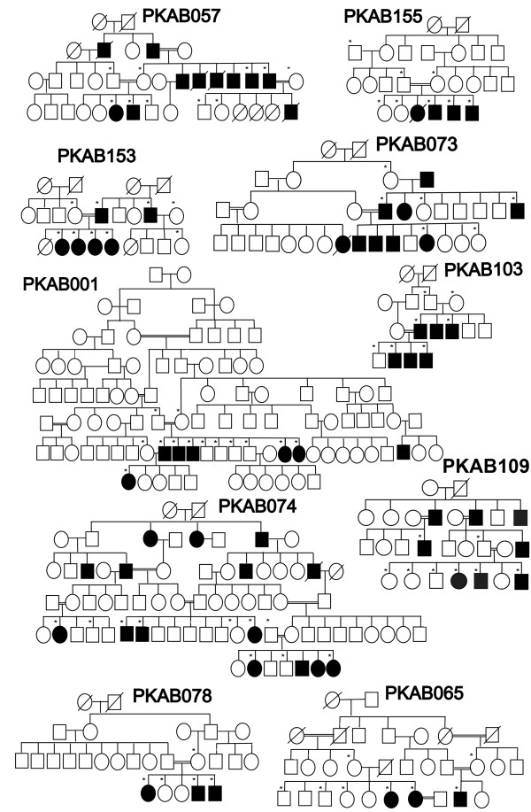
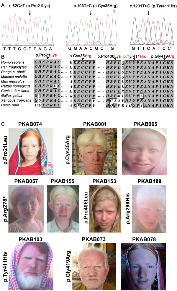
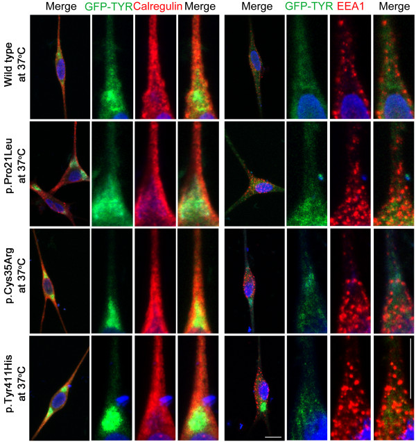
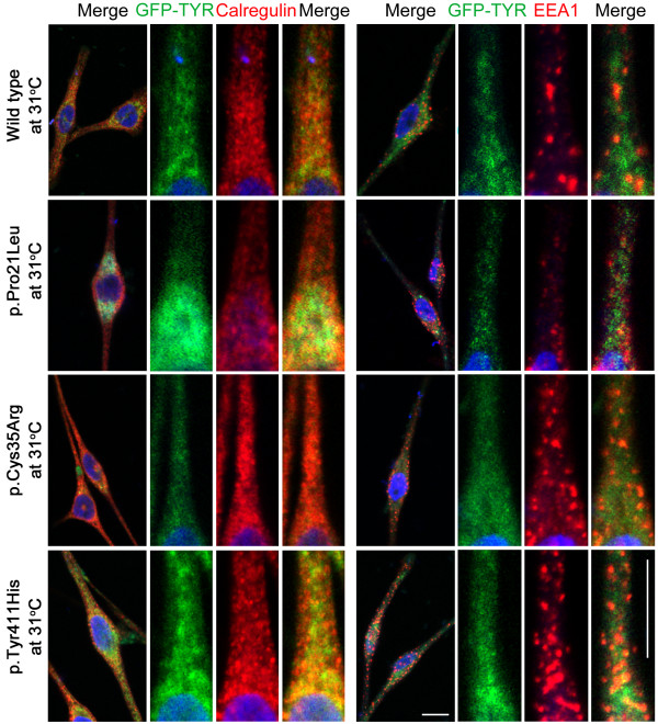
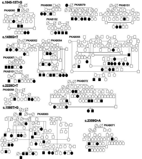
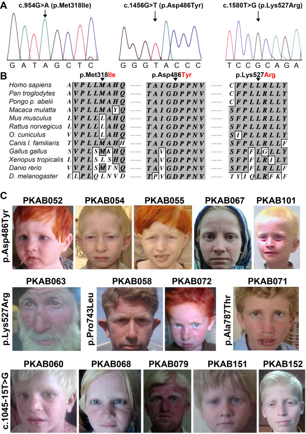
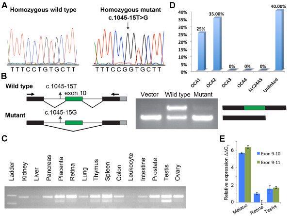
Results: Screening of the TYR gene revealed four known (p.Arg299His, p.Pro406Leu, p.Gly419Arg, p.Arg278*) and three novel mutations (p.Pro21Leu, p.Cys35Arg, p.Tyr411His) in ten families. Ex vivo studies revealed the retention of an EGFP-tagged mutant (p.Pro21Leu, p.Cys35Arg or p.Tyr411His) tyrosinase in the endoplasmic reticulum (ER) at 37°C, but a significant fraction of p.Cys35Arg and p.Tyr411His left the ER in cells grown at a permissive temperature (31°C). Three novel (p.Asp486Tyr, p.Leu527Arg, c.1045-15 T > G) and two known mutations (p.Pro743Leu, p.Ala787Thr) of OCA2 were found in fourteen families. Exon-trapping assays with a construct containing a novel c.1045-15 T > G mutation revealed an error in splicing. No mutation in TYRP1, SLC45A2, and SLC24A5 was found in the remaining 16 families. Clinical evaluation of the families segregating either TYR or OCA2 mutations showed nystagmus, photophobia, and loss of pigmentation in the skin or hair follicles. Most of the affected individuals had grayish-blue colored eyes.

Conclusions: Our results show that ten and fourteen families harbored mutations in the TYR and OCA2 genes, respectively. Our findings, along with the results of previous studies, indicate that the p.Cys35Arg, p.Arg278* and p.Gly419Arg alleles of TYR and the p.Asp486Tyr and c.1045-15 T > G alleles of OCA2 are the most common causes of OCA in Pakistani families. To the best of our knowledge, this study represents the first documentation of OCA2 alleles in the Pakistani population. A significant proportion of our cohort did not have mutations in known OCA genes. Overall, our study contributes to the development of genetic testing protocols and genetic counseling for OCA in Pakistani families.


Figures

Figure 1
Figure 1
Pedigrees of Pakistani families carrying TYR mutations. Pedigrees of ten multi-generational families segregating recessive nonsyndromic OCA due to mutations in the TYR gene. Filled and empty symbols represent affected and unaffected individuals, respectively. Double lines indicate consanguineous marriages. Asterisks indicate subjects enrolled in the protocol that contributed DNA samples.
Figure 2
Figure 2
Novel TYR mutations and resulting OCA1 phenotypes. A. Electropherograms of amplimers from genomic DNA templates illustrating homozygosity for the substitution mutations found in the affected individuals of the families. Arrows indicate the site of the mutations. All of the mutations described here are numbered from the ATG start codon (GenBank NM_000372). B. Clustal W alignment of tyrosinase proteins from various species that shows the conservation of residues at positions 21, 35 and 411 among ten species. The conserved amino acids are shown with a dark gray background, and the nonconserved amino acids are shown with a white background. C. Photographs of ten OCA1 probands. The family number and the mutation identified in the TYR gene are given for each proband; some of the probands have used hair dyes.
Figure 3
Figure 3
Subcellular distribution of wild-type and mutant tyrosinase in human melanocytes grown at 37°C. Subcellular distribution of wild-type and mutant (p.Pro21Leu, p.Cys35Arg and p.Tyr411His) tyrosinase proteins (green) in transiently transfected human melanocytes grown at 37°C. Calregulin and EEA1 were used as markers for the endoplasmic reticulum (red) and the early endosome (red), respectively. For each construct, boxed regions were magnified in the adjacent panels. Merged images show the co-localization of the p.Pro21Leu, p.Cys35Arg and p.Tyr411His tyrosinase mutations with calregulin, which indicates ER retention. The scale bar represents 10 μm for all panels.
Figure 4
Figure 4
Subcellular distribution of wild-type and mutant tyrosinase in human melanocytes grown at 31°C. The subcellular localization of GFP-tyrosinase in wild-type and p.Pro21Leu-transfected cells was not significantly different when cells were incubated at 31°C or at 37°C. After transfection with either p.Cys35Arg or p.Tyr411His mutant constructs, the melanocytes grown at 31°C showed an increase in the cytoplasmic vesicular co-localization of the mutant protein with EEA1. For each construct, boxed regions were magnified in the adjacent panels. The scale bar represents 10 um for all panels.
Figure 5
Figure 5
Allele frequency of the rs1042602 cSNP in the Pakistani population. The distribution of an ancestral C and derived A allele of TYR among Pakistani population. DNA samples from 200 individuals belonging to various ethnic groups within province of Punjab, Pakistan were genotyped for rs1042602. Also shown is the Human Genome Diversity Project data for comparison [55]. Details are available at the HGDP website (http://hgdp.uchicago.edu/).
Figure 6
Figure 6
Pedigrees of Pakistani families segregating OCA2 mutations. Pedigrees of fourteen multi-generational families with mutations identified in the OCA2 gene. Filled and empty symbols represent affected and unaffected individuals, respectively. Double lines indicate consanguineous marriages. Asterisks indicate subjects enrolled in the protocol that contributed DNA samples.
Figure 7
Figure 7
Novel OCA2 mutations and resulting OCA2 phenotypes. A.Electropherograms of amplimers from genomic DNA templates illustrating homozygosity for the substitution mutations found in the affected individuals of the families. Arrows indicate the site of the mutations. All mutations described here are numbered from the ATG start codon (GenBank NM_000275). B. Clustal W alignment of OCA2 proteins from various species shows conservation of the residues at positions 318, 486 and 527 among twelve species. The conserved amino acids are shown with a dark gray background, and the nonconserved amino acids are shown with a white background. C. Photographs of fourteen OCA2 probands. The family number and the mutation identified in the OCA2 gene are given for each proband; a number of the probands shown have used hair dyes.
Figure 8
Figure 8
Functional analysis of c.1045-15 T > G mutation. A. Electropherograms of amplimers from genomic DNA templates illustrating homozygosity for the wild-type and c.1045-15 T > G substitution mutation found in the affected individuals of the five OCA2 families. The arrow indicates the site of the mutation. B. To determine the effect of the c.1045-15 T > G mutation on splicing, exon 10 with 200 bp of the flanking intron of OCA2 was introduced into the pSPL3 vector and analyzed through an in vitro splicing assay. The transfected empty pSPL3 vector produced the expected product of 177 bp, whereas the wild-type exon 10 splice site produced two bands with (249 bp) and without (177 bp) exon-10 splicing when amplified with vector primers, which might indicate the presence of weak splice junctions around exon 10. The construct with the c.1045-15 T > G mutation produced a band of 177 bp, which upon sequencing, revealed the skipping of exon 10. With the mutant construct, a weak band of ~249 bp was occasionally observed, but sequencing revealed an aberrant splice product. C. Human OCA2 isoforms with and without exon 10 are expressed in many tissues. D. Molecular genetic analysis of known OCA genes in a cohort of forty Pakistani families indicates that (a) OCA2 mutations are the most common cause of OCA, and (b) a significant number of families do not have mutations in the known OCA genes. E. Real-time quantitative RT–PCR analysis of TYR mRNAs level in human melanocytes, retina and testis cDNA libraries. CT is the observed threshold number of PCR cycles required for detection of the amplification product; ΔCT is the calculated difference in CT between the TYR gene and an internal control standard (GAPDH) measured in the same sample. ΔΔCT is the calculated difference in ΔCT between the experimental and exon 9–11 isoform in retina. Compared to the retina and testis, melanocytes have a relatively high expression of both exon 9–10 and exon 9–11 isoforms of TYR.

Similar articles

Cited by

References

    1. Tomita Y, Suzuki T. Genetics of pigmentary disorders. Am J Med Genet C Semin Med Genet. 2004;131C:75–81. doi: 10.1002/ajmg.c.30036. - DOI - PubMed
    1. Li W, He M, Zhou H, Bourne JW, Liang P. Mutational data integration in gene-oriented files of the Hermansky-Pudlak Syndrome database. Hum Mutat. 2006;27:402–407. doi: 10.1002/humu.20309. - DOI - PubMed
    1. Seven M, Yosunkaya E, Yilmaz SB, Karaca E, Guven G, Yuksel A. A new syndrome presenting with dysmorphic facies, oculocutaneous albinism, glaucoma, cryptorchidism and mental retardation. Genet Couns. 2011;22:25–34. - PubMed
    1. Budisteanu M, Arghir A, Chirieac SM, Cardos G, Lungeanu A. Oculocutaneous albinism associated with multiple malformations and psychomotor retardation. Pediatr Dermatol. 2010;27:212–214. doi: 10.1111/j.1525-1470.2010.01115.x. - DOI - PubMed
    1. Tripathi RK, Hearing VJ, Urabe K, Aroca P, Spritz RA. Mutational mapping of the catalytic activities of human tyrosinase. J Biol Chem. 1992;267:23707–23712. - PubMed

Publication types

Substances

LinkOut - more resources