. 2012 Jun 26:3:925.
doi: 10.1038/ncomms1927.

ABCA4 is an N-retinylidene-phosphatidylethanolamine and phosphatidylethanolamine importer

Affiliations

ABCA4 is an N-retinylidene-phosphatidylethanolamine and phosphatidylethanolamine importer

Faraz Quazi et al. Nat Commun. .

Abstract

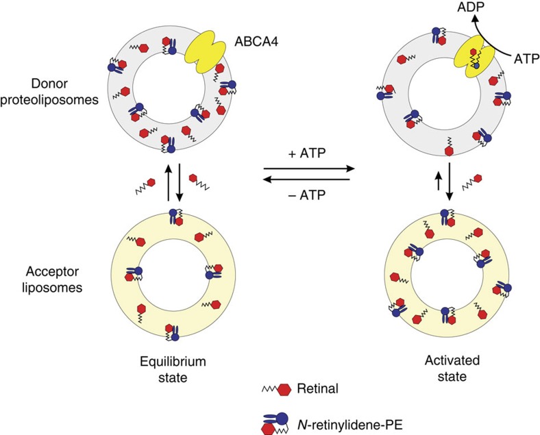
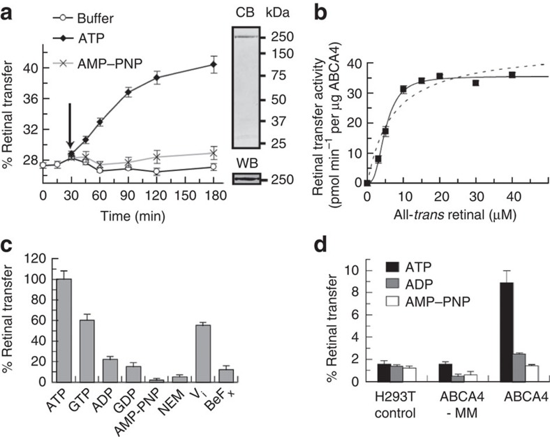
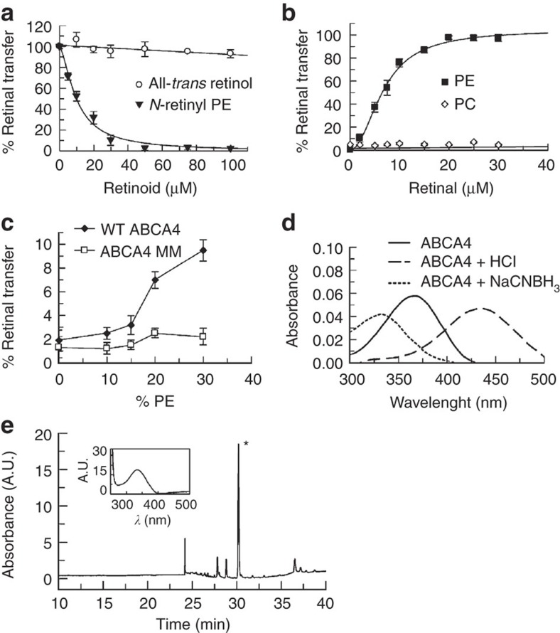
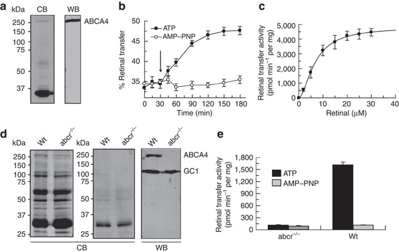
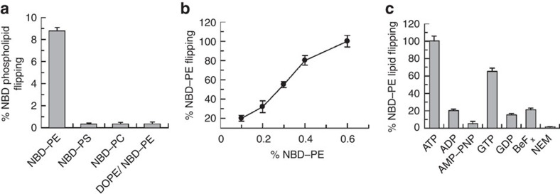
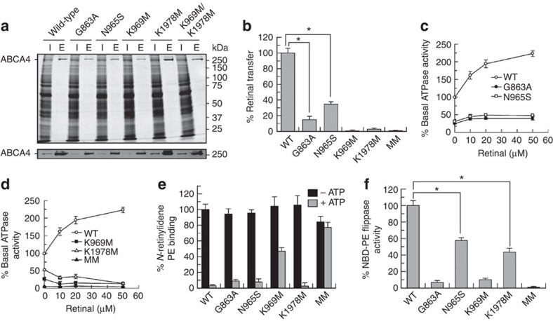
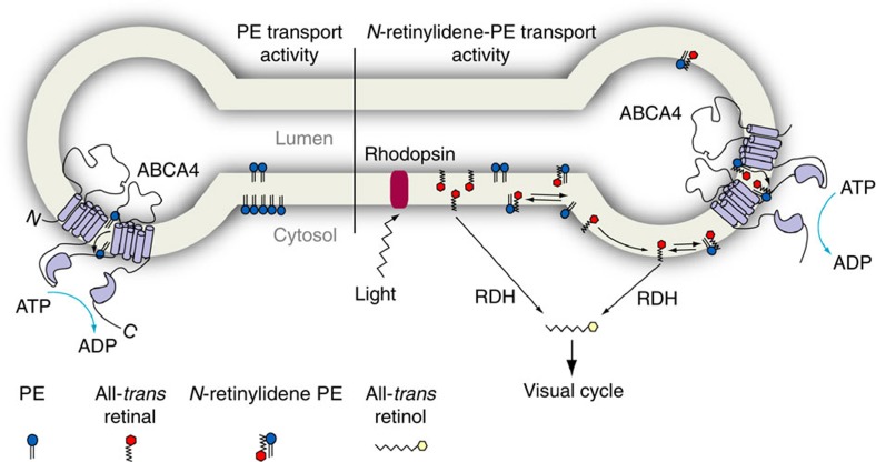
ATP-binding cassette (ABC) transporters comprise a superfamily of proteins, which actively transport a variety of compounds across cell membranes. Mammalian and most eukaryotic ABC transporters function as exporters, flipping or extruding substrates from the cytoplasmic to the extracellular or lumen side of cell membranes. Prokaryotic ABC transporters function either as exporters or importers. Here we show that ABCA4, an ABC transporter found in retinal photoreceptor cells and associated with Stargardt macular degeneration, is a novel importer that actively flips N-retinylidene-phosphatidylethanolamine from the lumen to the cytoplasmic leaflet of disc membranes, thereby facilitating the removal of potentially toxic retinoid compounds from photoreceptors. ABCA4 also actively transports phosphatidylethanolamine in the same direction. Mutations known to cause Stargardt disease decrease N-retinylidene-phosphatidylethanolamine and phosphatidylethanolamine transport activity of ABCA4. These studies provide the first direct evidence for a mammalian ABC transporter that functions as an importer and provide insight into mechanisms underlying substrate transport and the molecular basis of Stargardt disease.


Figures

Figure 1
Figure 1. Schematic diagram showing the rationale for ABCA4-catalysed retinoid transport activity between donor and acceptor vesicles.
In the absence of ATP (equilibrium state), retinal rapidly equilibrates between the donor proteoliposomes (or disc membranes) containing ABCA4 and acceptor liposomes. The distribution of retinal in each population is dependent on their phospholipid composition. Retinal freely diffuses in the lipid bilayer and reacts with PE to form an equilibrium mixture with N-retinylidene-PE, which is distributed on both sides of the lipid bilayer. Free PE is not shown for simplicity. Addition of ATP to the NBDs of ABCA4 results in the active transport or flipping of N-retinylidene-PE from the lumen side of the bilayer to the outside (equivalent to the cytoplasmic) of the bilayer resulting in an accumulation of N-retinylidene-PE on the outer leaflet. This increase coupled with its dissociation to PE and retinal results in a higher concentration of retinal on the outer leaflet and a net transfer of retinal to the acceptor liposomes. This can be measured by determining the accumulation of radiolabelled retinoid in the liposome after separation from donor proteoliposomes by centrifugation. The density of the liposomes is increased by encapsulated sucrose thereby facilitating separation by centrifugation. If ATP is removed such as by chelation of Mg2+ with EDTA or ABCA4 is inactivated by an inhibitor, retinal flows back to the proteoliposomes to generate an equilibrium state similar to the initial state.
Figure 2
Figure 2. ATP-dependent transfer of ATR from donor ABCA4 proteoliposomes to acceptor liposomes.
Donor proteoliposomes consisted of DOPC, DOPE and BPL at a weight ratio of 6:2:2 reconstituted with ABCA4 purified from either (ac) bovine ROS membranes or (d) ABCA4-transfected HEK293 cell extracts. Acceptor liposomes consisted of DOPC and DOPE at a weight ratio of 7:3. (a) Time course for the transfer of [3H] ATR from donor proteoliposomes to acceptor liposome vesicles. [3H] ATR rapidly distributed between donor proteoliposomes and acceptor liposomes with ~28% of the ATR partitioning into the acceptor liposomes at 37 °C. After 30 min, 2 mM ATP or AMP–PNP or control buffer was added (arrow) and radioactivity in the acceptor liposomes was measured at various times. Data are plotted as a mean with error bars representing±s.e.m. for n=3. Coomassie blue (CB)-stained gel and western blot (WB) shows the purity of the ABCA4 used for reconstitution. (b) ATP-dependent rate of [3H] ATR transfer as a function of ATR concentration. Solid line shows best-fit sigmoidal curve for K0.5 of 4.9±0.2 μM and a Vmax of 35.5±0.7 pmol min−1 per μg protein yielding a Hill coefficient of 2.3±0.3. Dotted line shows a best-fit Michaelis–Menten curve for comparison. (c) The effect of nucleotides (1 mM) and inhibitors (10 mM NEM, 1 mM orthovanadate (Vi) or 0.2 mM beryllium fluoride (BeFx)) on [3H] ATR transfer from proteoliposomes to liposomes. Percent relative retinal transfer activity data displayed as a mean with error bars representing±s.e.m for n=3. (d) Effect of ATP, ADP and AMP–PNP on [3H] ATR transfer from donor proteoliposomes reconstituted with WT ABCA4 or the ATPase-impaired K969M/K1978M double mutant (ABCA4-MM) purified from transfected HEK293 cells. ATR concentration was 10 μM. Data are plotted as a mean with error bars representing±s.e.m for n=3.
Figure 3
Figure 3. Effect of retinoids and PE on ATP-dependent transfer of ATR from donor proteoliposomes to acceptor liposomes.
(a) Inhibition of ATP-dependent [3H] ATR transfer (10 μM) from ABCA4-containing donor proteoliposomes to acceptor liposomes with increasing concentrations of all-trans retinol or N-retinyl-PE. Donor proteoliposomes were reconstituted with ABCA4 purified from bovine ROS membranes. Percent-relative retinal transfer plotted as a mean with error bars representing±s.d. for n=3. (b) ATP-dependent transfer of [3H] ATR from ABCA4-containing proteoliposomes composed of only DOPC (PC) or DOPC containing 30% DOPE (PE). Sigmoidal curve for PE was generated and yielded a K0.5=6.7 μM±0.3 with a Hill coefficient of 2.1±0.2. Percent relative retinal transfer plotted as a mean with error bars representing±s.d. for n=3. (c) Effect of increasing DOPE on the ATP-dependent transfer of [3H] ATR from DOPC/DOPE proteoliposomes reconstituted with either WT ABCA4 or ABCA4-MM mutant. Data was plotted as mean with error bars representing±s.e.m. for n=6. (d) Absorption spectra of purified ABCA4 with bound retinoid substrate. Spectrum at neutral pH (solid line) shows a maximum of 360 nm characteristic of non-protonated N-retinylidene-PE. Treatment with HCl resulted in an absorption maximum of 440 nm characteristic of protonated N-retinylidene-PE. Treatment with NaCNBH3 resulted in a shift to 330 nm characteristic of N-retinyl-PE. (e) HPLC chromatograph of NaCNBH3 reduced retinoid substrate in ABCA4 with y-axis in absorbance units (A.U.). Retention time and absorption spectrum (inset) of the predominant peak (*) showing a λ-max at 330 nm.
Figure 4
Figure 4. ATP-dependent transfer of ATR from ROS discs to acceptor vesicles.
(a) Coomassie blue stained SDS gel (CB) and western blot (WB) of bovine ROS disc vesicles used in the ATR transfer reactions. (b) Time course for the transfer of [3H] ATR from donor disc vesicles to acceptor vesicles. At 30 min, 5 mM ATP or AMP–PNP (arrow) was added to start the transfer reaction. A higher ATP concentration was required to compensate for the presence of non-specific ATPase in the disc membranes. Data plotted as a mean with error bars representing±s.d. for n=3. (c) ATP-dependent rate of [3H] ATR transfer (retinal transfer activity) from disc vesicles to acceptor liposomes as a function of ATR concentration. The best-fit sigmoidal curve was generated for a K0.5 of 6.8±0.3 μM and a Vmax of 4.9±0.1 nmol min−1 per mg protein, and yielded a Hill coefficient of 1.8±0.1. Data plotted as a mean with error bars representing±s.d. for n=3. (d) Coomassie blue (CB)-stained gels of ROS (left panel) and hypotonically lysed and washed discs (middle panel) from wild-type (Wt) and abca4 knockout (abca4−/−) mice. Western blot (right panel) labelled for ABCA4 and guanylate cyclase 1 (GC1). ROS and disc membranes from WT and abca4−/− mice display similar protein profiles except for the absence of ABCA4 in the abca4−/− preparations as revealed by western blotting. (e) ATP-dependent [3H] ATR transfer from disc membranes of WT and abca4−/− mice to acceptor vesicles. Data plotted as a mean with error bars representing±s.e.m. for n=9.
Figure 5
Figure 5. PE flippase activity of ABCA4.
(a) ABCA4 was reconstituted into DOPC vesicles containing 0.6% NBD-labelled-PE (NBD–PE), NBD-labelled-PS (NBD–PS), NBD-labelled-PC (NBD–PC) or 0.6% NBD-labelled-PE plus 30% unlabelled DOPE (DOPE/NBD–PE). Data plotted as a mean with error bars representing±s.e.m., n=9 for three independent experiments. (b) Dependence of NBD-labelled-PE flipping or transport across the bilayer as a function of % NBD-labelled-PE. Percent-relative PE flipping plotted as a mean with error bars representing±s.d. for n=3. (c) The effect of nucleotides (1 mM), beryllium fluoride (BeFx 0.2 mM) and NEM (10 mM) on the NBD-labelled-PE flippase activity. Percent-relative NBD-PE flipping plotted as a mean with error bars representing±s.d. for n=3.
Figure 6
Figure 6. Effect of Walker A and Stargardt mutations on ATR transfer activity.
(a) Wild-type and mutant ABCA4 containing a 1D4 epitope were expressed in HEK293T cells and purified on a Rho-1D4 immunoaffinity matrix. Commassie blue stained gel (upper) and western blot (lower) for the various ABCA4 mutants. Lanes labelled I (input) are the HEK293T detergent solubilized extract applied to the immunoaffinity matrix. Lanes labelled E (eluted) are fractions released from the immunoaffinity matrix with the 1D4 peptide. (b) ATP-dependent [3H] ATR transfer activity for the various mutants relative to wild-type ABCA4. Percent-relative retinal transfer plotted as a mean with error bars representing±s.e.m. for n=9 from three independent experiments. Statistically significant differences (P<0.05) are denoted with * for the indicated data sets as determined by analysis of variance (ANOVA). (c) ATPase activity of the Stargardt mutants as a function of ATR concentration and expressed as a % of WT activity in the absence of retinal. Data plotted as a mean with error bars representing ± s.e.m. for n=6 from two independent experiments. (d) ATPase activity of the Walker A mutants as a function of ATR concentration. Data plotted as a mean with error bars representing±s.e.m. for n=6 from two independent experiments. (e) Binding of N-retinylidene-PE to immobilized WT and mutant ABCA4 and release by 0.5 mM ATP. Binding normalized to WT ABCA4. Data plotted as a mean±s.e.m. for n=9 from three independent experiments. (f) ATP-dependent flippase activity of NBD-labelled-PE by mutant ABCA4. Percent-relative PE flipping plotted as a mean with error bars representing±s.e.m. for n=9 from three independent experiments. Statistically significant differences (P<0.05) are denoted with * as determined for the indicated data sets by ANOVA.
Figure 7
Figure 7. Schematic diagram showing ABCA4 functioning in the transport of N-retinylidene-PE and PE across the ROS disc membrane.
Left: ABCA4 shown to function in the transport of PE from the lumen to the cytoplasmic leaflet of the disc membrane. Right: ABCA4 shown to function in the transport of N-retinylidene-PE from the lumen to the cytoplasmic side of the disc membrane following the photobleaching of rhodopsin. N-retinylidene-PE dissociates into ATR and PE on the cytoplasmic side of the disc membrane with ATR subsequently being reduced to retinol by the retinol dehydrogenase (RDH) before entering the visual cycle for regeneration of 11-cis retinal.

References

    1. Hollenstein K., Dawson R. J. & Locher K. P. Structure and mechanism of ABC transporter proteins. Curr. Opin. Struct. Biol. 17, 412–418 (2007). - PubMed
    1. Yazaki K., Shitan N., Sugiyama A. & Takanashi K. Cell and molecular biology of ATP-binding cassette proteins in plants. Int. Rev. Cell. Mol. Biol. 276, 263–299 (2009). - PubMed
    1. Davidson A. L., Dassa E., Orelle C. & Chen J. Structure, function, and evolution of bacterial ATP-binding cassette systems. Microbiol. Mol. Biol. Rev. 72, 317–364 (2008). - PMC - PubMed
    1. Borst P. & Elferink R. O. Mammalian ABC transporters in health and disease. Annu. Rev. Biochem. 71, 537–592 (2002). - PubMed
    1. Dawson R. J., Hollenstein K. & Locher K. P. Uptake or extrusion: crystal structures of full ABC transporters suggest a common mechanism. Mol. Microbiol. 65, 250–257 (2007). - PubMed

Publication types

MeSH terms