. 2012 Jun 20;102(12):2782-90.
doi: 10.1016/j.bpj.2012.04.049. Epub 2012 Jun 19.

Cell-intrinsic functional effects of the α-cardiac myosin Arg-403-Gln mutation in familial hypertrophic cardiomyopathy

Affiliations

Cell-intrinsic functional effects of the α-cardiac myosin Arg-403-Gln mutation in familial hypertrophic cardiomyopathy

Peiying Chuan et al. Biophys J. .

Abstract

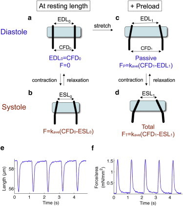
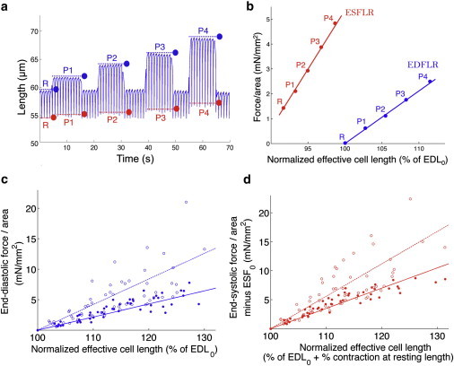
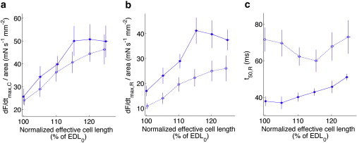
Human familial hypertrophic cardiomyopathy is the most common Mendelian cardiovascular disease worldwide. Among the most severe presentations of the disease are those in families heterozygous for the mutation R403Q in β-cardiac myosin. Mice heterozygous for this mutation in the α-cardiac myosin isoform display typical familial hypertrophic cardiomyopathy pathology. Here, we study cardiomyocytes from heterozygous 403/+ mice. The effects of the R403Q mutation on force-generating capabilities and dynamics of cardiomyocytes were investigated using a dual carbon nanofiber technique to measure single-cell parameters. We demonstrate the Frank-Starling effect at the single cardiomyocyte level by showing that cell stretch causes an increase in amplitude of contraction. Mutant 403/+ cardiomyocytes exhibit higher end-diastolic and end-systolic stiffness than +/+ cardiomyocytes, whereas active force generation capabilities remain unchanged. Additionally, 403/+ cardiomyocytes show slowed relaxation dynamics. These phenotypes are consistent with increased end-diastolic and end-systolic chamber elastance, as well as diastolic dysfunction seen at the level of the whole heart. Our results show that these functional effects of the R403Q mutation are cell-intrinsic, a property that may be a general phenomenon in familial hypertrophic cardiomyopathy.


Figures

Figure 1
Figure 1
Schematic of forces measured in cardiomyocyte-contractility experiments. (a) At resting length in diastole, the distance between two CFs at piezo translators (CFD0) and at the cell-attached CF tips (initial end-diastolic length, EDL0) are equal. Force is zero. (b) Upon contraction in systole, the distance between CF tips decreases (initial end-systolic length, ESL0). The force is the product of the average stiffness of the CFs (kave) and the distance that the CFs are bent (CFD0-ESL0). (c) Preload is applied by moving the piezo-translator mounted CFs apart to length CFD1. Cell-attached CF tips also move apart but to a lesser extent, to length EDL1. The passive force exerted by the cardiomyocyte is FP = kave(CFD1-EDL1). (d) Contraction under preload moves the CF tips to length ESL1, making the total force exerted FT = kave(CFD1-ESL1). (e) Representative trace of changes in distance between cell-attached CF tips (effective cell length) as the cardiomyocyte contracts at a rate of 1 Hz. (f) Normalization of force to cross-sectional area of the cardiomyocyte gives changes in contractile force per area, from which dynamic parameters can be determined.
Figure 2
Figure 2
403/+ cardiomyocytes have higher end-diastolic and end-systolic stiffness. (a) Representative data of effective cell length measurements (distance between the CF tips at the cell-attached end) of a cardiomyocyte undergoing voltage stimulation and contraction at 1 Hz, with the application of preload. Initially, the cell is at resting length (time segment R); increasing levels of preload are applied from time segments P1–P4. Dotted lines and dots indicate end-diastolic (blue) and end-systolic (red) lengths, respectively, and correspond to EDL and ESL in Fig. 1. (b) End-diastolic (blue) and end-systolic (red) points at rest (R) and per level of preload (P1–P4) are plotted as normalized force against normalized effective cell length. End-diastolic force-length relation (EDFLR, blue) and end-systolic force-length relation (ESFLR, red) are obtained from linear regressions of end-diastolic and end-systolic points, respectively. (c) EDFLRs of +/+ (solid circles, solid lines) and 403/+ (open circles, dotted lines) cardiomyocytes. The slope of EDFLR for 403/+ cardiomyocytes (0.36 mN/mm2) is steeper than that for +/+ cardiomyocytes (0.22 mN/mm2), indicating that 403/+ cardiomyocytes have higher end-diastolic stiffness. (d) Similarly, ESFLR of 403/+ cardiomyocytes (slope = 0.50 mN/mm2) is steeper than that for +/+ cardiomyocytes (slope = 0.37 mN/mm2), indicating higher end-systolic stiffness.
Figure 3
Figure 3
403/+ cardiomyocytes have slowed rates of relaxation compared to +/+ cardiomyocytes across all preloads studied. (a) Normalized maximum rates force development during contraction, dF/dtmax,C/area for +/+ (solid circles, solid lines) and 403/+ (open circles, dotted lines) cardiomyocytes are similar across all preloads. (b) Normalized maximum rates force reduction during relaxation, dF/dtmax,R/area are consistently slower for 403/+ compared to +/+ cardiomyocytes across all preloads. (c) The time taken to return halfway from systole to diastole, t50,R is greater for 403/+ cardiomyocytes than +/+ cardiomyocytes across all preloads.

References

    1. Maron B.J., Gardin J.M., Bild D.E. Prevalence of hypertrophic cardiomyopathy in a general population of young adults. Echocardiographic analysis of 4111 subjects in the CARDIA Study. Coronary Artery Risk Development in (Young) Adults. Circulation. 1995;92:785–789. - PubMed
    1. Maron B.J. Hypertrophic cardiomyopathy: a systematic review. JAMA. 2002;287:1308–1320. - PubMed
    1. Maron B.J., Spirito P., Arce J. Noninvasive assessment of left ventricular diastolic function by pulsed Doppler echocardiography in patients with hypertrophic cardiomyopathy. J. Am. Coll. Cardiol. 1987;10:733–742. - PubMed
    1. Epstein N.D., Cohn G.M., Fananapazir L. Differences in clinical expression of hypertrophic cardiomyopathy associated with two distinct mutations in the beta-myosin heavy chain gene. A 908-Leu----Val mutation and a 403-Arg----Gln mutation. Circulation. 1992;86:345–352. - PubMed
    1. Geisterfer-Lowrance A.A., Kass S., Seidman J.G. A molecular basis for familial hypertrophic cardiomyopathy: a beta cardiac myosin heavy chain gene missense mutation. Cell. 1990;62:999–1006. - PubMed

Publication types

Substances

LinkOut - more resources