Comparative Study
. 2012 Oct 11:222:269-80.
doi: 10.1016/j.neuroscience.2012.06.017. Epub 2012 Jun 23.

Strain differences in the effects of chronic corticosterone exposure in the hippocampus

Affiliations
Comparative Study

Strain differences in the effects of chronic corticosterone exposure in the hippocampus

G E Hodes et al. Neuroscience. .

Abstract

Stress hormones are thought to be involved in the etiology of depression, in part, because animal models show they cause morphological damage to the brain, an effect that can be reversed by chronic antidepressant treatment. The current study examined two mouse strains selected for naturalistic variation of tissue regeneration after injury for resistance to the effects of chronic corticosterone (CORT) exposure on cell proliferation and neurotrophin mobilization. The wound healer MRL/MpJ and control C57BL/6J mice were implanted subcutaneously with pellets that released CORT for 7 days. MRL/MpJ mice were resistant to reductions of hippocampal cell proliferation by chronic exposure to CORT when compared to vulnerable C57BL/6J mice. Chronic CORT exposure also reduced protein levels of brain-derived neurotrophic factor (BDNF) in the hippocampus of C57BL/6J but not MRL/MpJ mice. CORT pellet exposure increased circulating levels of CORT in the plasma of both strains in a dose-dependent manner although MRL/MpJ mice may have larger changes from baseline. The strains did not differ in circulating levels of corticosterone binding globulin (CBG). There were also no strain differences in CORT levels in the hippocampus, nor did CORT exposure alter glucocorticoid receptor or mineralocorticoid receptor expression in a strain-dependent manner. Strain differences were found in the N-methyl-D-aspartate (NMDA) receptor, and BDNF I and IV promoters. Strain and CORT exposure interacted to alter tropomyosine-receptor-kinase B (TrkB) expression and this may be a potential mechanism protecting MRL/MpJ mice. In addition, differences in the inflammatory response of matrix metalloproteinases (MMPs) may also contribute to these strain differences in resistance to the deleterious effects of CORT to the brain.


Figures

Figure 1
Figure 1. Schematic of experimental design
A. In experiment 1, C57BL/6J and MRL/MpJ mice were exposed to Placebo or CORT releasing pellets (1, 2 or 4 pellets) for 7 days. Mice were injected with 200 mg/kg of BrdU on day 7 and then sacrificed on day 8. Tissue was used to measure cell proliferation and BDNF levels in hippocampus. CORT and CBG levels were measured in plasma. B. In experiment 2 C57BL/6J and MRL/MpJ mice were exposed to placebo or CORT releasing pellets ( 2 pellets) for 7 days. Animals were sacrificed on Day 8 and mRNA and CORT was measured in the hippocampus. Circulating levels of CORT were measured in plasma.
Figure 2
Figure 2
Seven days of exposure to CORT pellets significantly reduced cell proliferation in C57BL/6J mice at all doses (Placebo: n = 11, 1 pellet: n = 8, 2 pellets: n = 10, 4 pellets: n = 10) but did not significantly reduce cell proliferation in MRL/MpJ mice (Placebo: n = 13, 1 pellet: n = 12, 2 pellets: n = 10, 4 pellets: n = 10) as indicated by a significant interaction of CORT exposure and strain [F3,74 = 3.92, p < 0.05].* denotes strain × CORT difference indicated by post-hoc test. ^ denotes a main effect of strain. # denotes a main effect of CORT
Figure 3
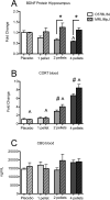
Figure 3. Dose response curve of protein levels of BDNF in hippocampus and CORT and CBG in plasma for C57BL/6J and MRL/MpJ mice
A. CORT pellet exposure reduced BDNF levels in the hippocampus of C57BL/6J (Placebo: n = 6, 1 pellet: n = 6, 2 pellets: n = 5, 4 pellets: n = 6) mice compared to MRL/MpJ (Placebo: n = 5, 1 pellet: n = 5, 2 pellets: n = 4, 4 pellets: n = 5) as indicated by a strain × CORT exposure interaction [F3,33 = 3.03, p < 0.05], Post-hoc analysis indicated that levels were lower in C57BL/6J mice given 2 or 4 CORT pellets than MRL/MpJ mice given the same number of pellets. Exposure to 4 pellets significantly reduced BDNF levels in C57BL/6J mice compared to their Placebo treated controls. B. CORT pellet exposure increased CORT levels in both strains [F3,36 = 109.05, p < 0.001], Post-hoc analysis indicated that mice given 2 or 4 pellets had higher circulating levels of CORT than those given placebo or 1 pellet. Mice given 4 pellets had higher levels of CORT than those given 2 pellets. MRL/MpJ mice (Placebo: n = 5, 1 pellet: n = 5, 2 pellets: n = 5, 4 pellets: n = 5) have higher overall normalized CORT values than C57BL/6J mice (Placebo: n = 6, 1 pellet: n = 6, 2 pellets: n = 6, 4 pellets: n = 6) when data was collapsed across dose [F3,20 = 6.25, p < 0.05] but there were no strain × CORT interactions [F3,20 = 2.28, p > 0.05]. C. C57BL/6J (Placebo: n = 5, 1 pellet: n = 5, 2 pellets: n = 5, 4 pellets: n = 5) and MRL/MpJ mice (Placebo: n = 5, 1 pellet: n = 5, 2 pellets: n = 5, 4 pellets: n = 5) did not differ in plasma levels of CBG. There was no strain × CORT exposure interaction [F1, 32 = 1.14, p > 0.05] and no main effects of CORT exposure [F1, 32 = 0.96, p > 0.05] or strain [F1, 32 = 0.88, p > 0.05]. * denotes strain × CORT differences indicated by post-hoc test. ^ denotes a main effect of strain. # denotes a main effect of CORT.
Figure 4
Figure 4. The effects of 7 days of CORT exposure with 2 CORT pellets on glucocorticoid receptor (GR) and mineralocorticoid receptor (MR) expression in the hippocampus and hypothalamus
A. Exposure to CORT pellets significantly reduced GR expression the hippocampus of C57BL/6J (Placebo: n = 7, CORT: n = 8) and MRL/MpJ mice (Placebo: n = 7, CORT: n = 8) as indicated by a main effect of CORT pellet implantation [F1,26 = 83.41, p < 0.01]. . A significant main effect of strain indicated MRL/MpJ mice had higher overall expression of GR[F1,26 = 14.71, p < 0.001]. B. CORT exposure significantly reduced GR expression in the hypothalamus of both C57BL/6J (Placebo: n = 8, CORT: n = 7) and MRL/MpJ mice (Placebo: n = 8, CORT: n = 8) as indicated by a main effect of CORT pellet implantation [F1,27 = 9.14, p < 0.01] , GR expression did not differ between strains in the hypothalamus [F1,27 = 1.76 , p > 0.05] and no strain × CORT exposure interaction [F1,27 = 0.55, p > 0.05]. C. CORT exposure significantly reduced MR expression the hippocampus of both C57BL/6J (Placebo: n = 7, CORT: n = 8) and MRL/MpJ mice (Placebo: n = 8, CORT: n = 8) [F1,26 = 10.28, p < 0.01]. MRL/MpJ mice had higher expression of MR in the hippocampus than C57BL/6J mice [F1,26 = 5.72, p < 0.05] with no significant interaction between strain × CORT exposure [ F1,26 = 2.69, p > 0.05]. D. There was no significant interaction between CORT exposure and strain for MR mRNA levels in the hypothalamus [F1,27 = 0.005, p > 0.05] of C57BL/6J (Placebo: n = 8, CORT: n = 8) and MRL/MpJ mice (Placebo: n = 7, CORT: n = 8) . There were no effects of strain [ F1,27 = 0.09, p > 0.05] or CORT pellet exposure[ F1,27 = 0.78, p > 0.05].* denotes strain × CORT differences indicated by post-hoc test. ^ denotes a main effect of strain.# denotes a main effect of CORT.
Figure 5
Figure 5. Potential molecular mechanisms of strain differences in resiliency to CORT
A. The effects of chronic exposure to 2 pellets of CORT on NMDA receptor subunit (NR) expression in the hippocampus of MRL/MpJ and C57BL/6J mice. MRL/MpJ mice (Placebo: n = 8, CORT: n = 8) had higher expression of the NR1 subunit than C57BL/6J mice (Placebo: n = 8, CORT: n = 8) as indicated by a main effect of strain [F1,28 = 28.70, p < 0.001]. There were no effects of CORT [F1,28 = 0.005, p > 0.05] and no strain by CORT exposure interactions [F1,27 = 0.001, p > 0.05] on NR1 expression. There were no effects of CORT exposure [F1,26 = 1.16, p > 0.05] or strain [F1,26 = 0.05, p > 0.05] or interactions [F1,26 = 0.15, p > 0.05] on NR2a expression in C57BL/6J (Placebo: n = 8, CORT: n = 8) or MRL/MpJ mice (Placebo: n = 7, CORT : n = 8). CORT exposure significantly reduced NR2b expression in both strains [F1,25 = 13.34, p < 0.01], but MRL/MpJ mice (Placebo: n = 8, CORT: n = 7) still had higher overall NR2b expression [F1,25 = 20.16, p < 0.001] than C57BL/6J mice (Placebo: n = 8, CORT: n = 6). There was no interaction between CORT exposure and strain on NR2b expression [F1,25 = 0.53, p > 0.05]. CORT exposure significantly reduced NR2c expression in both strains [F1,28 = 13.36, p < 0.01] but MRL/MpJ mice (Placebo: n = 8, CORT: n = 8) had significantly higher expression of NR2c [F1,28 = 4.43, p < 0.05]compared to C57BL/6J mice (Placebo: n = 8, CORT: n = 8). There was no strain × CORT exposure interaction on NR2c expression [F1,28 = 0.17, p > 0.05]. B. The effects of chronic CORT exposure on TrkB and BDNF promoter expression in the hippocampus. CORT exposure decreased TrkB expression in the hippocampus of C57BL/6J (Placebo: n = 8, CORT: n = 8) mice but did not reduce expression in MRL/MpJ mice (Placebo: n = 6, CORT: n = 7) as indicated by a significant interaction between CORT exposure and strain [F1,25 = 9.48, p < 0.01] . CORT exposure increased levels of BDNF promoter I [F1,?8 = 13.40 , p < 0.05] . The activity dependent promoter expression was higher in MRL/MpJ mice ( Placebo: n = 8, CORT: n = 8) compared to C57BL/6J mice (Placebo = 8, CORT: n = 8) as indicated by a main effect of strain [F1,28 = 13.19 , p < 0.05]. There was no interaction between CORT exposure and strain [F1,28 = 0.73 , p > 0.05]. CORT exposure increased levels of BDNF promoter IV [F1,28 = 20.86 , p < 0.05]. The activity dependent promoter expression of BDNF IV was higher in MRL/MpJ mice (Placebo: n = 8, CORT: n = 8) compared to C57BL/6J mice (Placebo: n = 8, CORT: n = 8) as indicated by a main effect of strain [F1,28 = 7.44 , p < 0.05]. There were no significant interactions between CORT exposure and strain [F1,28 = 0.73 , p > 0.05]. There were no significant effects of CORT exposure [F1,28 = 0.86, p > 0.05] or strain [F1,28 = 1.09 , p > 0.05] on BDNF promoter IX in C57BL/6J (Placebo: n = 8, CORT : n = 8) or MRL/MpJ mice (Placebo: n = 8, CORT: n = 8). There was no interaction between CORT exposure and strain [F1,28 = 0.05, p > 0.05]. C. The effects of chronic CORT exposure on expression of MMPs. MMP2 was elevated in the hippocampus of MRL/MpJ mice ( Placebo: n = 7, CORT: n = 7) given both placebo and CORT pellets whereas CORT decreased MMP2 in the hippocampus of C57BL/6J mice (Placebo: n = 6, CORT: n = 7) as indicated by a significant interaction [F1,23 = 4.30, p < 0.05]. MMP3 levels were elevated in the hippocampus of MRL/MpJ mice (Placebo: n = 8, CORT: n = 8) compared to C57BL/6J mice (Placebo: n = 7, CORT: n = 8) as indicated by a main effect of strain [F1,27 = 7.36, p < 0.05]. There was also a trend towards an interaction between CORT exposure and strain [F1,27 = 3.17 p = 0.08] but there was no main effect of CORT [F1,27 = 0.04, p > 0.05]. * denotes strain × CORT differences indicated by post-hoc test. ^ denotes a main effect of strain.# denotes a main effect of CORT.

Similar articles

Cited by

References

    1. Adachi M, Barrot M, Autry AE, Theobald D, Monteggia LM. Selective loss of brain-derived neurotrophic factor in the dentate gyrus attenuates antidepressant efficacy. Biol Psychiatry. 2008;63:642–649. - PMC - PubMed
    1. Alahmed S, Herbert J. Strain differences in proliferation of progenitor cells in the dentate gyrus of the adult rat and the response to fluoxetine are dependent on corticosterone. Neuroscience. 2008;157:677–682. - PMC - PubMed
    1. Balu DT, Hodes GE, Anderson BT, Lucki I. Enhanced sensitivity of the MRL/MpJ mouse to the neuroplastic and behavioral effects of chronic antidepressant treatments. Neuropsychopharmacology. 2009a;34:1764–1773. - PMC - PubMed
    1. Balu DT, Hodes GE, Hill TE, Ho N, Rahman Z, Bender CN, Ring RH, Dwyer JM, Rosenzweig-Lipson S, Hughes ZA, Schechter LE, Lucki I. Flow cytometric analysis of BrdU incorporation as a high-throughput method for measuring adult neurogenesis in the mouse. J Pharmacol Toxicol Methods. 2009b;59:100–107. - PMC - PubMed
    1. Benekareddy M, Mehrotra P, Kulkarni VA, Ramakrishnan P, Dias BG, Vaidya VA. Antidepressant treatments regulate matrix metalloproteinases-2 and -9 (MMP-2/MMP-9) and tissue inhibitors of the metalloproteinases (TIMPS 1-4) in the adult rat hippocampus. Synapse. 2008;62:590–600. - PubMed

Publication types

MeSH terms