. 2012 Oct 1;21(19):4201-13.
doi: 10.1093/hmg/dds244. Epub 2012 Jun 26.

G2019S leucine-rich repeat kinase 2 causes uncoupling protein-mediated mitochondrial depolarization

Affiliations

G2019S leucine-rich repeat kinase 2 causes uncoupling protein-mediated mitochondrial depolarization

Tatiana D Papkovskaia et al. Hum Mol Genet. .

Abstract

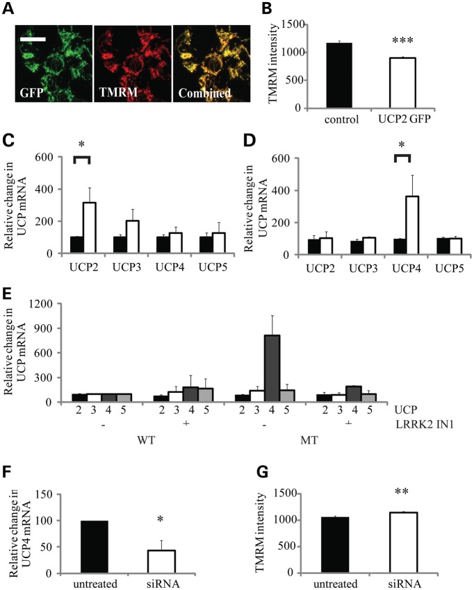
The G2019S leucine rich repeat kinase 2 (LRRK2) mutation is the most common genetic cause of Parkinson's disease (PD), clinically and pathologically indistinguishable from idiopathic PD. Mitochondrial abnormalities are a common feature in PD pathogenesis and we have investigated the impact of G2019S mutant LRRK2 expression on mitochondrial bioenergetics. LRRK2 protein expression was detected in fibroblasts and lymphoblasts at levels higher than those observed in the mouse brain. The presence of G2019S LRRK2 mutation did not influence LRRK2 expression in fibroblasts. However, the expression of the G2019S LRRK2 mutation in both fibroblast and neuroblastoma cells was associated with mitochondrial uncoupling. This was characterized by decreased mitochondrial membrane potential and increased oxygen utilization under basal and oligomycin-inhibited conditions. This resulted in a decrease in cellular ATP levels consistent with compromised cellular function. This uncoupling of mitochondrial oxidative phosphorylation was associated with a cell-specific increase in uncoupling protein (UCP) 2 and 4 expression. Restoration of mitochondrial membrane potential by the UCP inhibitor genipin confirmed the role of UCPs in this mechanism. The G2019S LRRK2-induced mitochondrial uncoupling and UCP4 mRNA up-regulation were LRRK2 kinase-dependent, whereas endogenous LRRK2 levels were required for constitutive UCP expression. We propose that normal mitochondrial function was deregulated by the expression of G2019S LRRK2 in a kinase-dependent mechanism that is a modification of the normal LRRK2 function, and this leads to the vulnerability of selected neuronal populations in PD.


Figures

Figure 1.
Figure 1.
Analysis of LRRK2 protein expression. LRRK2 protein levels were analysed using western blot analysis (3514-1 antibody) of LRRK2 immunoprecipitates (IP, 100–500 antibody) from 1 mg of protein. (A) The relative LRRK2 levels were highest in control human lymphoblasts (l), which were greater than in fibroblasts (f), which were greater than in control SHSY5Y cells (control). However LRRK2 levels were much greater in SHSY5Y cells over-expressing WT LRRK2 (1:10 denotes loading of 10% of the IP sample). (B) LRRK2 expression levels were comparable in fibroblasts from control (c) and G2019S LRRK2 PD patients (p), with GAPDH demonstrating equivalent protein input.
Figure 2.
Figure 2.
Mitochondrial function in G2019S LRRK2 patient fibroblasts. Mitochondrial function was assessed in fibroblast cell lines expressing MT G2019S LRRK2 (G2019S) and controls (control). Rates of cellular oxygen consumption in the presence of glucose were increased in the MT cells as determined by (A) an extracellular phosphorescent oxygen probe [relative fluorescence unit (RFU)/min/µg protein] and (B) a Clark-type oxygen electrode (nmole O/min/µg protein). (C) Mitochondrial content was comparable between control and MT fibroblasts as determined by western blot analysis, probing for mitochondrial markers (TFAM, mitochondrial transcription factor A; Core, complex III core subunit) relative to actin in fibroblasts from control and G2019S LRRK2 PD patients. (D) Mitochondrial membrane potential was reduced in the G2019S cells as assessed by live-cell imaging of TMRM fluorescence (RFU/cell, n = 1600 cells), with typical images of TMRM staining in control and G2019S fibroblasts showing the reduction in TMRM intensity. Cellular oxygen utilization measurements for control (open bars) and G2019S (closed bars) fibroblasts with a Clark-type oxygen electrode (nmole O/min/µg protein) in the absence or presence of: (E) oligomycin (2 µg/ml) showed an increased proton leak in G2019S LRRK2 fibroblasts; (F) FCCP (0.5 µm) demonstrated comparable maximal respiratory chain capacities between the two cohorts (n = 12,). (G) Cellular ATP content was decreased in the G2019S MT fibroblasts (RLU/µg protein) and (H) rates of cellular ROS generation were reduced in the MT cells as determined by evaluating the rate of DHE (100 nm) oxidation (RFU/min/cell, n = 300 cells). All values are expressed as mean ± SEM, using fibroblasts from eight patients with the G2019S LRRK2 mutation and seven controls, n = 3 per cell line unless stated otherwise. Statistical analyses were performed using unpaired Student's t-test; statistical significance: *P < 0.05, ***P < 0.0001 for G2019S versus control fibroblasts.
Figure 3.
Figure 3.
Analysis of SHSY5Y cells over-expressing WT or G2019S LRRK2. (A) Western blot analysis of ectopic LRRK2 level (V5 antibody) in SHSY5Y cells with stable LRRK2 expression confirmed that the levels of WT LRRK2 was ∼2-fold greater than the MT G2019S (MT) LRRK2. (B) Rates of cellular oxygen utilization were increased in G2019S-over-expressing cells as revealed by oxygen consumption measurements, using a Clark-type oxygen electrode (nmole O/min/µg protein, n = 6). (C) Mitochondrial membrane potential was reduced in the MT cells as determined by live-cell imaging of TMRM fluorescence [relative fluorescence unit (RFU)/cell, n = 200 cells]. (D) Inhibition of LRRK2 kinase by IN-1 (1 µm, 90 min) restored oxygen consumption rates in the MT clones without affecting the rates in control or WT cells (nmole O/min/µg protein, n = 4); (E) LRRK2 kinase inhibition was confirmed at the end of the experiment in (D), as determined by the decrease in the phosphorylation of LRRK2 at serine 935 [IP followed by western blot analysis with P-Ser 935 relative to total LRRK2 (3514-1)]; (F) Mitochondrial membrane potential was restored in G2019S cells following kinase inhibition as determined by TMRM staining (RFU/cell, n = 200 cells). All values are expressed as mean ± SEM, unless stated otherwise. Statistical analyses were performed using ANOVA (the Bonferonni post hoc test); statistical significance: *P < 0.05, ***P < 0.0001 for G2019S versus control and WT over-expressing SHSY5Y cells or untreated versus LRRK2 IN1-treated cells.
Figure 4.
Figure 4.
LRRK2 intracellular and mitochondrial localization. The distribution of LRRK2 was analysed in the subcellular fractions isolated from SHSY5Y cells over-expressing WT LRRK2 (A) and control lymphoblasts (B). Disrupted cells were fractionated by differential centrifugation (1–250 000g) and 10% of each pellet and cytosol (cyt) fraction analysed by western blotting. LRRK2 distribution was determined by (A) V5 or (B) NT2 LRRK2 immunoreactivity. Subcellular distribution was characterized by probing blots with mitochondrial complex III core protein (Core), lysosomal (GCase), ER calreticulin (Cal) and vesicular synaptophysin (Synapto) markers. LRRK2 was most abundant in the cytosolic and vesicular fractions, with lower levels in the other subcellular compartments. Affinity-purified mitochondria (Mito) were isolated from the postnuclear supernatant (PNS) from WT and G2019S (MT) LRRK2-over-expressing SHSY5Y cells. These highly purified mitochondria were essentially free from other subcellular components, and similar levels of LRRK2 (V5 antibody) were detected in the mitochondria from WT (C) and MT (D) LRRK2-expressing cells. (E) LRRK2 was found to be associated with mitochondrial membranes as determined by mitochondrial fractionation experiments. Affinity-purified mitochondria from WT SHSY5Y cells were subjected to fractionation in hypotonic and sodium carbonate treatment (Na2CO3) with equivalent percentages of the soluble (s) and pellet (p) fractions analysed on western blots, using LRRK2 (V5) and SDHA as an inner-membrane and cytochrome c (cytc) as an inter-membrane space marker.
Figure 5.
Figure 5.
Analysis of the role of UCP expression in G2019S LRRK2-induced mitochondrial uncoupling. Confocal microscopy of SHSY5Y cells transiently transfected with murine GFP-UCP2 demonstrated: (A) co-localization of UCP2 with mitochondrial TMRM staining and (B) a decrease in mitochondrial membrane potential as quantified by TMRM fluorescence in GFP-positive cells [relative fluorescence unit (RFU)/cell, n = 100 cells]. G2019S LRRK2 expression was associated with increased UCP mRNA levels. mRNA for UCPs2-5 was analysed by RT-PCR in (C), control (black bar) and G2019S LRRK2 (open bar) fibroblasts and (D) SHSY5Y cells over-expressing WT LRRK2 (black bar) and G2019S LRRK2 (MT, open bar). Data are expressed relative to GAPDH mRNA levels and normalized to the control cells. (E) Inhibition of LRRK2 kinase by IN1 (1 µm, 90 min) decreased the significant increase in UCP4 mRNA levels (n = 4) observed in SHSY5Y cells expressing G2019S (MT) LRRK2. Data are expressed relative to GAPDH mRNA levels and normalized to the ratios detected in WT untreated cells. (F) Knockdown of LRRK2 in control SHSY5Y cells by siRNA decreased UCP4 mRNA levels as assessed by RT PCR (n = 3), which was (G) associated with an increased mitochondrial membrane potential as quantified by the TMRM signal intensity (RFU/cell, n = 200 cells). All values are expressed as mean ± SEM (n = 6), unless stated otherwise. Statistical analyses were performed using unpaired Student's t-test; statistical significance: *P < 0.05, **P < 0.005, ***P < 0.0005 for G2019S versus WT over-expressing SHSY5Y cells or control versus siRNA-transfected SHSY5Y cells.
Figure 6.
Figure 6.
Influence of genipin upon mitochondrial membrane potential. The influence of genipin upon the mitochondrial membrane potential as determined by live-cell imaging of TMRM fluorescence. (A) UCP2 transfected SHSY5Y cells were treated with increasing concentrations of genipin (solid line 1.375 nm, dashed line 2.5 nm, dotted line 5 nm).There was a time-dependent increase in mitochondrial membrane potential of cells. (B) Genipin treatment (1.375 nm) increased the mitochondrial membrane potential in the G2019S LRRK2 (solid line) expressing SHSY5Y cells, with only mild influence upon untransfected (dotted line) and WT LRRK2 (dashed line) over-expressing SHSY5Y cells. (C) Genipin treatment (1.375 nm) of control (C2, C4, solid lines) and G2019S LRRK2 fibroblasts (P1, P2, P7, dotted lines) caused an increase in mitochondrial membrane potential in all cell lines. This was more pronounced for the G2019S fibroblasts. The TMRM intensity data were expressed as the percentage of the untreated controls cells at t = 0. Data represent the mean values of four independent experiments focusing on a field containing approximately 30 cells.

References

    1. Lees A.J., Hardy J., Revesz T. Parkinson's disease. Lancet. 2009;373:2055–2066. doi:10.1016/S0140-6736(09)60492-X. - DOI - PubMed
    1. Healy D.G., Wood N.W., Schapira A.H. Test for LRRK2 mutations in patients with Parkinson's disease. Pract. Neurol. 2008;8:381–385. doi:10.1136/jnnp.2008.162420. - DOI - PubMed
    1. Marin I. The Parkinson disease gene LRRK2: evolutionary and structural insights. Mol. Biol. Evol. 2006;23:2423–2433. doi:10.1093/molbev/msl114. - DOI - PubMed
    1. Greggio E., Jain S., Kingsbury A., Bandopadhyay R., Lewis P., Kaganovich A., van der Brug M.P., Beilina A., Blackinton J., Thomas K.J., et al. Kinase activity is required for the toxic effects of mutant LRRK2/dardarin. Neurobiol. Dis. 2006;23:329–341. doi:10.1016/j.nbd.2006.04.001. - DOI - PubMed
    1. Ito G., Okai T., Fujino G., Takeda K., Ichijo H., Katada T., Iwatsubo T. GTP binding is essential to the protein kinase activity of LRRK2, a causative gene product for familial Parkinson's disease. Biochemistry. 2007;46:1380–1388. doi:10.1021/bi061960m. - DOI - PubMed

Publication types

MeSH terms

Substances