. 2013 Jan 1;119(1):61-71.
doi: 10.1002/cncr.27661. Epub 2012 Jun 26.

C-X-C chemokine receptor 7: a functionally associated molecular marker for bladder cancer

Affiliations

C-X-C chemokine receptor 7: a functionally associated molecular marker for bladder cancer

Travis J Yates et al. Cancer. .

Abstract

Background: C-X-C chemokine receptor 4 (CXCR4) and CXCR7 are 7-transmembrane chemokine receptors of the stroma-derived factor (SDF-1). CXCR4, but not CXCR7, has been examined in bladder cancer (BCa). This study examined the functional and clinical significance of CXCR7 in BCa.

Methods: CXCR4 and CXCR7 levels were measured in BCa cell lines, tissues (normal = 25; BCa = 44), and urine specimens (n = 186) by quantitative polymerase chain reaction and/or immunohistochemistry. CXCR7 function in BCa cells were examined by transient transfections using a CXCR7 expression vector or small interfering RNA.

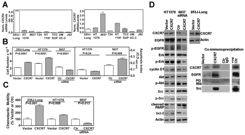
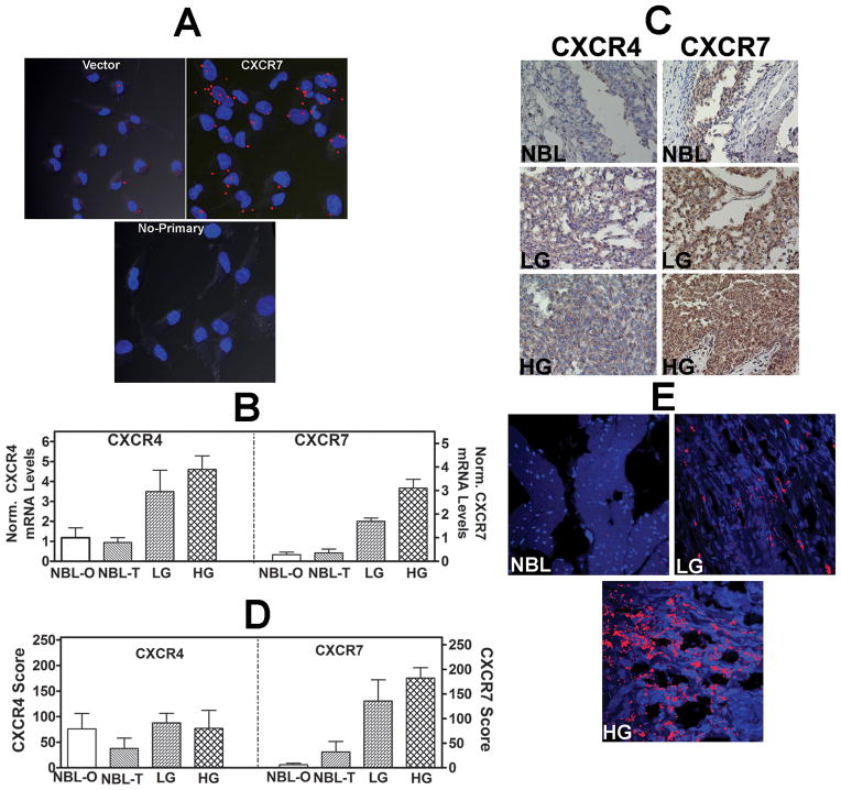
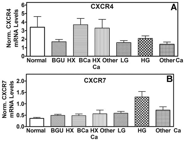
Results: In BCa cell lines, CXCR7 messenger RNA levels were 5- to 37-fold higher than those for CXCR4. Transient overexpression of CXCR7 in BCa cell lines promoted growth and chemotactic motility. CXCR7 colocalized and formed a functional complex with epidermal growth factor receptor, phosphoinositide 3-kinase/Akt, Erk, and src and induced their phosphorylation. CXCR7 also induced up-regulation of cyclin-D1 and bcl-2. Suppression of CXCR7 expression reversed these effects and induced apoptosis. CXCR7 messenger RNA levels and CXCR7 staining scores were significantly (5- to 10-fold) higher in BCa tissues than in normal tissues (P < .001). CXCR7 expression independently associated with metastasis (P = .019) and disease-specific mortality (P = .03). CXCR7 was highly expressed in endothelial cells in high-grade BCa tissues when compared to low-grade BCa and normal bladder. CXCR7 levels were elevated in exfoliated urothelial cells from high-grade BCa patients (P = .0001; 90% sensitivity; 75% specificity); CXCR4 levels were unaltered.

Conclusions: CXCR7 promotes BCa cell proliferation and motility plausibly through epidermal growth factor receptor receptor and Akt signaling. CXCR7 expression is elevated in BCa tissues and exfoliated cells and is associated with high-grade and metastasis.


Figures

Figure 1
Figure 1. CXCR7 expression and function in bladder cancer cells
A: Analysis of CXCR4, CXCR7 mRNA levels in BCa cells, by Q-PCR. Data: Mean±sd. CXCR4 and CXCR7 values: 23J-Lung: 6.2±0.4, 0.4±0.1; HT1376: 0.1±0.02 0.6±0.02; 5637: 0.06±0.001, 2.2±0.3; T24: 0.03±0.006, 0.2±0.01; HT1197: 0.04±0.005, 1.1±0.1; TCCSUP: 0.01±0.007, 0.5±0.1; UMUC-3: 0.03±0.01, 0.1±0.007; J82: 0.03±0.002, 0.3±0.007 B: Effect of CXCR7 expression on growth and apoptosis. Left panel: Cell counting data at 72 hours following transfection either with vector, CXCR7 (253J-Lung and HT1376) or CXCR7 siRNA (5637). 253J-Lung: vector: 2.2±0.12, CXCR7: 3.7±0.17; HT1376: vector: 3.5±0.21, CXCR7: 4.9±0.22. 5637: control siRNA: 2.4±0.22, CXCR7 siRNA: 1.6±0.14. Right panel: Transfectants were assayed for apoptosis; Apoptosis index for each transfectant is presented. Data: Mean ± sd. HT136: vector: 0.09±0.02, CXCR7: 0.07±0.005; 5637: control siRNA: 0.16±0.02, CXCR7 siRNA: 0.26±0.03. C: Determination of the chemotactic motility of the CXCR7 overexpressing (HT1376, 253J-Lung) and CXCR7 siRNA (5637) transfectants. Data: Mean±sd. HT1376: vector: 100±33, CXCR7: 311±25; 253J-Lung: 100±22, CXCR7: 167±10; 5637: control siRNA: 100±7, CXCR7 siRNA: 37±7 D: Analysis of CXCR7 transfectants. HT1376 and 253J-Lung cells were transiently transfected with a CXCR7 plasmid. 5637 cells were transfected with CXCR7 siRNA. Cell lysates were analyzed by immunoblotting; loading control: actin. Right panel: Cell lysates of the 253J-Lung vector and CXCR7 transfectants were immunoprecipitated using an anti-CXCR7 antibody and the immunoprecipitates were immunoblotted for various antigens.
Figure 2
Figure 2. Analysis of CXCR7 transfectants and expression of CXCR4 and CXCR7 in bladder tissues
A: In-cell co-IP of 253J-Lung vector and CXCR7 transfectants using the anti-CXCR7 and anti-EGFR antibodies; lower panel, no primary antibody control for the CXCR7 transfectant. Red dots indicate co-localization of CXCR7 and EGF-R. Magnification: 400×. B: CXCR4 and CXCR7 transcript levels in bladder tissues. NBL: normal bladder; NBL-O: NBL tissue obtained from organ donors; NBL-T: NBL tissue obtained from BCa patients at the time of cystectomy. LG: low-grade BCa; HG: high-grade BCa. The mean±sd levels are shown. C: CXCR4 and CXCR71 were localized in normal bladder, LG and HG BCa tissues by IHC. Representative specimens from each category are shown. D: Mean±sd staining scores of CXCR4 and CXCR7 in bladder specimens are shown. Due to poor fixation which resulted in the loss of tissues during staining, 22 high-grade, 7 low-grade tissues and 15 NBL tissues could be stained. E: Localization of CXCR7 in bladder endothelial cells. Bladder tissues were stained with anti-CD31 (endothelial cell marker) and CXCR7 using the in-cell co-IP technique. Red staining represents co-localization of CD31 and CXCR7, i.e., localization of CXCR7 in endothelial cells. Magnification: 400×.
Figure 3
Figure 3. CXCR4 and CXCR7 levels in bladder tissues and exfoliated urothelial cells. A and B
The mean±sd mRNA levels of CXCR4 (B) and CXCR7 (C) among different categories are shown. Mean levels of CXCR4 and CXCR7 among Group 1 (2.6±1.8; 0.38±0.26) and Group 2 (4.9±10.6; 0.33±0.12; P>0.05) of normal individuals, explained in Table 2, were not significantly different.

Similar articles

Cited by

References

    1. Kaufman DS, Shipley WU, Feldman AS. Bladder Cancer. Lancet. 2009;374:239–249. - PubMed
    1. Vandercappellen J, Van Damme J, Struyf S. The role of CXC chemokines and their receptors in cancer. Cancer Lett. 2008;267:226–44. - PubMed
    1. Maksym RB, Tarnowski M, Grymula K, Tarnowska J, Wysoczynski M, Liu R, et al. The role of stromal-derived factor-1--CXCR7 axis in development and cancer. Eur J Pharmacol. 2009;625:31–40. - PMC - PubMed
    1. Sun X, Cheng G, Hao M, Zheng J, Zhou X, Zhang J, et al. CXCL12/CXCR4/CXCR7 chemokine axis and cancer progression. Cancer Metastasis Rev. 2010;29:709–22. - PMC - PubMed
    1. Teicher BA, Fricker SP. CXCL12 (SDF-1)/CXCR4 pathway in cancer. Clin Cancer Res. 2010;16:2927–31. - PubMed

Publication types

MeSH terms