Comparative Study
. 2013 Jan;83(1):114-26.
doi: 10.1002/cyto.a.22090. Epub 2012 Jun 26.

Efficient and simultaneous generation of hematopoietic and vascular progenitors from human induced pluripotent stem cells

Affiliations
Comparative Study

Efficient and simultaneous generation of hematopoietic and vascular progenitors from human induced pluripotent stem cells

Tea Soon Park et al. Cytometry A. 2013 Jan.

Abstract

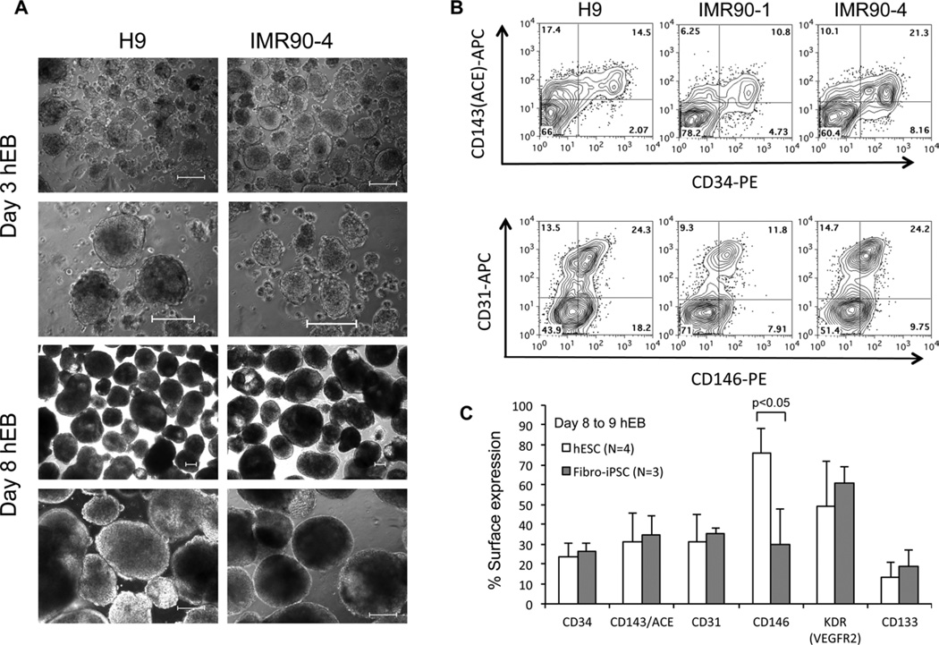
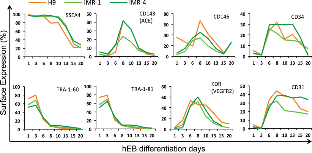
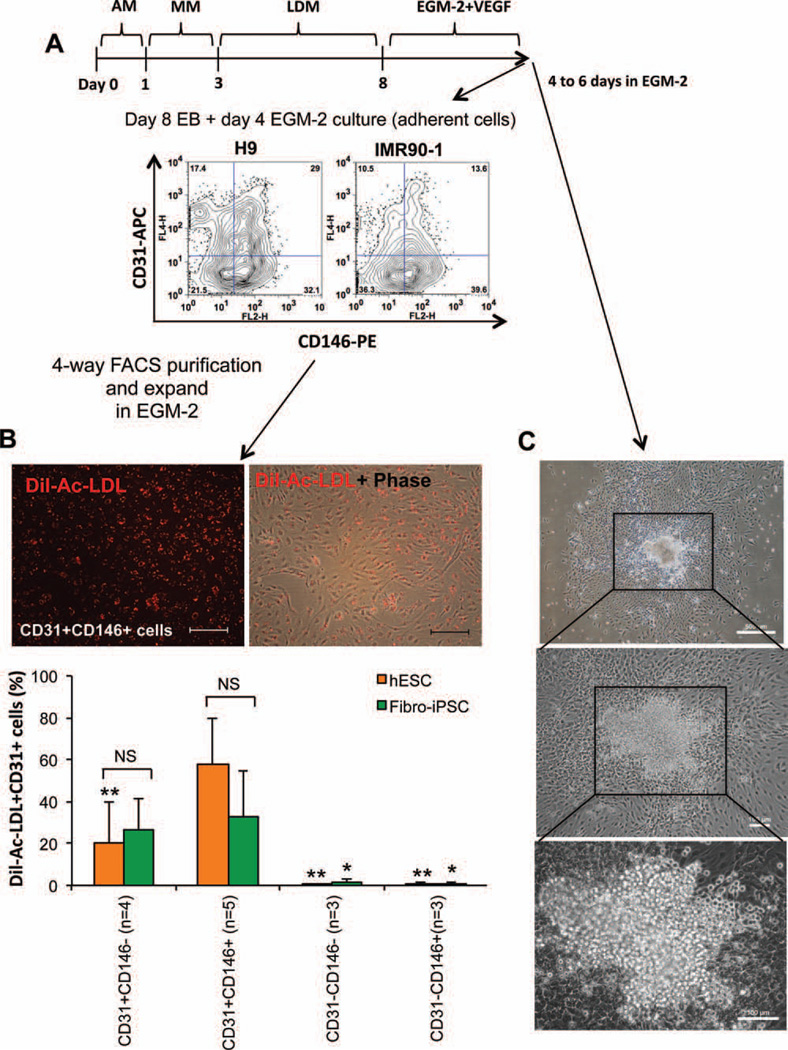
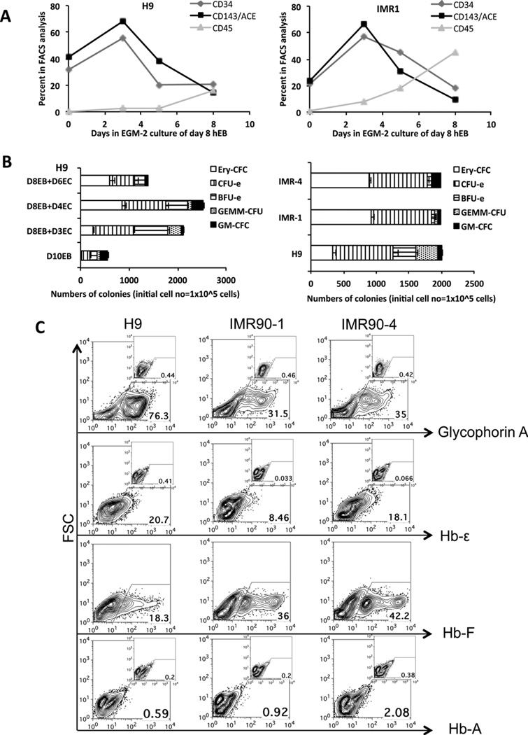
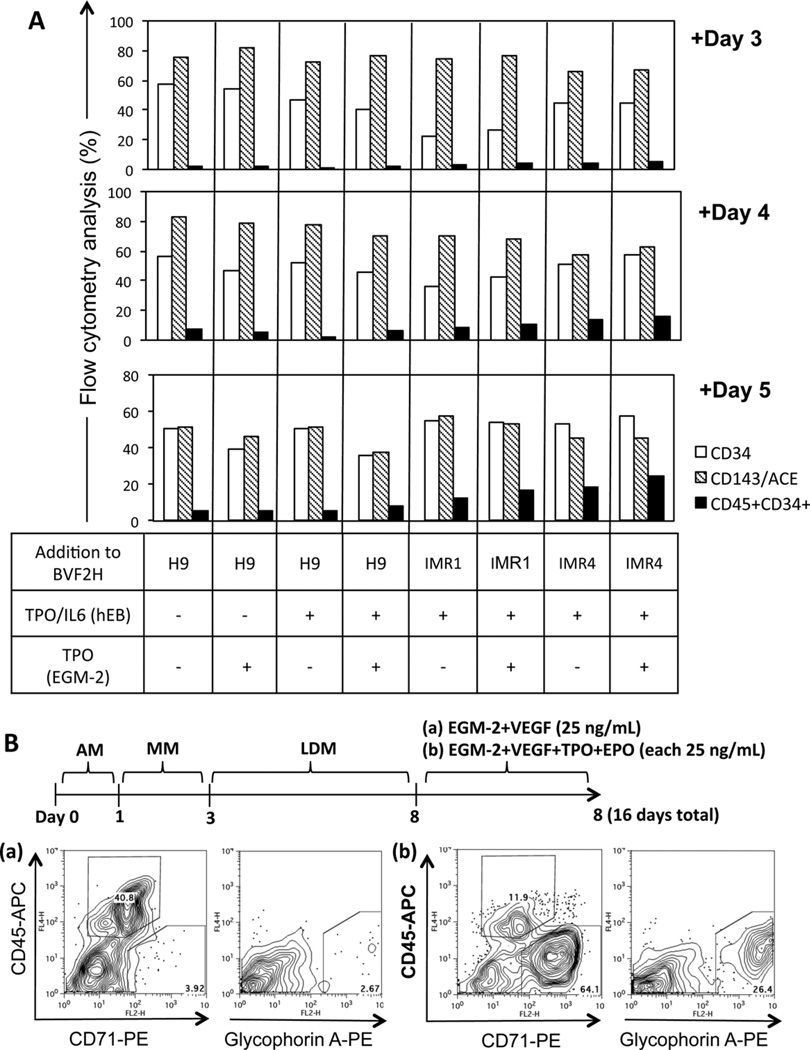
The hematopoietic and vascular lineages are intimately entwined as they arise together from bipotent hemangioblasts and hemogenic endothelial precursors during human embryonic development. In vitro differentiation of human pluripotent stem cells toward these lineages provides opportunities for elucidating the mechanisms of hematopoietic genesis. We previously demonstrated the stepwise in vitro differentiation of human embryonic stem cells (hESC) to definitive erythromyelopoiesis through clonogenic bipotent primitive hemangioblasts. This system recapitulates an orderly hematopoiesis similar to human yolk sac development via the generation of mesodermal-hematoendothelial progenitor cells that give rise to endothelium followed by embryonic primitive and definitive hematopoietic cells. Here, we report that under modified feeder-free endothelial culture conditions, multipotent CD34⁺ CD45⁺ hematopoietic progenitors arise in mass quantities from differentiated hESC and human induced pluripotent stem cells (hiPSC). These hematopoietic progenitors arose directly from adherent endothelial/stromal cell layers in a manner resembling in vivo hematopoiesis from embryonic hemogenic endothelium. Although fibroblast-derived hiPSC lines were previously found inefficient in hemato-endothelial differentiation capacity, our culture system also supported robust hiPSC hemato-vascular differentiation at levels comparable to hESC. We present comparative differentiation results for simultaneously generating hematopoietic and vascular progenitors from both hESC and fibroblast-hiPSC. This defined, optimized, and low-density differentiation system will be ideal for direct single-cell time course studies of the earliest hematopoietic events using time-lapse videography, or bulk kinetics using flow cytometry analyses on emerging hematopoietic progenitors.


Figures

Figure 1
Figure 1. Hemato-vascular differentiation of hESC and fibroblast-iPSC using a serum free hEB system supplemented with BMP4, VEGF, FGF2, and heparan sulfate (BVF2H)
(A) Phase contrast pictures of hEB from H9 and IMR90-4 at day 3 of hEB differentiation (first 2 rows). Day 8 hEBs (bottom 2 rows) are larger and become cystic. The scale bars represent 100 µm. (B) Flow cytometry analysis of dissociated day 10 H9, IMR90-1, and IMR90-4 hEB cells demonstrates similar expressions and percentages of CD143/ACE, CD34, CD31, and CD146. Shown are representative experiments performed at least three times. (C) Flow cytometry analysis of hESC and fibroblast-derived hiPSC (FibroiPSC) and the average percentages of cells with surface expression of CD34, CD143/ACE, CD31, CD146, KDR (VEGF receptor 2, VEGFR2), and CD133. Number of times experiments were performed and P values are indicated.
Figure 2
Figure 2. Kinetics of hemato-endothelial surface marker expression of differentiated H9, IMR90-1 (IMR-1) and IMR90-4 (IMR-4) hEB cells
Using our standard hEB differentiation protocol, day 1, 3, 6, 8, 10, 13, 15, and 20 hEBs were harvested, enzymatically digested to single cells, and analyzed for the expressions of SSEA4, TRA-1-60, TRA-1-81, CD143/ACE, CD146, KDR (VEGF receptor 2, VEGFR2), CD34, and CD31.
Figure 3
Figure 3. Endothelial differentiation of hESC and hiPSC
(A) Day 8 hEB were disaggregated using collagenase type-IV and plated onto the fibronectin coated plates in EGM-2 medium. Day 4 EGM-2-cultured hEB cells were analyzed and purified by FACS for surface expression of CD31 and CD146. (B) Day 8 hEB cells differentiated in EGM-2 medium were FACS-purified into four populations (CD31+CD146, CD31+CD146+, CD31CD146, and CD31CD146+ cells), and expanded for additional 7—12 days prior to assay of Dil-Acetylated-LDL (Dil-Ac-LDL) uptake to demonstrate endothelial cell function. Representative picture of Dil-Ac-LDL uptaking cells of CD31+CD146+ cells (Left) and the merged image of phase contrast picture (Right) derived from IMR90-4. Scale bars represent 100 µm. Quantitative comparisons of expression of Dil-Ac-LDL uptaking cells from FACS purified four populations (bottom panel). (C) Day 8 hEB clumps differentiated from IMR90-4 generated hematopoietic cobblestones that emerged directly from adherent endothelial layers. Shown are averaged experiments performed three to five times with n, standard deviation, and P values indicated for the values of the CD31+CD146+ group compared to the other three categories of sorted populations (* = P < 0.05 for fibro-iPSC; ** = P < 0.05 for hESC; NS = comparison was not significant).
Figure 4
Figure 4. Hematopoietic cells generated from EGM-2 culture condition
(A) Floating, non-adherent cells were collected from EGM-2 hEB cultures. Day 8 hEB were cultured as described in the text for an additional 3, 5, and 8 days in EGM-2 culture, and analyzed by flow cytometry for surface expression of CD34, CD143/ACE, and CD45. (B) Hematopoietic CFU of floating, non-adherent cells generated in EGM-2 cultures from differentiated H9 cells at different time points (left panel). Floating cells from day 8 EB with additional day 3 in EGM-2 culture (total day 11) of H9, IMR90-1 (IMR-1), and IMR90-4 (IMR-4) were compared for hematopoietic CFU assay (right panel). (C) Hematopoietic colonies from methylcellulose cultures were pooled and analyzed by flow cytometry for expression of glycophorin A (CD235a) and embryonic (Hb-ε), fetal (Hb-F), and adult (Hemoglobin β chain, HbA) hemoglobin expression. Inserts are appropriate isotype controls. Shown are representative experiments performed three times.
Figure 5
Figure 5. Supplementation of adherent EGM-2 hEB cultures with additional hematopoietic growth factors
(A) Effects of supplementation with TPO and IL6 during day 3 to 8 of BMP4, VEGF, FGF2, and heparan sulfate (BVF2H) hEB differentiation, and addition of TPO during EGM-2 culture (i.e., day 8 hEB followed by an additional 3, 4, or 5 days in EGM-2 culture). Supplementation with hematopoietic growth factors altered the production of CD34, CD143/ACE, and CD45 expressing hEB cells. The addition of TPO and IL6 during hEB BVF2H differentiation, and the addition of TPO during EGM-2 culture generated the highest percentages of CD34+CD45+ hEB cells in IMR90-4 cells from day 8 of hEB plus day 5 of EGM-2 culture (bottom graph, far right). (B) Day 8 hEB with addition of day 8 in EGM-2 culture supplemented with VEGF (25 ng/mL) of IMR90-1 cells in expression of CD45, CD71, and glycophorin A (a). Addition of TPO and erythropoietin (EPO) (25 ng/mL each) to the EGM-2 medium supplemented with VEGF (25 ng/mL) directed hematopoietic cells to differentiate to the erythroid lineage (b).

Similar articles

Cited by

References

    1. Takahashi K, Tanabe K, Ohnuki M, Narita M, Ichisaka T, Tomoda K, Yamanaka S. Induction of pluripotent stem cells from adult human fibroblasts by defined factors. Cell. 2007;131:861–872. - PubMed
    1. Yu J, Vodyanik MA, Smuga-Otto K, Antosiewicz-Bourget J, Frane JL, Tian S, Nie J, Jonsdottir GA, Ruotti V, Stewart R, et al. Induced pluripotent stem cell lines derived from human somatic cells. Science. 2007;318:1917–1920. - PubMed
    1. Park IH, Arora N, Huo H, Maherali N, Ahfeldt T, Shimamura A, Lensch MW, Cowan C, Hochedlinger K, Daley GQ. Disease-specific induced pluripotent stem cells. Cell. 2008;134:877–886. - PMC - PubMed
    1. Lengerke C, Daley GQ. Disease models from pluripotent stem cells. Annals of the New York Academy of Sciences. 2009;1176:191–196. - PubMed
    1. Grskovic M, Javaherian A, Strulovici B, Daley GQ. Induced pluripotent stem cells—Opportunities for disease modelling and drug discovery. Nature reviews. Drug Discov. 2011;10:915–929. - PubMed

Publication types

MeSH terms