

Inclined selective plane illumination microscopy adaptor for conventional microscopes
- PMID: 22736488
- PMCID: PMC3742098
- DOI: 10.1002/jemt.22089
Inclined selective plane illumination microscopy adaptor for conventional microscopes
Abstract
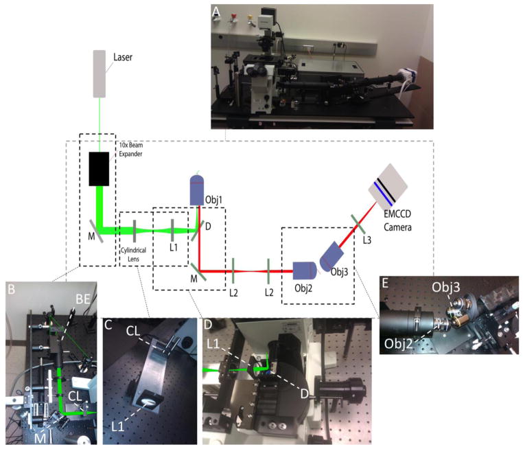
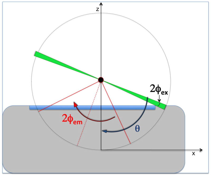
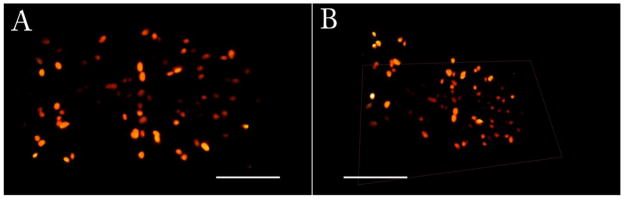
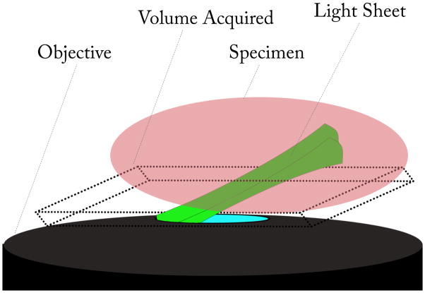
Driven by the biological sciences, there is an increased need for imaging modalities capable of live cell imaging with high spatial and temporal resolution. To achieve this goal in a comprehensive manner, three-dimensional acquisitions are necessary. Ideal features of a modern microscope system should include high imaging speed, high contrast ratio, low photo-bleaching and photo-toxicity, good resolution in a 3D context, and mosaic acquisition for large samples. Given the importance of collecting data in live sample further increases the technical challenges required to solve these issues. This work presents a practical version of a microscopy method, Selective Plane Illumination Microscopy re-introduced by Huisken et al. (Science2004,305,1007-1009). This method is gaining importance in the biomedical field, but its use is limited by difficulties associated with unconventional microscope design which employs two objectives and a particular kind of sample preparation needed to insert the sample between the objectives. Based on the selective plane illumination principle but with a design similar to the Total Internal Reflection Fluorescence microscope, Dunsby (Dunsby, Opt Express 2008,16,20306-20316) demonstrated the oblique plane microscope (OPM) using a single objective which uses conventional sample preparation protocols. However, the Dunsby instrument was not intended to be part of a commercial microscope. In this work, we describe a system with the advantages of OPM and that can be used as an adaptor to commonly used microscopes, such as IX-71 Olympus, simplifying the construction of the OPM and increasing performance of a conventional microscope. We named our design inclined selective plane illumination microscope (iSPIM).
Copyright © 2012 Wiley Periodicals, Inc.
Figures
References
-
- Botcherby EJ, Juskaitis R, Booth MJ, Wilson T. An optical technique for remote focusing in microscopy. Optics Communications. 2008;281(4):880–887.
-
- Buytaert JA, Dirckx JJ. Design and quantitative resolution measurements of an optical virtual sectioning three-dimensional imaging technique for biomedical specimens, featuring two-micrometer slicing resolution. J Biomed Opt. 2007;12(1):014039. - PubMed
-
- Dodt HU, Leischner U, Schierloh A, Jahrling N, Mauch CP, Deininger K, Deussing JM, Eder M, Zieglgansberger W, Becker K. Ultramicroscopy: three-dimensional visualization of neuronal networks in the whole mouse brain. Nat Methods. 2007;4(4):331–6. - PubMed
-
- Dunsby C. Optically sectioned imaging by oblique plane microscopy. Opt Express. 2008;16(25):20306–16. - PubMed
-
- Engelbrecht CJ, Stelzer EH. Resolution enhancement in a light-sheet-based microscope (SPIM) Opt Lett. 2006;31(10):1477–9. - PubMed
Publication types
MeSH terms
Substances
Grants and funding
LinkOut - more resources
Full Text Sources
Other Literature Sources
