

Microarray analysis of murine retinal light damage reveals changes in iron regulatory, complement, and antioxidant genes in the neurosensory retina and isolated RPE
- PMID: 22736611
- PMCID: PMC4159963
- DOI: 10.1167/iovs.12-10204
Microarray analysis of murine retinal light damage reveals changes in iron regulatory, complement, and antioxidant genes in the neurosensory retina and isolated RPE
Abstract
Purpose: The purpose of this study was to investigate light damage-induced transcript changes within neurosensory retina (NSR) and isolated retinal pigment epithelium (RPE). Similar studies have been conducted previously, but were usually limited to the NSR and only a portion of the transcriptome. Herein most of the transcriptome, not just in the NSR but also in isolated RPE, was queried.
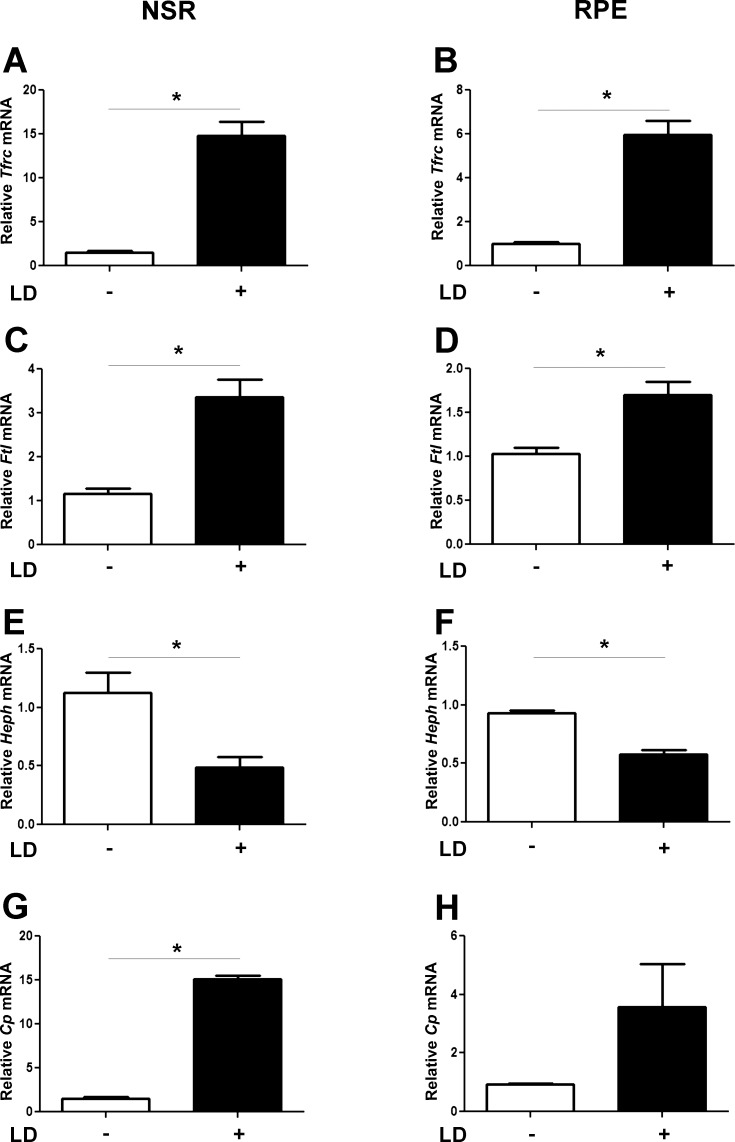
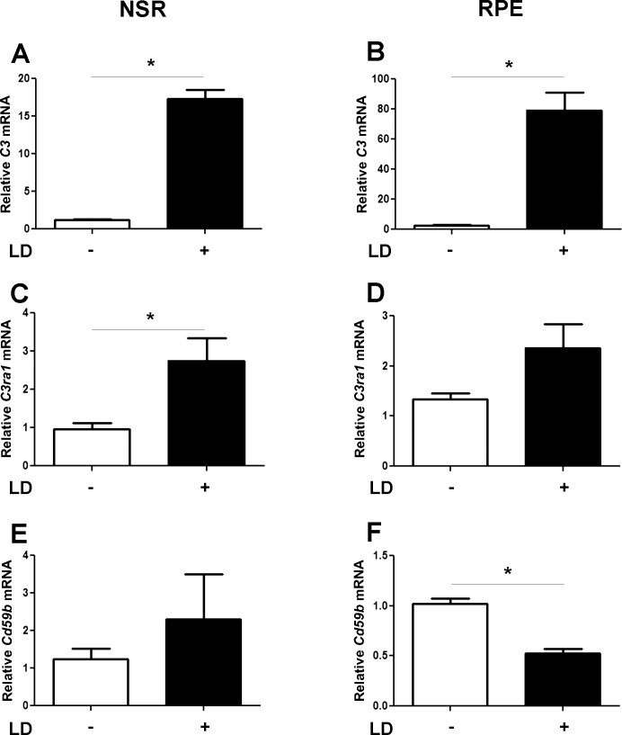
Methods: Mice were exposed to 10,000 lux cool white fluorescent light for 18 hours and euthanized 4 hours after photic injury. NSR and isolated RPE were collected, and RNA was isolated. DNA microarray hybridization was conducted as described in the Affymetrix GeneChip Expression Analysis Technical Manual. Microarray analysis was performed using probe intensity data derived from the Mouse Gene 1.0 ST Array. For the genes of interest, confirmation of gene expression was done using quantitative real-time PCR. Immunofluorescence assessed protein levels and localization.
Results: Numerous iron regulatory genes were significantly changed in the light-exposed NSR and RPE. Several of these gene expression changes favored an iron-overloaded state. For example, the transferrin receptor was upregulated in both light-exposed NSR and RPE. Consistent with this, there was stronger transferrin receptor immunoreactivity in the light-exposed retinas. Significant changes in gene expression following light damage were also observed in oxidative stress and complement system genes.
Conclusions: The concept of a photooxidative stress-induced vicious cycle of increased iron uptake leading to further oxidative stress was introduced.
Conflict of interest statement
Disclosure:
Figures


Similar articles
-
AMD-like retinopathy associated with intravenous iron.Exp Eye Res. 2016 Oct;151:122-33. doi: 10.1016/j.exer.2016.08.008. Epub 2016 Aug 23. Exp Eye Res. 2016. PMID: 27565570 Free PMC article.
-
Polarized distribution of heme transporters in retinal pigment epithelium and their regulation in the iron-overload disease hemochromatosis.Invest Ophthalmol Vis Sci. 2011 Nov 29;52(12):9279-86. doi: 10.1167/iovs.11-8264. Invest Ophthalmol Vis Sci. 2011. PMID: 22058337 Free PMC article.
-
Light damage induced changes in mouse retinal gene expression.Exp Eye Res. 2004 Aug;79(2):239-47. doi: 10.1016/j.exer.2004.05.002. Exp Eye Res. 2004. PMID: 15325571
-
CD1 Mouse Retina Is Shielded From Iron Overload Caused by a High Iron Diet.Invest Ophthalmol Vis Sci. 2015 Aug;56(9):5344-52. doi: 10.1167/iovs.15-17026. Invest Ophthalmol Vis Sci. 2015. PMID: 26275132 Free PMC article.
-
Endogenous and Exogenous Regulation of Redox Homeostasis in Retinal Pigment Epithelium Cells: An Updated Antioxidant Perspective.Int J Mol Sci. 2023 Jun 28;24(13):10776. doi: 10.3390/ijms241310776. Int J Mol Sci. 2023. PMID: 37445953 Free PMC article. Review.
Cited by
-
Degeneration modulates retinal response to transient exogenous oxidative injury.PLoS One. 2014 Feb 21;9(2):e87751. doi: 10.1371/journal.pone.0087751. eCollection 2014. PLoS One. 2014. PMID: 24586289 Free PMC article.
-
Correlation between peripapillary retinal nerve fiber layer thickness and fundus autofluorescence in primary open-angle glaucoma.Clin Ophthalmol. 2013;7:1883-8. doi: 10.2147/OPTH.S49112. Epub 2013 Sep 19. Clin Ophthalmol. 2013. PMID: 24092967 Free PMC article.
-
Iron importers Zip8 and Zip14 are expressed in retina and regulated by retinal iron levels.Exp Eye Res. 2017 Feb;155:15-23. doi: 10.1016/j.exer.2016.12.008. Epub 2017 Jan 3. Exp Eye Res. 2017. PMID: 28057442 Free PMC article.
-
EP300 Protects from Light-Induced Retinopathy in Zebrafish.Front Pharmacol. 2016 May 19;7:126. doi: 10.3389/fphar.2016.00126. eCollection 2016. Front Pharmacol. 2016. PMID: 27242532 Free PMC article.
-
Changes in gene expression associated with retinal degeneration in the rd3 mouse.Mol Vis. 2013 May 6;19:955-69. Print 2013. Mol Vis. 2013. PMID: 23687432 Free PMC article.
References
-
- Gitlin JD. Aceruloplasminemia. Pediatr Res. 1998;44:271–276 - PubMed
-
- Dunaief JL, Richa C, Franks EP, et al. Macular degeneration in a patient with aceruloplasminemia, a disease associated with retinal iron overload. Ophthalmology. 2005;112:1062–1065 - PubMed
-
- Fortuna F, Barboni P, Liguori R, et al. Visual system involvement in patients with Friedreich's ataxia. Brain. 2009;132:116–123 - PubMed
Publication types
MeSH terms
Substances
Grants and funding
LinkOut - more resources
Full Text Sources
Other Literature Sources
Medical
Molecular Biology Databases
