. 2012 Aug 10;287(33):27629-36.
doi: 10.1074/jbc.M112.391722. Epub 2012 Jun 26.

Modular dispensability of dysferlin C2 domains reveals rational design for mini-dysferlin molecules

Affiliations

Modular dispensability of dysferlin C2 domains reveals rational design for mini-dysferlin molecules

Bilal A Azakir et al. J Biol Chem. .

Retraction in

Abstract

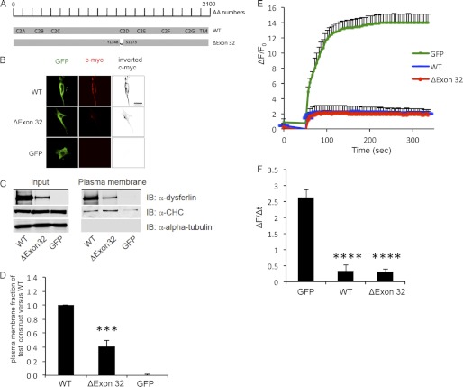
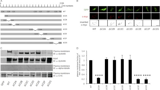
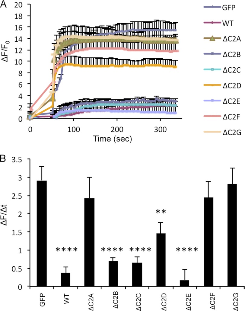
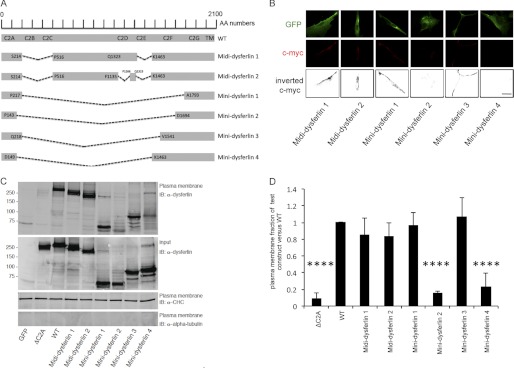
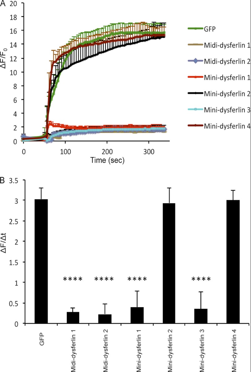
Dysferlin is a large transmembrane protein composed of a C-terminal transmembrane domain, two DysF domains, and seven C2 domains that mediate lipid- and protein-binding interactions. Recessive loss-of-function mutations in dysferlin lead to muscular dystrophies, for which no treatment is currently available. The large size of dysferlin precludes its encapsulation into an adeno-associated virus (AAV), the vector of choice for gene delivery to muscle. To design mini-dysferlin molecules suitable for AAV-mediated gene transfer, we tested internally truncated dysferlin constructs, each lacking one of the seven C2 domains, for their ability to localize to the plasma membrane and to repair laser-induced plasmalemmal wounds in dysferlin-deficient human myoblasts. We demonstrate that the dysferlin C2B, C2C, C2D, and C2E domains are dispensable for correct plasmalemmal localization. Furthermore, we show that the C2B, C2C, and C2E domains and, to a lesser extent, the C2D domain are dispensable for dysferlin membrane repair function. On the basis of these results, we designed small dysferlin molecules that can localize to the plasma membrane and reseal laser-induced plasmalemmal injuries and that are small enough to be incorporated into AAV. These results lay the groundwork for AAV-mediated gene therapy experiments in dysferlin-deficient mouse models.


Figures

FIGURE 1.
FIGURE 1.
DysferlinΔexon32 retains its biological function. A, schematic representation of the dysferlinΔexon32 mutation in the dysferlin protein sequence, compared with WT dysferlin. AA numbers, amino acid enumeration; TM, transmembrane domain. B, immunostaining using an extracellular c-Myc dysferlin epitope (red) in C2C12 myoblasts demonstrates plasma membrane localization of the overexpressed GFP-dysferlinΔexon32-Myc construct (ΔExon 32) and GFP-dysferlin-Myc (WT), but not the GFP vector alone (GFP). Immunostaining using GFP (green) and the GFP tag demonstrates total cellular expression of each construct. Inverted black and white images of c-Myc are shown in the right panels to better visualize the plasma membrane staining with anti-c-Myc antibody. Scale bar = 20 μm. C, COS-7 cells were transfected with GFP, WT dysferlin, or dysferlinΔexon32. Western blots of plasma membrane protein extracts were stained with anti-dysferlin antibodies, with anti-α-tubulin antibodies as a negative control, and with anti-clathrin heavy chain antibodies (α-CHC) as a positive control (n = 4). IB, immunoblot. D, graphical representation of C showing the ratio between the amount of the plasmalemmal dysferlin construct versus the total dysferlin construct compared with the ratio between the amount of plasmalemmal WT dysferlin versus total WT dysferlin. E, plasma membrane repair assay was performed on the dysferlin-deficient myoblast culture (ULM1/01) transfected with GFP, WT dysferlin, or dysferlinΔexon32. The relative fluorescence intensity (ΔF/F0) over time following laser-induced injury is represented as means ± S.D. Numbers of individual measurements are as follows: for GFP, n = 12; for dysferlinΔexon32, n = 16; and for WT dysferlin, n = 16. F, graphical representation of E depicting the change in relative fluorescence intensity at 5 min post-injury. ***, p < 0.001; ****, p < 0.0001.
FIGURE 2.
FIGURE 2.
GFP-tagged dysferlinΔC2B, dysferlinΔC2C, dysferlinΔC2D, and dysferlinΔC2E localize to the plasma membrane. A, schematic representation of the deleted C2 domain regions in the dysferlin protein sequence compared with WT dysferlin. AA numbers, amino acid enumeration; TM, transmembrane domain. B, immunostaining using the extracellular c-Myc dysferlin epitope (red) in C2C12 myoblasts transfected with the indicated plasmids. Immunostaining using GFP (green) demonstrates cellular expression of each construct. Inverted black and white images of c-Myc are represented in the lower panels to better visualize the plasma membrane staining with the anti-c-Myc antibody. Scale bar = 20 μm. C, COS-7 cells were transfected with the indicated plasmids. Western blots of plasma membrane protein extracts were stained with anti-dysferlin antibodies, with anti-α-tubulin antibodies as a negative control, and with anti-clathrin heavy chain antibodies (α-CHC) as a positive control (n = 4). IB, immunoblot. D, graphical representation of C showing the ratio of the amount of the plasmalemmal dysferlin construct versus the total dysferlin construct compared with the ratio of the amount of plasmalemmal WT dysferlin versus total WT dysferlin. ****, p < 0.0001.
FIGURE 3.
FIGURE 3.
GFP-tagged dysferlinΔC2B, dysferlinΔC2C, and dysferlinΔC2E restore defect in the membrane repair of dysferlin-deficient myoblasts. A, plasma membrane repair assay was performed on the dysferlin-deficient myoblast culture (ULM1/01) transfected with the indicated plasmids. The relative fluorescence intensity (ΔF/F0) over time following laser-induced injury is presented as means ± S.D. Numbers of individual measurements are as follows: for GFP, n = 12; and for WT dysferlin and dysferlinΔC2A–ΔC2G, n = 15 for each construct. B, graphical representation of A depicting the change in relative fluorescence intensity at 5 min post-injury. **, p < 0.01; ****, p < 0.0001.
FIGURE 4.
FIGURE 4.
Midi-dysferlins 1 and 2 and mini-dysferlins 1 and 3 localize to the plasma membrane. A, schematic representation of the deleted amino acid regions in the dysferlin protein sequence compared with WT dysferlin. A short amino acid sequence adjacent to the C2A domain was used as the linker sequence in mini-dysferlins 1 and 3. A short amino sequence adjacent to the C2G or C2F domain was used as the linker sequence in mini-dysferlins 2 and 4, respectively. AA numbers, amino acid enumeration; TM, transmembrane domain. B, immunostaining using the extracellular c-Myc dysferlin epitope (red) in C2C12 myoblasts transfected with the indicated plasmids. Immunostaining using GFP (green) demonstrates total cellular expression of each construct. Inverted black and white images of c-Myc are represented in the lower panels to better visualize the plasma membrane staining with the anti-c-Myc antibody. Scale bar = 20 μm. C, COS-7 cells were transfected with the indicated plasmids. Western blots of plasma membrane protein extracts were stained with anti-dysferlin antibodies, with anti-α-tubulin antibodies as a negative control, and with anti-clathrin heavy chain antibodies (α-CHC) as a positive control (n = 4). IB, immunoblot. D, graphical representation of C showing the ratio of the amount of plasmalemmal dysferlin construct versus the total dysferlin construct compared with the ratio of the amount of plasmalemmal WT dysferlin versus total WT dysferlin. ****, p < 0.0001.
FIGURE 5.
FIGURE 5.
Midi-dysferlins 1 and 2 and mini-dysferlins 1 and 3 restore the defect in membrane repair. A, plasma membrane repair assay was performed on the dysferlin-deficient myoblast culture (ULM1/01) transfected with the indicated plasmids. The relative fluorescence intensity (ΔF/F0) over time following laser-induced injury is presented as means ± S.D. Numbers of individual measurements are as follows: for GFP, n = 15; for midi-dysferlins 1 and 2, n = 20 for each construct; for mini-dysferlin 1, n = 24; for mini-dysferlin 2, n = 20; for mini-dysferlin 3, n = 21; and for mini-dysferlin 4, n = 18. B, graphical representation of A depicting the change in relative fluorescence intensity at 5 min post-injury. ****, p < 0.0001.

Similar articles

Cited by

References

    1. Therrien C., Di Fulvio S., Pickles S., Sinnreich M. (2009) Characterization of lipid binding specificities of dysferlin C2 domains reveals novel interactions with phosphoinositides. Biochemistry 48, 2377–2384 - PubMed
    1. Davis D. B., Doherty K. R., Delmonte A. J., McNally E. M. (2002) Calcium-sensitive phospholipid binding properties of normal and mutant ferlin C2 domains. J. Biol. Chem. 277, 22883–22888 - PubMed
    1. Lennon N. J., Kho A., Bacskai B. J., Perlmutter S. L., Hyman B. T., Brown R. H., Jr. (2003) Dysferlin interacts with annexins A1 and A2 and mediates sarcolemmal wound-healing. J. Biol. Chem. 278, 50466–50473 - PubMed
    1. Azakir B. A., Di Fulvio S., Therrien C., Sinnreich M. (2010) Dysferlin interacts with tubulin and microtubules in mouse skeletal muscle. PLoS ONE 5, e10122. - PMC - PubMed
    1. Cai C., Weisleder N., Ko J. K., Komazaki S., Sunada Y., Nishi M., Takeshima H., Ma J. (2009) Membrane repair defects in muscular dystrophy are linked to altered interaction between MG53, caveolin-3, and dysferlin. J. Biol. Chem. 284, 15894–15902 - PMC - PubMed

Publication types

MeSH terms