. 2012 Aug 17;287(34):28362-77.
doi: 10.1074/jbc.M111.335679. Epub 2012 Jun 26.

Rapid uptake and degradation of CXCL12 depend on CXCR7 carboxyl-terminal serine/threonine residues

Affiliations

Rapid uptake and degradation of CXCL12 depend on CXCR7 carboxyl-terminal serine/threonine residues

Frauke Hoffmann et al. J Biol Chem. .

Abstract

CXCL12 signaling through G protein-coupled CXCR4 regulates cell migration during ontogenesis and disease states including cancer and inflammation. The second CXCL12-receptor CXCR7 modulates the CXCL12/CXCR4 pathway by acting as a CXCL12 scavenger and exerts G protein-independent functions. Given the distinct properties of CXCR4 and CXCR7, we hypothesized that the distinct C-terminal domains differently regulate receptor trafficking and stability. Here, we examined epitope-tagged wild type and C-terminal mutant receptors in human embryonic kidney cells (HEK293) with respect to trafficking, stability, (125)I-CXCL12 degradation, and G protein-coupling. The 24 CXCR7 C-terminal residues were sufficient to promote rapid spontaneous internalization. Replacement of the CXCR7 C terminus with that of CXCR4 (CXCR7-4tail mutant) abolished spontaneous internalization but permitted ligand-induced internalization and phosphorylation at the heterologous domain. The reverse tail-swap caused ligand-independent internalization of the resulting CXCR4-7tail mutant. Receptor-mediated (125)I-CXCL12 uptake and release of (125)I-CXCL12 degradation products were accelerated with receptors bearing the CXCR7 C terminus and impaired after conversion of CXCR7 C-terminal serine/threonine residues into alanines. C-terminal lysine residues were dispensable for plasma membrane targeting and the CXCL12 scavenger function but involved in constitutive degradation of CXCR7. Although the CXCR7 C terminus abolished G protein coupling in the CXCR4-7tail mutant, replacement of the CXCR7 C terminus, CXCR7 second intracellular loop, or both domains with the corresponding CXCR4 domain did not result in a G protein-coupled CXCR7 chimera. Taken together, we provide evidence that the CXCR7 C terminus influences the ligand-uptake/degradation rate, G protein coupling, and receptor stability. Regulatory pathways targeting CXCR7 C-terminal serine/threonine sites may control the CXCL12 scavenger activity of CXCR7.


Figures

FIGURE 1.
FIGURE 1.
Differences in ligand-independent internalization of wild type and tail-swap mutant CXCR4 and CXCR7 receptors. HEK293 cells were transiently transfected with constructs for HA-tagged receptors as indicated. A–H, surface receptors were pulse labeled with anti-HA antibody applied to live HEK293 cells at 4 °C. Confocal images show the subcellular localization of the labeled receptors after the anti-HA pulse (A–D) or after a 30-min ligand-free interval at 37 °C (E–H). I, quantitative analysis of receptor internalization by ELISA. Surface receptor levels after different ligand-free intervals are given as percentage of the surface receptor level immediately after pulse labeling (starting value). Data represent mean ± S.E. calculated from 4 independent experiments with 4 repeats each. Two-way ANOVA comparing wild type receptor and tail-swap mutant. Scale bar = 10 μm in A–H.
FIGURE 2.
FIGURE 2.
Motifs in the CXCR7 C-terminal domain regulating ligand-independent receptor internalization. HEK293 cells were transiently transfected with constructs for HA-tagged receptors carrying mutations in putative internalization regulating C-terminal motifs. In CXCR7–4-7 and CXCR7-Δ19 residues 320–338 of CXCR7 (putative helix eight-containing region) were replaced by the corresponding CXCR4 sequence or deleted. In CXCR4–7(C9) the last nine residues of CXCR4 were replaced by those of CXCR7. In CXCR7-Y/A and CXCR7-K/R all C-terminal tyrosine and lysine residues were replaced by alanines and arginines, respectively. A–J, surface receptors were pulse labeled by anti-HA antibody applied to live cells at 4 °C. Confocal images show the subcellular localization of label receptors immediately after the anti-HA pulse (0 min, A–E) or after a 30-min ligand-free interval at 37 °C (F–J). A, B, F, and G, CXCR7 residues 320–338 are not involved in ligand-independent CXCR7 internalization. C and H, the last nine CXCR7 C-terminal residues containing a YSAL motif are not sufficient for ligand-independent CXCR4 internalization. D, E, I, and J, C-terminal tyrosine and lysine residues are not critical for plasma membrane targeting and ligand-independent internalization of CXCR7. K, quantitative analysis of receptor internalization by ELISA. For each receptor, the surface receptor level after the 30-min ligand-free interval is expressed as percentage of its surface receptor level after 0 min (nonpermeabilized cells). L-O, fixed permeabilized and nonpermeabilized (nonperm.) transfectants of CXCR7-WT and CXCR7-K/R were immunostained with anti-HA antibody. Both receptors are present at the plasma membrane. Permeabilization reveals preponderant intracellular localization of CXCR7-WT and CXCR7-K/R. P, analysis of CXCR7-WT and CXCR7-K/R surface receptor expression in nonpermeabilized cells subjected to 3-h vehicle (veh.) or 3-h CHX treatments using ELISA. Student's t test comparing signals after 30 versus 0 min (*), vehicle versus CHX (§), and CXCR7-WT versus CXCR7-K/R ($). Data represent mean ± S.E. calculated from 3 independent experiments with 4 repeats each. Scale bar = 10 μm in A–J and L–O.
FIGURE 3.
FIGURE 3.
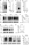
The CXCR7 C-terminal domain regulates receptor degradation. A–J, immunoblots with wheat germ lectin agarose-purified lysates of HEK293 cells transiently transfected with empty vector (mock) or N-terminal HA-epitope-tagged wild type (7WT, 4WT), tail-swap mutant (7–4tail, 4–7tail) receptors, and CXCR7-K/R (7-K/R). The recombinantly expressed chemokine receptors were identified with an anti-HA antibody. The endogenous transferrin receptor (TFR) was labeled after stripping and served as loading control. For quantitative analysis of chemokine receptor expression levels, the HA/TFR-ratio was determined in at least 3 independent experiments using CCD camera-based densitometry and expressed as percentage of the group indicated in the figure. A, the anti HA-antibody recognizes the CXCR7-WT and CXCR7–4tail glycoproteins as a smear ranging from 50 to 70 kDa and produces a nonspecific signal at 80 kDa. B, the expression level of CXCR7–4tail is 6.6-fold higher than that of CXCR7-WT. C and D, CXCR4-WT and CXCR4–7tail migrate at 47 kDa with CXCR4-WT showing a 5-fold stronger signal than CXCR4–7tail. E-H, kinetics of receptor degradation in nonstimulated cultures receiving CHX for 0, 1, and 3 h to block protein synthesis. Blots with receptors carrying the CXCR7 C terminus were imaged with longer exposure times. Receptor degradation is attenuated in CXCR7-K/R and CXCR7–4tail as compared with CXCR7-WT and accelerated in CXCR4–7tail as compared with CXCR4-WT. I and J, influence of long term CXCL12 treatment (16 h) on receptor expression levels (CHX-free condition). CXCL12 treatment causes a 70% decrease of recombinant CXCR4 in CXCR4-WT transfectants. Levels of recombinant receptors are not affected by CXCL12 in CXCR7-WT, CXCR7–4tail, and CXCR4–7tail transfectants. *, Student's t test; #, two-way ANOVA; §, one-way ANOVA. Data represent mean ± S.E. calculated from three to five independent experiments.
FIGURE 4.
FIGURE 4.
G protein coupling is absent in wild type CXCR7 and CXCR7/CXCR4-chimeric receptors. A–H, HEK293 cells were transiently transfected with wild type receptors (CXCR7-WT, CXCR4-WT) and tail-swap mutants (CXCR7–4tail, CXCR4–7tail). A and B, homologous radioligand competition binding using 125I-CXCL12 (25 pm) and increasing concentrations of nonlabeled CXCL12. A and B, transfection with CXCR7-WT and CXCR7–4tail chimeras in which the second cytosolic domain (2cD) was replaced with the corresponding region of CXCR4 (mutants are referred to as CXCR7–2cD4 and CXCR7–2cD4–4tail). Results in A and B represent 4 independent experiments (3 repeats each) after normalization with the calculated top binding obtained for CXCR7-WT (A) and CXCR7–2cD4 (B). Potency of CXCR7-WT is 10-fold higher than potency of CXCR4-WT and CXCR7–4tail. Note that replacement of the 2cD does not affect potency of CXCR7-WT and CXCR7–4tail. C-F, CXCR7–2cD4 and CXCR7–2cD4–4tail transfectants were pulse-labeled with anti-HA antibody (4 °C). Cells were washed, fixed, permeabilized, and detected immediately after loading with the HA antibody (0 min) or after a 30-min ligand-free interval at 37 °C. C and E, both chimeras are present at the plasma membrane. D and F, pulse-labeled CXCR7–2cD4 surface receptors are rapidly internalized, whereas the CXCR7–2cD4–4tail receptors remain at the plasma membrane. G and H, analysis of G protein coupling. G, cells were transfected with the qi5 chimera alone or with CXCR4-WT in combination with qi5. Stimulation occurred at t = 30 s with 80 nm CXCL12. AMD3100 was applied 15 min prior to recording. Data were baseline corrected and traces were calculated as averaged traces from a single experiment with each trace representing data from three to four wells. H, mean maximum fluorescence amplitudes (± S.D.) were obtained. Experimental set up was as described for G, only that cells were transfected with qi5 in combination with different receptor constructs. Data were derived from two to three independent transfections with a minimum of three repeats (wells) per receptor. *, Student's t test.
FIGURE 5.
FIGURE 5.
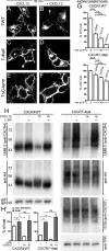
Ligand-dependent activation and internalization of the CXCR7–4tail mutant. HEK293 cells were transiently transfected with CXCR7-WT, CXCR7–4tail, CXCR7ΔC-term, or CXCR4-WT as indicated. A–G, surface receptors were pulse labeled with anti-HA (A–D and G) or 11G8 anti-CXCR7 (E and F) antibody at 4 °C. A–F, confocal images show the subcellular receptor localization after 30 min at 37 °C in the absence (A, C, and E) or presence of CXCL12 (B, D, and F). CXCR7–4tail undergoes only ligand-dependent internalization (C and D), whereas CXCR7ΔC-term is internalization defective (E and F) and CXCR7-WT internalizes ligand independently (A and B). G, quantitative analysis of ligand-induced receptor internalization by ELISA. Cultures were incubated for 30 min at 37 °C with vehicle, CCX733 or CXCL12. Surface receptor levels are given as percentage of the surface receptor level immediately after pulse labeling (0 min). Results represent mean ± S.E. calculated from three to four independent experiments with 4 repeats each. Note that CCX733 and CXCL12 significantly increase receptor internalization. Asterisks indicate differences between the indicated groups (one-way ANOVA). H, immunoblots of wheat germ lectin-agarose (WGA)-purified cell lysates. Cultures were stimulated with CXCL12 and lysates were dephosphorylated with λ-PP as indicated. Upper panel, detection of CXCR4-WT and CXCR7–4tail protein using the anti-CXCR4 antibody UMB-2, which recognizes only the nonphosphorylated C-terminal CXCR4 epitope. Comparison of λ-PP-treated and λ-PP-untreated samples of nonstimulated cultures (lanes 1 and 2) indicates little constitutive phosphorylation of the UMB-2 epitope in CXCR4-WT but strong constitutive phosphorylation in CXCR7–4tail. CXCL12 treatment causes almost complete phosphorylation of the UMB-2 epitope both in CXCR4-WT and CXCR7–4tail (lanes 3 and 4). Middle and lower panels, aliquots of the samples shown in the upper panel were detected with anti-HA, 11G8, and anti-transferrin receptor (TFR) antibodies as indicated to confirm equal protein loading. H′, quantitative analyses of UMB-2 signal intensities in immunoblots from CXCR4-WT and CXCR7–4tail-transfected HEK293 cells using CCD camera-based densitometry. Relative amounts of nonphosphorylated (non-phospho) and total receptors were determined by measuring the UMB-2/HA ratios in λ-PP-untreated and λ-PP-treated samples, respectively. Results from 3 independent experiments were averaged and expressed as percentage of total receptor in cultures not receiving CXCL12 (*, p < 0.05, paired Student's t test).
FIGURE 6.
FIGURE 6.
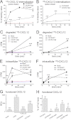
Rapid endocytosis and degradation of 125I-CXCL12 depend on the CXCR7 C-terminal domain. HEK293 cells were transiently transfected with empty vector (mock), wild type receptors (CXCR7-WT, CXCR4-WT), tail-swap mutants (CXCR7–4tail, CXCR4–7tail), or CXCR7ΔC-term as indicated. A and B, internalization kinetics of receptor-radioligand complexes. Cells were loaded with radioligand at 4 °C (pulse), washed, and lysed immediately (starting value) or incubated for the indicated time intervals at 37 °C and lysed after an acidic wash (chase). Data show internalized 125I-CXCL12 of acid-washed groups as percent of the starting value. Receptors carrying the CXCR7 C-terminal domain mediate faster radioligand uptake than receptors carrying the CXCR4 C-terminal domain. CXCR7ΔC-term fails to internalize 125I-CXCL12. Results represent mean ± S.E. from 2 independent experiments with 5 repeats each. C and D, scavenging activity. Cells were incubated with 125I-CXCL12 for the indicated time intervals at 37 °C. Then, supernatants were subjected to TCA precipitation to separate degraded 125I-CXCL12 from precipitated 125I-CXCL12. Note faster accumulation of 125I-CXCL12 degradation products in culture supernatants of CXCR7-WT and CXCR4–7tail transfectants than in culture supernatants of CXCR7–4tail and CXCR4-WT transfectants, respectively. There is no significant increase in extracellular125I-CXCL12 degradation products in culture supernatants of CXCR7ΔC-term transfectants. E and F, intracellular accumulation of 125I-CXCL12. Transfectants were incubated with radioligand for the indicated time at 37 °C and subjected to an acidic wash to strip residual surface-bound 125I-CXCL12 before lysis and measurement of intracellular 125I. In cultures transfected with receptors carrying the CXCR7 C terminus, intracellular 125I-CXCL12 levels increase rapidly before decreasing (CXCR7-WT) or reaching a steady state (CXCR4–7tail). Cells overexpressing receptors with the CXCR4 C terminus continue to accumulate intracellular 125I-CXCL12 during the entire experimental period. CXCR7ΔC-term shows no intracellular accumulation of 125I-CXCL12. C–F, data are mean ± S.E. from 2 independent experiments with 4 repeats each. A–F, two-way ANOVA. Asterisks indicate significant differences between wild type receptors and corresponding tail-swap mutants. G and H, influence of receptor-mediated scavenger activity on functional CXCL12. CXCL12 (20 nm) was incubated overnight with HEK293 cells transfected with the indicated receptors. Supernatants were used to stimulate transwell migration of Jurkat T cells. G, graph shows fluorescence intensities of calcein-AM-stained migrated Jurkat T cells. CXCL12-supplemented medium incubated with mock-transfected cells causes a 9-fold increase in cell migration as compared with nonsupplemented medium. Preincubation with CXCR7-WT abolishes and preincubation with CXCR7ΔC-term reduces the migratory response. *, Student's t tests versus mock + CXCL12. H, migratory response with supernatants from CXCR7ΔC-term was used to normalize responses with supernatants from wild type and tail-swap mutant receptors. *, §, Student's t tests versus corresponding wild type receptor (*) or versus CXCR7ΔC-term (§). Data in G and H are 2–4 independent experiments with 2–6 repeats each.
FIGURE 7.
FIGURE 7.
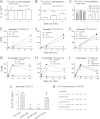
Influence of C-terminal lysine and serine/threonine residues on CXCR7-mediated endocytosis and degradation of CXCL12. HEK293 cells were transiently transfected with empty vector (mock), CXCR7-WT, CXCR7-K/R, a CXCR7-ST/A mutant lacking all C-terminal serine and threonine residues, CXCR7-STT/A with S335A, T338A, and T341A conversions, and CXCR7-STS/A with S350A, T352A, and S355A conversions. A–C, receptor-mediated internalization of 125I-CXCL12. Cells were loaded with radioligand at 4 °C (pulse), PBS washed, and lysed immediately (starting value) or incubated for the indicated time intervals at 37 °C and lysed after an acidic wash (chase). Percent internalization was calculated by dividing counts of internalized 125I-CXCL12 in the acid-washed groups by the starting value. Internalization data were then normalized to CXCR7-WT-transfected sister cultures (100% line). A, CXCR7-K/R and CXCR7-WT mediate similar 125I-CXCL12 internalization. B and C, as compared with CXCR7-WT, 125I-CXCL12 internalization via CXCR7-ST/A is reduced at 5, 15, and 30 min and 125I-CXCL12 internalization via CXCR7-STT/A and CXCR7-STS/A is reduced at 5 min after pulse labeling. D–F, release of degraded 125I-CXCL12. Cells were incubated with 125I-CXCL12 as indicated. Supernatants were subjected to TCA precipitation before degraded 125I-CXCL12 was counted. As compared with CXCR7-WT, CXCR7-K/R, CXCR7-ST/A, and CXCR7-STS/A cause slower accumulation of extracellular 125I-CXCL12 degradation products. G and H, intracellular accumulation of 125I-CXCL12. Cells were incubated with 125I-CXCL12 as indicated, subjected to acidic wash, and lysed. As compared with CXCR7-WT, CXCR7-K/R transfectants accumulate more, whereas CXCR7-ST/A and CXCR7-STS/A transfectants accumulate less 125I-CXCL12. A–I, two-way ANOVA. Asterisks indicate significant differences between wild type and mutant receptors. Data represent mean ± S.E. calculated from 2 independent experiments with 4 repeats each. J, influence of receptor-mediated scavenger activity on functional CXCL12. CXCL12 (20 nm) was incubated overnight with HEK293 cells transfected with the indicated receptors. Supernatants were used to stimulate transwell migration of Jurkat T cells. Migratory response with supernatants from CXCR7ΔC-term was used for normalization. * and §, Student's t tests versus CXCR7-WT (*) or versus CXCR7ΔC-term (§). Data are 3 independent experiments with 2–6 repeats each. K, schematic representation of the CXCR7 C terminus showing C-terminal serine (S) and threonine (T) residues and ST/A conversions in the mutant receptors.

Similar articles

Cited by

References

    1. Sierro F., Biben C., Martínez-Muñoz L., Mellado M., Ransohoff R. M., Li M., Woehl B., Leung H., Groom J., Batten M., Harvey R. P., Martínez-A C., Mackay C. R., Mackay F. (2007) Disrupted cardiac development but normal hematopoiesis in mice deficient in the second CXCL12/SDF-1 receptor, CXCR7. Proc. Natl. Acad. Sci. U.S.A. 104, 14759–14764 - PMC - PubMed
    1. Sun X., Cheng G., Hao M., Zheng J., Zhou X., Zhang J., Taichman R. S., Pienta K. J., Wang J. (2010) CXCL12/CXCR4/CXCR7 chemokine axis and cancer progression. Cancer Metastasis Rev. 29, 709–722 - PMC - PubMed
    1. Zou Y. R., Kottmann A. H., Kuroda M., Taniuchi I., Littman D. R. (1998) Function of the chemokine receptor CXCR4 in hematopoiesis and in cerebellar development. Nature 393, 595–599 - PubMed
    1. Nagasawa T., Hirota S., Tachibana K., Takakura N., Nishikawa S., Kitamura Y., Yoshida N., Kikutani H., Kishimoto T. (1996) Defects of B-cell lymphopoiesis and bone-marrow myelopoiesis in mice lacking the CXC chemokine PBSF/SDF-1. Nature 382, 635–638 - PubMed
    1. Ma Q., Jones D., Borghesani P. R., Segal R. A., Nagasawa T., Kishimoto T., Bronson R. T., Springer T. A. (1998) Impaired B-lymphopoiesis, myelopoiesis, and derailed cerebellar neuron migration in CXCR4- and SDF-1-deficient mice. Proc. Natl. Acad. Sci. U.S.A. 95, 9448–9453 - PMC - PubMed

Publication types

LinkOut - more resources