. 2012;8(6):e1002755.
doi: 10.1371/journal.ppat.1002755. Epub 2012 Jun 21.

Structural and functional insights into the malaria parasite moving junction complex

Affiliations

Structural and functional insights into the malaria parasite moving junction complex

Brigitte Vulliez-Le Normand et al. PLoS Pathog. 2012.

Abstract

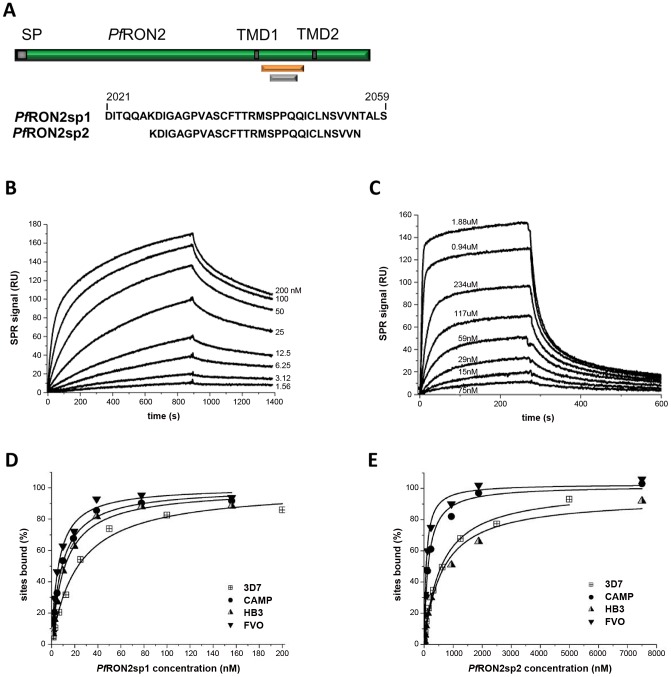
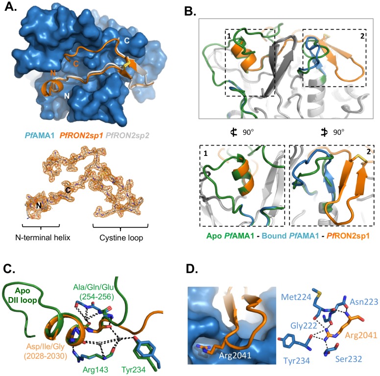
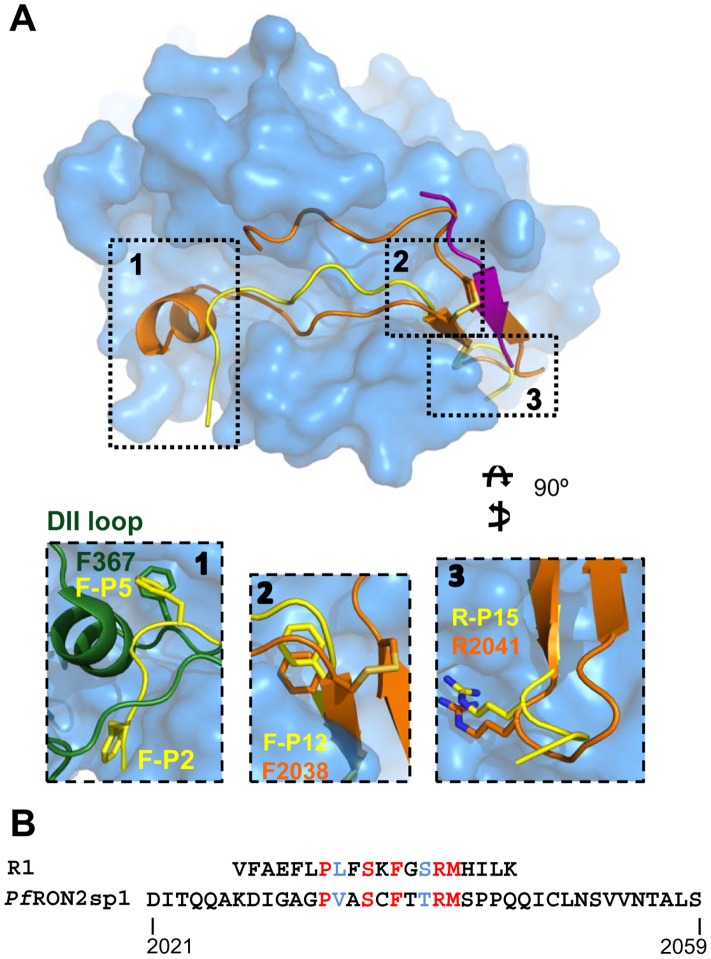
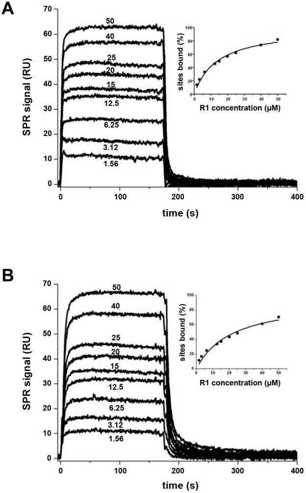
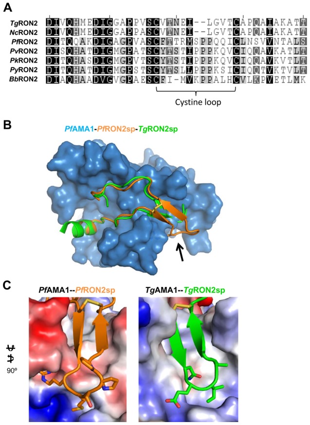
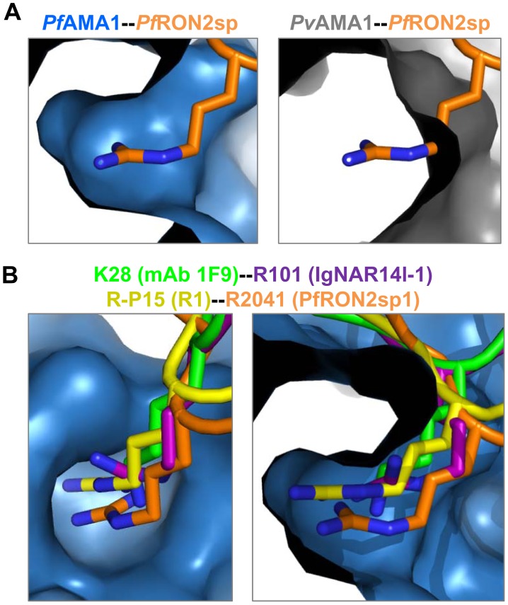
Members of the phylum Apicomplexa, which include the malaria parasite Plasmodium, share many features in their invasion mechanism in spite of their diverse host cell specificities and life cycle characteristics. The formation of a moving junction (MJ) between the membranes of the invading apicomplexan parasite and the host cell is common to these intracellular pathogens. The MJ contains two key parasite components: the surface protein Apical Membrane Antigen 1 (AMA1) and its receptor, the Rhoptry Neck Protein (RON) complex, which is targeted to the host cell membrane during invasion. In particular, RON2, a transmembrane component of the RON complex, interacts directly with AMA1. Here, we report the crystal structure of AMA1 from Plasmodium falciparum in complex with a peptide derived from the extracellular region of PfRON2, highlighting clear specificities of the P. falciparum RON2-AMA1 interaction. The receptor-binding site of PfAMA1 comprises the hydrophobic groove and a region that becomes exposed by displacement of the flexible Domain II loop. Mutations of key contact residues of PfRON2 and PfAMA1 abrogate binding between the recombinant proteins. Although PfRON2 contacts some polymorphic residues, binding studies with PfAMA1 from different strains show that these have little effect on affinity. Moreover, we demonstrate that the PfRON2 peptide inhibits erythrocyte invasion by P. falciparum merozoites and that this strong inhibitory potency is not affected by AMA1 polymorphisms. In parallel, we have determined the crystal structure of PfAMA1 in complex with the invasion-inhibitory peptide R1 derived by phage display, revealing an unexpected structural mimicry of the PfRON2 peptide. These results identify the key residues governing the interactions between AMA1 and RON2 in P. falciparum and suggest novel approaches to antimalarial therapeutics.


Conflict of interest statement

I have read the journal's policy and have the following conflicts: Patent as a conflict of interest (The content of this manuscript has been the object of a European Patent Application filed on May 6, 2011 under the application number EP 11305540.4).

Figures

Figure 1
Figure 1. Surface Plasmon Resonance studies of peptides PfRON2sp1 and PfRON2sp2 binding to recombinant PfAMA1 from multiple strains reveal that PfRON2sp1 has a consistently higher affinity.
(A) PfRON2sp1 (orange) and PfRON2sp2 (grey) represent peptides of PfRON2 (green). SP, signal peptide. TMD, putative transmembrane domain. (B). Sensorgrams showing PfRON2sp1 (analyte) binding to PfAMA1 3D7 (immobilized). The PfRON2sp1 concentrations are indicated for each curve (nM). (C). Sensorgrams showing PfRON2sp2 (analyte) binding to PfAMA1 CAMP (immobilized), with PfRON2sp2 concentrations indicated. (D, E). Variation percentage of bound sites (deduced from the steady-state response) with respect to analyte concentration (D, PfRON2sp1; E, PfRON2sp2) obtained from binding to immobilized recombinant PfAMA1 from strains 3D7 (shown in B), CAMP (shown in C), FVO and HB3. The derived apparent equilibrium dissociation constants KD are given in Table 1.
Figure 2
Figure 2. Structure of PfAMA1 complexed with PfRON2-derived peptides.
(A) Top - Co-crystal structures of PfAMA1 (blue surface) with PfRON2sp1 (orange) and PfRON2sp2 (grey), show a disulfide-anchored U-shaped conformation in the apical groove of PfAMA1. Bottom - Electron density map (orange) for PfRON2sp1 contoured at 1.0 σ, highlighting well ordered density from the N-terminal helix, through the cystine loop, to the C-terminal coil. (B) Notable changes in the structure of PfAMA1 between the apo structure (green; PDB ID 1Z40) and the PfAMA1-PfRON2sp1 co-structure (blue-orange) as observed from a side view. Box 1 - The DII loop of apo PfAMA1 is ejected from the apical groove during binding to PfRON2sp1, leaving room for the PfRON2sp1 N-terminal helix to occupy the space vacated by the DII loop helix. Box 2 - The β-strands of the PfRON2sp1 cystine loop order a PfAMA1 surface loop, generating a contiguous three-stranded β-sheet. (C) In the region of the PfRON2sp1 N-terminal helix, there is notable structural mimicry to the PfAMA1 apo DII loop, including several conserved residues, and a conserved hydrogen bonding network incorporating three buried water molecules. (D) Arg2041, specific to P. falciparum, fits snugly into a deep pocket in the surface of PfAMA1 and is stabilized through a complex network of seven hydrogen bonds.
Figure 3
Figure 3. Structure of PfAMA1 complexed with R1 peptide.
(A). The co-crystal structure of PfAMA1 (blue surface) with R1 reveals two bound peptides, R1 major (yellow) and R1 minor (purple). (B). Detailed analysis of interactions at the PfAMA1–R1-major, PfAMA1–R1-minor, and R1-major–R1-minor interfaces. Surface representation of PfAMA1 (blue), with R1-major (yellow) and R1-minor (purple) shown as cartoons. Box 1 – R1-major anchors its N-terminus to PfAMA1 through 3 backbone hydrogen bonds. Box 2 – the central region of the PfAMA1 apical groove is occupied by R1-major through both hydrophobic and polar interactions. Box 3 – R1-minor forms most of its anchor points to PfAMA1 through the apical loops and does not contact the base of the groove, which is occupied by R1-major. Panel 4 – Backbone hydrogen bonds between R1-minor and R1-major generate a β-sheet, while R1-major is further pinned to the PfAMA1 groove through 3 hydrogen bonds. Panel 5 – R1-major integrates into PfAMA1 with the use of an arginine knob-in-hole interaction stabilized by 6 hydrogen bonds, which is also exploited byPfRON2sp.
Figure 4
Figure 4. Structural mimicry of PfRON2 by peptide R1 in binding to PfAMA1.
(A) Top (left) and end-on (right) views of PfAMA1-PfRON2sp1 (orange cartoon) overlayed on PfAMA1-R1-major (yellow)/R1-minor (purple), show that the PfAMA1 groove is capable of accepting only PfRON2sp1 or the two R1 peptides at one time. Box 1 shows that Phe-P5 of R1 mimics Phe367 of the DII loop, while boxes 2 and 3 highlight spatial conservation of a phenylalanine anchor at the center of the groove, and a knob-in-hole interaction incorporating the peptide Arg-P15. R1-major is shown in yellow, PfRON2sp1 in orange and apo PfAMA1 in green. (B). Comparison of the R1 and PfRON2sp1 sequences reveals five identical (red) and two similar (blue) residues.
Figure 5
Figure 5. Highly potent cross-strain inhibition of red blood cell invasion of PfRON2sp1.
Comparison of PfRON2sp1 and R1 peptides (concentrations 0.2 to 20 µM) in inhibiting red blood cell invasion by P. falciparum 3D7 or HB3 highlights the higher inhibitory efficiency and cross-strain reactivity of PfRON2sp1. Parasitemia of control infected red blood cells (IRBC) 16 hours post-invasion was used as the 100% invasion reference. Means (± SD for N = 3) are shown.
Figure 6
Figure 6. Surface Plasmon Resonance studies of peptide R1 binding to PfAMA1 mutants 3D7mut and Dico3.
(A). Left - sensorgrams, showing R1 (analyte) binding to PfAMA1 3D7mut (immobilized). R1 concentrations are indicated for each curve (µM). Right - the variation in percentage of bound sites (deduced from the steady-state response) with respect to analyte concentration. (B). Left - sensorgrams, showing R1 (analyte) binding to Dico3 (immobilized), with R1 concentrations indicated. Right - the variation in percentage of bound sites (deduced from the steady-state response) with respect to analyte concentration. The equilibrium dissociation constant KD derived from the steady state binding curves is 15.2 µM for 3D7mut and 22.3 µM for Dico3.
Figure 7
Figure 7. Mutations of PfAMA1 and PfRON2-5 reveal residues critical for high affinity interaction.
(A) Interface between PfAMA1 and PfRON2sp1 shown in open-book presentation. Residues of both components that were mutated are labeled. (B). Binding characteristics of recombinant GST-PfRON2-5 mutants to dissect hot-spot residues in PfRON2. PfAMA1-expressing BHK-21 cells were incubated with 10 µg/ml of PfRON2 or mutated proteins (GST-fusion proteins), washed and the binding of recombinant PfRON2 fragment was revealed with anti-GST antibody. PfAMA1 was detected with mAb F8.12.19, which recognizes extracellular Domain III. (C). Binding consequences of PfAMA1 mutations. Mutated versions of PfAMA1 were expressed on the surface of BHK-21 cells and incubated with wild-type PfRON2 recombinant proteins at 10 and 1 µg/ml.
Figure 8
Figure 8. The RON2 cystine loop governs specificity.
(A). Alignment of RON2 sequences truncated to correlate PfRON2sp1 with RON2 sequences from the following accession numbers: TgRON2 - TGME49_100100, NcRON2 - NCLIV_064620, PfRON2 - PF14_0495, PvRON2 - PVX_117880, PyRON2 – PY_06813, BbRON2 (BBOV_I001630). (B). Overlay of TgRON2sp (green; PDB ID 2Y8T) onto PfAMA1-PfRON2sp (blue-orange) shows that both peptides adopt a helix/coil/cystine loop/coil architecture in the AMA1 groove, with the highest divergence localized to the cystine loop (black arrow). (C). Electrostatic surface renderings of PfAMA1 (left) and TgAMA1 (right), with the secondary structure of the RON2 binding partner and residues defining the base of cystine loop shown, illustrates that both interactions are highly complementary, but highly genus specific.
Figure 9
Figure 9. The Arg knob-in-hole interaction is critical for species selectivity and interaction with invasion inhibitory antibodies and peptides.
(A). Left - A cut-away surface of PfAMA1 (blue), reveals that Arg2041 of PfRON2sp1 (orange) integrates deeply into a well-defined pocket. Right - However, no analogous pocket is observed in PvAMA1 (grey; PDB ID 1W8K). (B). Peptides and antibodies known to be invasion inhibitory for P. falciparum occupy the key Arg binding site, as shown by orthogonal views of the PfAMA1-PfRON2sp1 co-structure (blue-orange) overlayed with the mAb 1F9 co-structure (1F9, green; PDB ID 2Q8B), IgNAR14l-1 co-structure (IgNAR, purple; PDB ID 2Z8V), and R1 co-structure (R1, yellow; reported here).

References

    1. WHO. Guidelines for the treatment of malaria. 2nd edition. Geneva: World Health Organization Press. 2010.
    1. Aikawa M, Miller LH, Johnson J, Rabbege J. Erythrocyte entry by malarial parasites. A moving junction between erythrocyte and parasite. J Cell Biol. 1978;77:72–82. - PMC - PubMed
    1. Baum J, Papenfuss AT, Baum B, Speed TP, Cowman AF. Regulation of apicomplexan actin-based motility. Nat Rev Microbiol. 2006;4:621–628. - PubMed
    1. Cowman AF, Crabb BS. Invasion of red blood cells by malaria parasites. Cell. 2006;124:755–766. - PubMed
    1. Riglar DT, Richard D, Wilson DW, Boyle MJ, Dekiwadia C, et al. Super-resolution dissection of coordinated events during malaria parasite invasion of the human erythrocyte. Cell Host Microbe. 2011;9:9–20. - PubMed

Publication types

MeSH terms

LinkOut - more resources