. 2012;8(6):e1002777.
doi: 10.1371/journal.pgen.1002777. Epub 2012 Jun 21.

Stretching the rules: monocentric chromosomes with multiple centromere domains

Affiliations

Stretching the rules: monocentric chromosomes with multiple centromere domains

Pavel Neumann et al. PLoS Genet. 2012.

Abstract

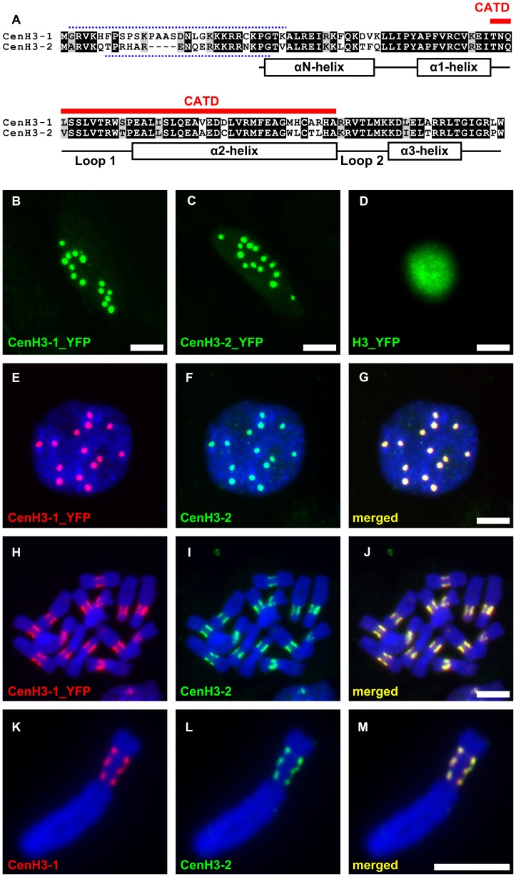
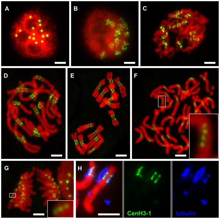
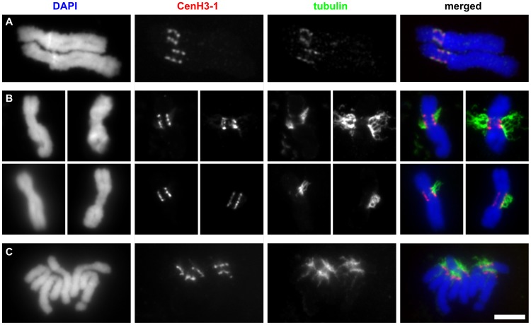
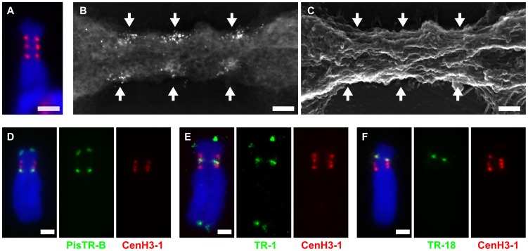
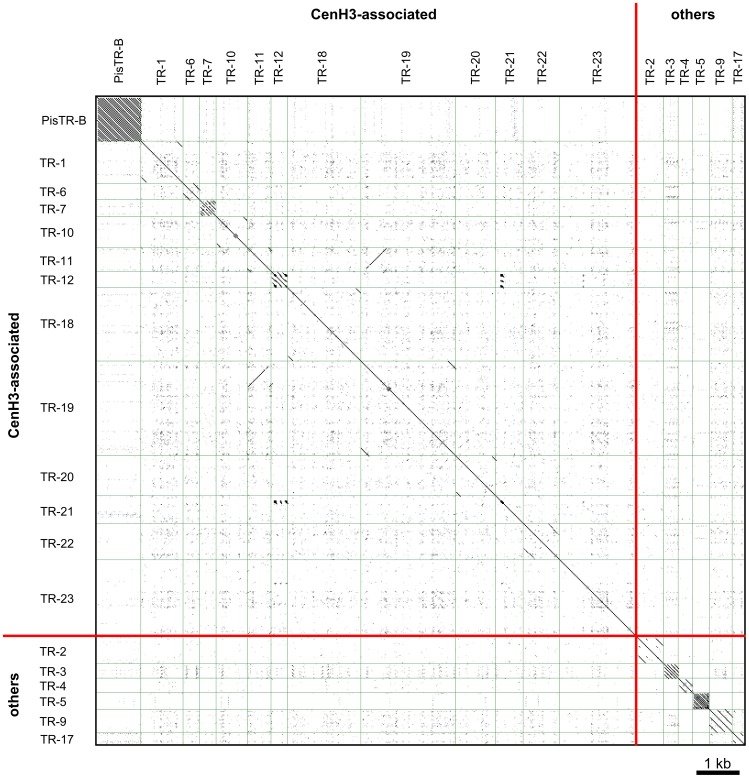
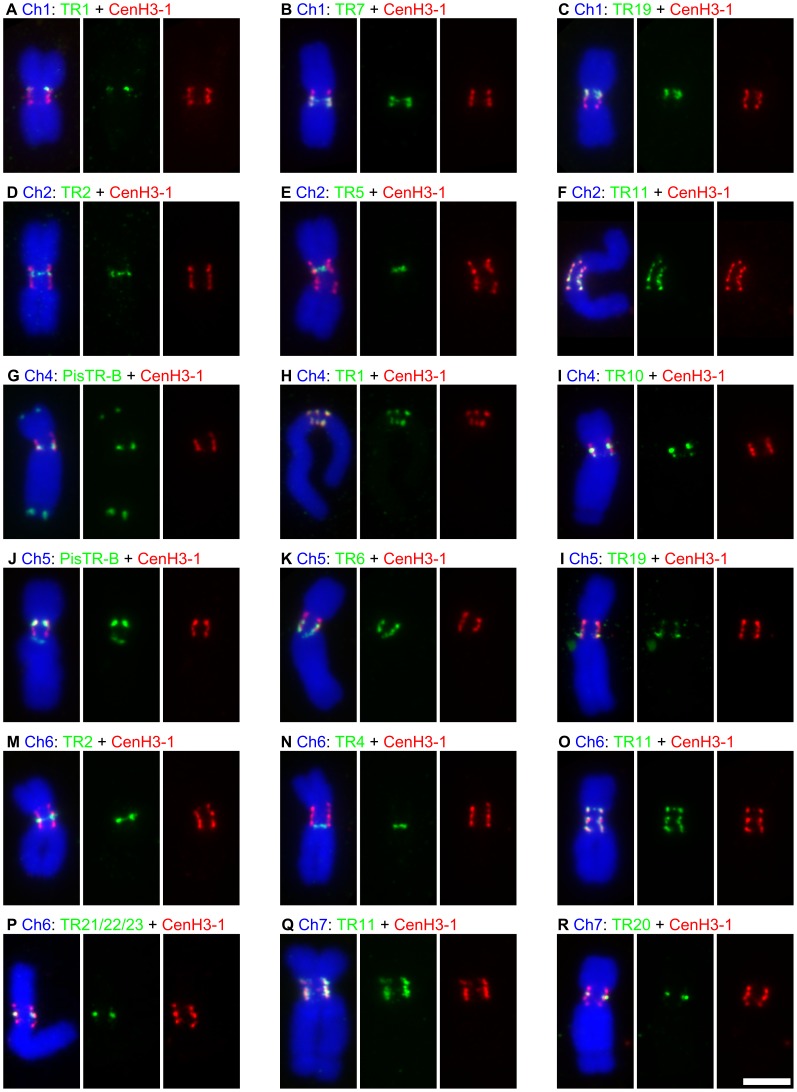
The centromere is a functional chromosome domain that is essential for faithful chromosome segregation during cell division and that can be reliably identified by the presence of the centromere-specific histone H3 variant CenH3. In monocentric chromosomes, the centromere is characterized by a single CenH3-containing region within a morphologically distinct primary constriction. This region usually spans up to a few Mbp composed mainly of centromere-specific satellite DNA common to all chromosomes of a given species. In holocentric chromosomes, there is no primary constriction; the centromere is composed of many CenH3 loci distributed along the entire length of a chromosome. Using correlative fluorescence light microscopy and high-resolution electron microscopy, we show that pea (Pisum sativum) chromosomes exhibit remarkably long primary constrictions that contain 3-5 explicit CenH3-containing regions, a novelty in centromere organization. In addition, we estimate that the size of the chromosome segment delimited by two outermost domains varies between 69 Mbp and 107 Mbp, several factors larger than any known centromere length. These domains are almost entirely composed of repetitive DNA sequences belonging to 13 distinct families of satellite DNA and one family of centromeric retrotransposons, all of which are unevenly distributed among pea chromosomes. We present the centromeres of Pisum as novel "meta-polycentric" functional domains. Our results demonstrate that the organization and DNA composition of functional centromere domains can be far more complex than previously thought, do not require single repetitive elements, and do not require single centromere domains in order to segregate properly. Based on these findings, we propose Pisum as a useful model for investigation of centromere architecture and the still poorly understood role of repetitive DNA in centromere evolution, determination, and function.


Conflict of interest statement

The authors have declared that no competing interests exist.

Figures

Figure 1
Figure 1. Pea has two variants of the CenH3 that fully colocalize in centromeres of all chromosomes.
A: Alignment of protein sequences of the pea CenH3 histones. Red line above the alignment marks a putative centromere targeting domain (CATD). Dotted lines above and below the alignment show the peptide sequences which were used as antigens to produce antibody to CenH3-1 and CenH3-2, respectively. Secondary structure of the histone fold domain is depicted below the alignment. B–C: Direct visualization of fusion proteins of CenH3-1 or CenH3-2 with YFP revealed 14 foci in the interphase nucleus, corresponding to the number of chromosomes in diploid cells. D: Fusion protein of canonical H3 with YFP is localized in whole nucleus. ELISA assays of the two CenH3 antibodies revealed low level of cross-reaction of the CenH3-1 antibody to the peptide designed from the CenH3-2 (data not shown). As we could not determine if the cross-reactivity was sufficient to produce signal after detection in situ, the colocalization experiments were performed using highly-specific antibodies to YFP and CenH3-2 in hairy root lines expressing CenH3-1_YFP. E–J: Detection of CenH3-1_YFP (red) and CenH3-2 (green) revealed full colocalization of the two proteins both in interphase nucleus (E–G) and metaphase chromosomes (H–J). K–M: Fully overlapping signals were observed also using simultaneous detection of CenH3 proteins with antibodies to CenH3-1 (red) and CenH3-2 (green) as shown on the example of chromosome 3 possessing three distinct domains containing CenH3. This indicates that either of the two antibodies was capable of detecting all functional centromere domains and that the gaps between individual domains lack CenH3 of any type. Bar = 5 µm.
Figure 2
Figure 2. Organization of CenH3-containing domains during the cell cycle.
A: Number of CenH3-containing domains in interphase nucleus corresponds to the number of chromosomes in diploid cells (2n = 14). B–D: Mitotic chromosomes at the early prophase (B), prophase (C) and prometaphase (D) show multiple domains containing the CenH3 which are clearly separated with chromatin segments lacking the CenH3. E–G: The separation of CenH3-containing domains becomes less apparent with the progress of chromatin condensation: metaphase (E), spread of single-chromatid anaphase chromosomes (F), anaphase (G). However, the multiple domain structure can be observed in some cases even in the highly condensed anaphase chromosomes (detail windows in F and G). H: Simultaneous detection of CenH3-1 (green) and tubulin (blue) revealed that microtubules are attached to all CenH3 containing domains as shown for chromosome 3. All chromosomes were counterstained with DAPI (pseudocolored in red). Bar = 5 µm.
Figure 3
Figure 3. The CenH3-containing domains are fully colocalized with tubulin.
A: Immunodetection of tubulin and CenH3-1 on two isolated metaphase chromosomes 3. Although isolated chromosomes never remained attached to microtubules they rarely exhibited weak tubulin signals which fully colocalized with CenH3-1. B–C: Detection of tubulin and CenH3-1 on chromosomes prepared using squash technique. The squash technique allowed some chromosomes to remain attached to microtubules. Whenever present, the remnants of mitotic spindle attached to chromosomes at all CenH3-containing domains on both metaphase (B) and anaphase (C) chromosomes. Bar = 5 µm.
Figure 4
Figure 4. Organization and DNA sequence composition of CenH3-containing domains in chromosome 3.
A–B: Primary constriction of chromosome 3 contains three functional centromere domains as defined by the presence of CenH3-1. Correlative fluorescence and scanning electron microscopy images of the same chromosome using FluoroNanogold showed that the three domains recognized with fluorescence (A, red signals) are composed of multiple foci from markers (bright spots) near the surface of the primary constriction (B, backscattered electron micrograph). C: Secondary electron micrograph image of the same chromosome. The primary constriction exhibits few chromomeres and a typical longitudinal orientation of fibrillar substructures, to which the CenH3 domains roughly correspond. The arrows mark the CenH3-1 containing regions. D–F: Detection of three different families of satellite DNA by FISH (green) combined with immunodection of CenH3-1 (red). Each of the functional centromere domains is composed of different family of satellite DNA; the domain closest to the long arm is composed of PisTR-B (D), the middle one of TR-1 (E) and the one closest to the short arm of TR-18 (F). Chromosomes were counterstained with DAPI (blue). Bar = 2 µm (A and D–F) or 0.2 µm (B–C).
Figure 5
Figure 5. All to all dot-plot comparison of the pea satellite repeats.
With exception of TR-11 and TR-19, the sequences of different satellite DNA families show no similarity. A fragment of long monomer of TR-19 shows high similarity to TR-11. FISH experiments revealed that all loci of TR-19 repeat contain also TR-11, but only some of TR-11 loci contain TR-19 (Figure 6 and data not shown), indicating that these two repeats should be considered as different families. Sequences used for dot-plot comparison are provided in Dataset S1.
Figure 6
Figure 6. Association of satellite DNA sequences with CenH3-containing domains.
The figure shows results of simultaneous detection of different families of satellite DNA by FISH (green) and immunodection of CenH3-1 (red). A–C, F–L and O–R: All families of satellite DNA showing high ChiP enrichment were found to be colocalized with regions containing CenH3-1. D–E and M–N: The DNA families with no ChIP enrichment but present in primary constrictions were indeed found outside of the CenH3-containing domains. Colocalization of CenH3-1 and satellite DNA families on chromosome 3 is shown in the Figure 4. Bar = 5 µm.

References

    1. Jiang JM, Birchler JA, Parrott WA, Dawe RK. A molecular view of plant centromeres. Trends Plant Sci. 2003;8:570–575. - PubMed
    1. Malik HS, Henikoff S. Major evolutionary transitions in centromere complexity. Cell. 2009;138:1067–1082. - PubMed
    1. Black BE, Bassett EA. The histone variant CENP-A and centromere specification. Curr Opin Cell Biol. 2008;20:91–100. - PubMed
    1. Sullivan BA, Karpen GH. Centromeric chromatin exhibits a histone modification pattern that is distinct from both euchromatin and heterochromatin. Nat Struct Mol Biol. 2004;11:1076–1083. - PMC - PubMed
    1. Ribeiro SA, Vagnarelli P, Dong Y, Hori T, McEwen BF, et al. A super-resolution map of the vertebrate kinetochore. Proc Natl Acad Sci U S A. 2010;107:10484–10489. - PMC - PubMed

Publication types