. 2011 Dec 1;51(1):103-134.
doi: 10.1007/s13225-011-0145-6. Epub 2011 Nov 16.

Capnodiaceae

Affiliations

Capnodiaceae

Putarak Chomnunti et al. Fungal Divers. .

Abstract

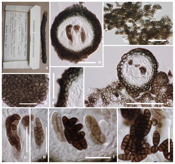
In this paper we revisit the Capnodiaceae with notes on selected genera. Type specimens of the ascomycetous genera Aithaloderma, Anopeltis, Callebaea, Capnodaria, Echinothecium, Phragmocapnias and Scorias were re-examined, described and illustrated. Leptoxyphium is anamorphic Capnodiaceae and Polychaeton is a legitimate and earlier name for Capnodium, but in order to maintain nomenclatural stability we propose that the teleomorphic name should be conisdered for the approved lists of names currently in preparation for fungi. Notes are provided on the ascomycetous genus Scoriadopsis. However, we were unable to locate the type of this genus during the time frame of this study. The ascomycetous genera Aithaloderma, Ceramoclasteropsis, Hyaloscolecostroma and Trichomerium are excluded from Capnodiaceae on the basis of having ascostromata and trans-septate hyaline ascospores and should be accommodated in Chaetothyriaceae. Callebaea is excluded as the ascomata are thyriothecia and the genus is placed in Micropeltidaceae. Echinothecium is excluded as synonym of Sphaerellothecium and is transferred to Mycosphaerellaceae. The type specimen of Capnophaeum is lost and this should be considered as a doubtful genus. The coelomycetous Microxiphium is polyphyletic, while the status of Fumiglobus, Polychaetella and Tripospermum is unclear. Fourteen new collections of sooty moulds made in Thailand were isolated and sequenced. The nuclear large and small rDNA was partially sequenced and compared in a phylogeny used to build a more complete understanding of the relationships of genera in Capnodiaceae. Four new species are described and illustrated, while Phragmocapnias and Scorias are epitypified with fresh collections.


Figures

Fig. 1
Fig. 1
A RAxML maximum likelihood tree from obtained from a data set of 51 taxa including representatives of Capnodiales, focused on Capnodiaceae, comparing two genes (SSU, LSU). The first set of numbers above the nodes are bootstraps from 1000 pseudorepetitions and the second represent Bayesian posterior probabilities expressed as percentages. Only values above 50% are shown. Culture and voucher numbers are indicated after species names and names of isolates with newly obtained sequences for this study are bolded.
Fig. 2
Fig. 2
A–E Sooty moulds growing on host leaves. A. Unidentified tree. B. Bischofia javanica. c. Euphorbia sp. D. Psidium guajava. E. Cestrum diurnum. 2FI Representative pycnidia of Capnidiaceae genera accepted in this paper. F. Phragmocapnias. G. Capnodium. H. Leptoxyphium. I. Scorias.
Fig. 3
Fig. 3. A–I Phragmocapnias betle (holotype of Capnodium betle)
A, B. Ascomata on leaves. C, E. Vertical section through stalked ascoma. D. Ascomata exterior wall with setae. F–H. Asci with short pedicel. I. Cylindric–clavate ascospores. Bars: E = 100 μm, C, D = 50 μm, F, H, G = 20 μm, I = 10 μm.
Fig. 4
Fig. 4. A–L Phragmocapnias betle (epitype)
A. Ascomata on living leaf. B, C. Ascomata which are subglobose with setae. D, F. Bitunicate asci. E. Mycelium beneath ascomata. G–L. Ascospores surrounded by a mucilaginous sheath. Bars: B, C = 100 μm, D, F = 50 μm, E = 20 μm, G–L = 10 μm.
Fig. 5
Fig. 5. A–K Phragmocapnias asiaticus (holotype)
A. Black mycelium covering the leaf surface. B. Pycnidium with long stalks on the host. C, D. Mycelial network. E, F. Conical pycnidium and pycnidium wall. Note the conida in the swollen part in F. G. Pycnidia. H. Ostiole. I–K. Conidia. Bars: C, E = 50 μm, D–F = 20 μm, H, K = 10 μm, I, J = 5 μm.
Fig. 6
Fig. 6. A–G Phragmocapnias siamensis (holotype)
A. Black mycelium covering the leaf surface. B. Pycnidia on host. C, D. Mycelial network. E, F. Conical pycnidia and pycnidia wall. G. Conidia. Bars: F = 100 μm, C–E = 50 μm, G = 20 μm.
Fig. 7
Fig. 7. A–LLeptoxyphium cacuminum (holotype)
A. Gregarious pycnidia on host surface. B, D, G. Stalked pycnidia with wider base. E. Formation of pycnidia from aggregated hyphae. C, F. Black stalked funnel cupulate apex. H–L. Conidia, conidiogenous boundary with hyaline hyphae surrounding the ostiole. Bars: B, D, E, G = 200 μm, C–F = 50 μm, H–L = 20 μm.
Fig. 8
Fig. 8. A–L Leptoxyphium cacuminum (holotype) in culture
A. Colony on PDA. C, D. Conidial mass at the apex of conidia. D, E. Olive–green stalked funnel shaped at apex. F–H. Septate hyphae. I–L. Conidia produced from the apex of conidiophores. Bars: A = 2 cm, D = 200 μm, E–L = 50 μm.
Fig. 9
Fig. 9. A–GCapnodium citri (type?)
A. Superficial mycelium on host. B, F. Cylindrical, septate mycelium. C–E. Elongate pycnidia. G. Conidia. Bars: B, C = 200 μm, D, E = 100 μm, F = 50 μm, G = 20 μm.
Fig. 10
Fig. 10. A–MCapnodium coartatum (holotype)
A–D. Stalked pycnidia. E. Immature pycnidia. F. Mycelia. G, H. Pycnidia rising from mycelium. I. Abundant conidia at apex pycnidia and pycnidia wall. J, K. Ostiole surround by hyaline hyphae. L, M = Conidia. Bars: A–D = 200 μm, F = 100 μm, E, G, I–K = 50 μm, L, M = 20 μm.
Fig. 11
Fig. 11. A–JCapnodium tiliae (exsicata of Fumago tiliae)
A. Label data. B. Vertical section through ascoma. C. Dark brown mycelium. D. Peridium. E. Peridium with cells of textura angularis. G–I. Cylindrical to cylindric–clavate asci. J. Ascospores 3–4 septate. Scale bars: F = 200 μm, B–D = 100 μm, E, G–I = 50 μm, J = 20 μm.
Fig. 12
Fig. 12. A–PScorias spongiosa (syntype)
A, B. Ascomata on hyphal mass on host. C, D. Squash of ascomata. E–J. Vertical section through ascomata on hyphal mass. K: Stalked ascomata. L–N: Asci with thick wall. F, G. Pale brown ascospores. Bars: D–H, J = 100 μm, C, I = 50 μm, K–N = 20 μm, O, P = 10 μm.
Fig. 13
Fig. 13. A–HScorias spongiosa (epitype)
A. Ascomata and pycnidia on surface of leaf. B, D–F. Immature ascomata and pycnidia. C. Pycnidia arising from mycelium. H. Conida. Bars A = 200 μm, D, E = 100 μm, B, F = 50 μm, H = 20 μm.
Fig. 14
Fig. 14. A–KScorias spongiosa (epitype)
A. Hyphae. B. Immature pycnidia. C. Mycelium bearing pycnidium. D–G. Mature pycnidia. H. Septate hyphae. I–K. Hyaline conidia. Bars: A = 200 μm, E–G = 100 μm, B–D = 50 μm, H–J = 20 μm, K = 10 μm.
Fig. 15
Fig. 15. A–LSphaerellothecium reticulatum (syntype of Echinothecium reticulatum)
A. Ascomata on Parmelia sulcata. B–E. Ascoma with eternal hyphal appendages. F, G. Mature hyphae. H–J. Bitunicate asci. K, L. Ascospores. Bars: A, C, D, F = 50 μm, B, E, G–I = 20 μm, J, K = 10 μm.
Fig. 16
Fig. 16. A–KSphaerellothecium araneosum (holotype of Sphaerella araneosa)
A. Fruiting bodies on host tissue. B. Ascomata with hyphae. C. Section through ascoma. D. Section through peridium. E–G. Ascus. H–J. Ascospores. K. Mature ascospore. Bars: A = 200 μm, B = 100 μm, C = 50 μm, D = 20 μm, E–K = 10 μm.
Fig. 17
Fig. 17. A–LAithaloderma clavatisporum (isotype)
A. Sooty mold and ascostromata on surface of host. B. Ascostromata with short setae. C–E. Section through ascostromata. F. Network of hyphae.. G–I. Asci. J–L. Ascospores. Bars: B–D= 100 μm, E–G= 50 μm, H–L= 20 μm.
Fig. 18
Fig. 18. A–KMicrocallis rutideae (holotype)
A, B. Ascomata on leaf surface. C, D. Circular ascomata comprising meandering hyphae. E. Vertical section of ascostomata. F–K. Young asci. Bars: C, E = 100 μm, D, J = 50 μm, F–I, K = 20 μm.
Fig. 19
Fig. 19. A–JAnopeltis venezuelensis (holotype)
A. Appearance of leaves colonized by the fungus. B. Ascostromata with mycelium and conidia. C. Pale brown angular cells of ascostromata edge. D. Section of ascostomata with angular cells. E. Young locule/asci. F. Vertical section of ascostromata with young asci. G. Angular cells of ascostromata. H–J. Brown conidia with 1–septa. Bars: B–D = 100 μm, C, E–G = 20 μm, H–J = 10 μm.

References

    1. Andrew JH. Biological control in the phyllosphere. Ann Rev Phytopathol. 1982;30:603–635. - PubMed
    1. Barr ME. Amherst. University of Massachusetts; Massachusetts: 1987. Prodomus to class Loculoascomycetes.
    1. Batista AC, Ciferri R. Capnodiales. Capnodiales. Saccardoa. 1963;2:1–296.
    1. Batista AC, Ciferri R. The Chaetothyriales. Sydowia. 1962;3:1–129.
    1. Batista AC, Ciferri R. The sooty–molds of the family Asbolisiaceae. Quad Ist Bot Univ Lab Crittogam Pavia. 1963;31:1–229.

LinkOut - more resources