. 2012 Jun 21:6:189.
doi: 10.3389/fnhum.2012.00189. eCollection 2012.

On the relationship between the "default mode network" and the "social brain"

Affiliations

On the relationship between the "default mode network" and the "social brain"

Rogier B Mars et al. Front Hum Neurosci. .

Abstract

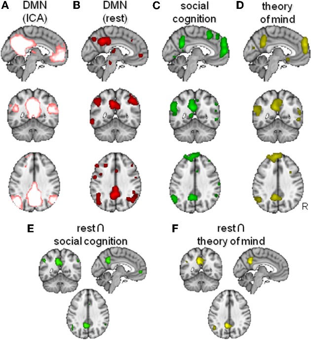
The default mode network (DMN) of the brain consists of areas that are typically more active during rest than during active task performance. Recently however, this network has been shown to be activated by certain types of tasks. Social cognition, particularly higher-order tasks such as attributing mental states to others, has been suggested to activate a network of areas at least partly overlapping with the DMN. Here, we explore this claim, drawing on evidence from meta-analyses of functional MRI data and recent studies investigating the structural and functional connectivity of the social brain. In addition, we discuss recent evidence for the existence of a DMN in non-human primates. We conclude by discussing some of the implications of these observations.

Keywords: TPJ; default mode network; fMRI; medial frontal cortex; mentalizing; posterior cingulate; social cognition; theory of mind.


Figures

Figure 1
Figure 1
Overlap between the default mode network (DMN) and areas activated by social cognition paradigms. (A) DMN as found using model-free analysis of resting state fMRI data (Smith et al., 2009). (B,C) Activation likelihood maps of activity during passive “rest” conditions (B), social cognition (C) and theory of mind (D). (E,F) Conjunction maps of rest and social cognition (E) and of rest and theory of mind (F).
Figure 2
Figure 2
Overlap between connectivity-based subregions of the inferior parietal lobule and temporoparietal junction area and the DMN. Top: Overlap between resting state function connectivity of the posterior TPJ as defined by Mars et al. (in press) in green and the DMN as defined using independent component analysis in pink. Bottom: Overlap between the DMN in pink and the anterior (TPJa) and posterior (TPJp) areas from Mars et al. (in press) and the anterior and posterior angular gyrus from Mars et al. (2011).
Figure 3
Figure 3
Social brain in the macaque? (A) Areas in the macaque showing resting state functional connectivity with a region in the middle superior temporal sulcus that, in turn, showed increases in gray matter density in individuals living in larger social groups (data from Sallet et al., 2011). (B) Group independent component capturing the default mode network. (C) Dual regression results showing a region of the medial frontal cortex (in blue) that in increasingly recruited into the DMN (in red) when animals live in bigger groups.

References

    1. Amodio D. M., Frith C. D. (2006). Meeting of minds: the medial frontal cortex and social cognition. Nat. Rev. Neurosci. 7, 268–277 10.1038/nrn1884 - DOI - PubMed
    1. Anderson J. S., Nielsen J. A., Froehlich A. L., DuBray M. B., Druzgal T. J., Cariello A. N., Cooperrider J. R., Zielinski B. A., Ravichandran C., Fletcher P. T., Alexander A. L., Bigler E. D., Lange N., Lainhart J. E. (2011). Functional connectivity magnetic resonance imaging classification of autism. Brain 134(Pt 12), 3742–3754 10.1093/brain/awr263 - DOI - PMC - PubMed
    1. Andreasen N. C., O'Leary D. S., Cizadlo T., Arndt S., Rezai K., Watkins G. L., Ponto L. L., Hichwa R. D. (1995). Remembering the past: two facets of episodic memory explored with positron emission tomography. Am. J. Psychiatry 152, 1576–1585 - PubMed
    1. Andrews-Hanna J. R., Reidler J. S., Sepulcre J., Poulin R., Buckner R. L. (2010). Functional-anatomic fractionation of the brain's default network. Neuron 65, 550–562 10.1016/j.neuron.2010.02.005 - DOI - PMC - PubMed
    1. Beckmann C. F., DeLuca M., Devlin J. T., Smith S. M. (2005). Investigations into resting-state connectivity using independent component analysis. Philos. Trans. R. Soc. B Biol. Sci. 360, 1001–1013 10.1098/rstb.2005.1634 - DOI - PMC - PubMed

LinkOut - more resources