. 2012;7(6):e38635.
doi: 10.1371/journal.pone.0038635. Epub 2012 Jun 21.

Augmented TLR2 expression on monocytes in both human Kawasaki disease and a mouse model of coronary arteritis

Affiliations

Augmented TLR2 expression on monocytes in both human Kawasaki disease and a mouse model of coronary arteritis

I-Chun Lin et al. PLoS One. 2012.

Abstract

Background: Kawasaki disease (KD) of unknown immunopathogenesis is an acute febrile systemic vasculitis and the leading cause of acquired heart diseases in childhood. To search for a better strategy for the prevention and treatment of KD, this study compared and validated human KD immunopathogenesis in a mouse model of Lactobacillus casei cell wall extract (LCWE)-induced coronary arteritis.

Methods: Recruited subjects fulfilled the criteria of KD and were admitted for intravenous gamma globulin (IVIG) treatment at the Kaohsiung Chang Gung Memorial Hospital from 2001 to 2009. Blood samples from KD patients were collected before and after IVIG treatment, and cardiovascular abnormalities were examined by transthoracic echocardiography. Wild-type male BALB/c mice (4-week-old) were intraperitoneally injected with LCWE (1 mg/mL) to induce coronary arteritis. The induced immune response in mice was examined on days 1, 3, 7, and 14 post injections, and histopathology studies were performed on days 7 and 14.

Results: Both human KD patients and LCWE-treated mice developed coronary arteritis, myocarditis, valvulitis, and pericarditis, as well as elevated plasma levels of interleukin (IL)-2, IL-6, IL-10, monocyte chemoattractant protein (MCP)-1, and tumor necrosis factor (TNF)-α in acute phase. Most of these proinflammatory cytokines declined to normal levels in mice, whereas normal levels were achieved in patients only after IVIG treatment, with a few exceptions. Toll-like receptor (TLR)-2, but not TLR4 surface enhancement on circulating CD14+ monocytes, was augmented in KD patients before IVIG treatment and in LCWE-treated mice, which declined in patients after IVIG treatment.

Conclusion: This result suggests that that not only TLR2 augmentation on CD14+ monocytes might be an inflammatory marker for both human KD patients and LCWE-induced CAL mouse model but also this model is feasible for studying therapeutic strategies of coronary arteritis in human KD by modulating TLR2-mediated immune activation on CD14+ monocytes.


Conflict of interest statement

Competing Interests: The authors have declared that no competing interests exist.

Figures

Figure 1
Figure 1. Immune abnormalities in human patients with Kawasaki disease (KD).
Plasma levels of interleukin (IL)-6, monocyte chemoattractant protein (MCP)-1, tumor necrosis factor (TNF)-α, IL-2, and IL-10, assessed by Luminex technology, were significantly increased in KD patients before intravenous gamma globulin (Pre-IVIG), when compared to those in controls. IL-6, MCP-1, and TNF-α were significantly suppressed after IVIG treatment (Post-IVIG), but IL-2 and IL-10 responded differently. Values are expressed as mean±SEM; n = 39 in paired KD patients, n = 15 in control patients. *P<0.05; **P<0.01.
Figure 2
Figure 2. TLR2 and TLR4 surface expression on peripheral blood leukocytes (PBLs) in human KD patients.
(A) Representative histograms showed surface expression of TLR2 and TLR4 by mean fluorescence intensity (MFI) on peripheral monocytes, neutrophils, and lymphocytes in KD patients before IVIG treatment and control patients. (B) The representative histogram displayed surface expression of TLR2 by MFI on peripheral CD14+ monocytes in human KD patients before and after IVIG treatment as well as those in control patients. (C) TLR2 expression over CD14+ monocytes are expressed as mean±SEM by MFI. n = 6 in controls; n = 12 Pre-IVIG; n = 8 Post-IVIG. **P<0.01.
Figure 3
Figure 3. Immune abnormalities in the Lactobacillus casei cell wall extract (LCWE)-treated mice.
Plasma levels of IL-6, MCP-1, TNF-α, IL-2, and IL-10 were assessed by Luminex technology. IL-2, IL-6, MCP-1 and TNF-α were significantly increased early after LCWE stimulation and spontaneously declined to basal levels during days 3 and 7 post injection. In contrast, IL-10 was significantly elevated at day 3 post injection. Values are expressed as mean±SEM; n = 9 in LCWE-treated mice; n≥7 in PBS-treated mice, per time point for all, except n = 5 for IL-2 per group per time point. *P<0.05; **P<0.01.
Figure 4
Figure 4. TLR2 and TLR4/MD2 surface expression in LCWE-treated mice.
(A) Representative histograms showed surface expression of TLR2 and TLR4/MD2 by MFI on monocytes, neutrophils, and lymphocytes in the LCWE-treated mice and PBS-treated control mice on post-injection day 7. The numbers labeled over the right upper quadrants indicated the representative percentage of TLR2+ or TLR4/MD2+ cells, corresponding to each cell subpopulation. (B) The surface expression of TLR2 on circulating CD14+ monocytes were analyzed by relative TLR2 MFI (rMFI = TLR2 MFI/isotype MFI) at the indicated time points post-injection. (C) Circulating CD14+ monocytes were significantly increased in LCWE-treated mice on days 3, 7, and 14 post injections. Values are expressed as mean±SME, n = 7 mice/group per time point; *P<0.05; **P<0.01 versus PBS controls.
Figure 5
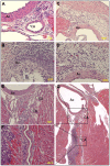
Figure 5. Histopathology of cardiac tissues in LCWE-treated mice.
(A–G) Arteritis developed as infiltration of mononuclear cells and neutrophils in the perivascular/adventitial regions of the aortic vessel/aortic valve (Ao) and coronary artery (CA) (D is the higher magnification of C). Myocarditis (B, D, F and G), valvulitis (B), and pericarditis (G) were complicated with the infiltration invading into the myocardium, aortic valve, and pericardium, respectively. (E, F) Higher magnification of black boxes on the serial section next to (G) showed infiltration around and along the vessel walls of the CA and myocardium in detail. Original magnification: ×200 in (A, B, D and F), ×100 in (C, E) and ×10 in (G). Each yellow bar  = 100 µm. Black asterisks indicate the lumen of the CA.

Similar articles

Cited by

References

    1. Kawasaki T, Kosaki F, Okawa S, Shigematsu I, Yanagawa H. A new infantile acute febrile mucocutaneous lymph node syndrome (MLNS) prevailing in Japan. Pediatrics. 1974;54:271–276. - PubMed
    1. Barron KS. Kawasaki disease in children. Curr Opin Rheumatol. 1998;10:29–37. - PubMed
    1. Taubert KA, Rowley AH, Shulman ST. Seven-year national survey of Kawasaki disease and acute rheumatic fever. Pediatr Infect Dis J. 1994;13:704–708. - PubMed
    1. Nakamura Y, Yashiro M, Uehara R, Oki I, Kayaba K, et al. Increasing incidence of Kawasaki disease in Japan: nationwide survey. Pediatr Int. 2008;50:287–290. - PubMed
    1. Suzuki A, Miyagawa-Tomita S, Komatsu K, Nishikawa T, Sakomura Y, et al. Active remodeling of the coronary arterial lesions in the late phase of Kawasaki disease: immunohistochemical study. Circulation. 2000;101:2935–2941. - PubMed

Publication types

MeSH terms