. 2012;7(6):e39338.
doi: 10.1371/journal.pone.0039338. Epub 2012 Jun 21.

Fragile X mental retardation protein interacts with the RNA-binding protein Caprin1 in neuronal RiboNucleoProtein complexes [corrected]

Affiliations

Fragile X mental retardation protein interacts with the RNA-binding protein Caprin1 in neuronal RiboNucleoProtein complexes [corrected]

Rachid El Fatimy et al. PLoS One. 2012.

Erratum in

  • PLoS One. 2012;7(9). doi:10.1371/annotation/05374d07-34cf-483f-80f4-ec87374cbeb6

Abstract

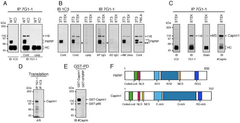
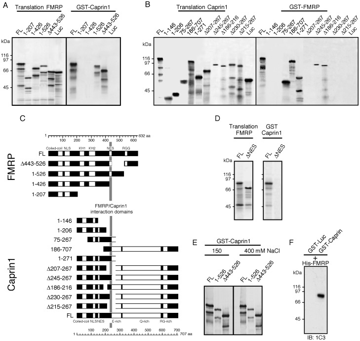
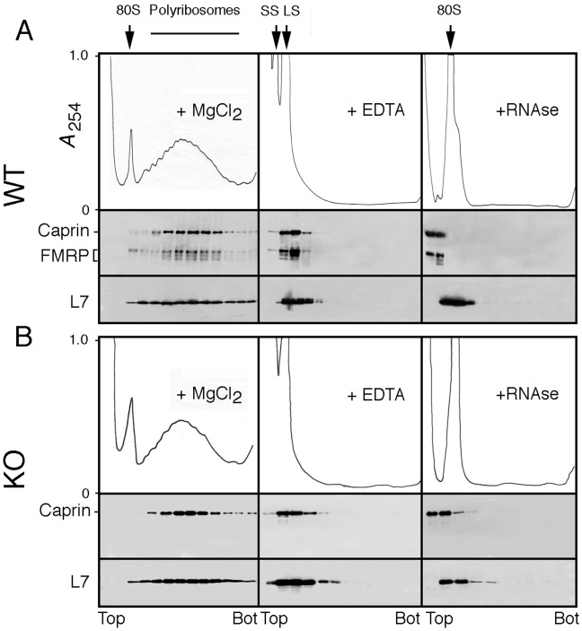
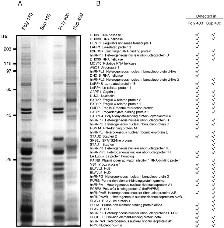
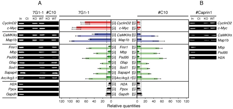
Fragile X syndrome is caused by the absence of the Fragile X Mental Retardation Protein (FMRP), an RNA-binding protein. FMRP is associated with messenger RiboNucleoParticles (mRNPs) present in polyribosomes and its absence in neurons leads to alteration in synaptic plasticity as a result of translation regulation defects. The molecular mechanisms by which FMRP plays a role in translation regulation remain elusive. Using immunoprecipitation approaches with monoclonal Ab7G1-1 and a new generation of chicken antibodies, we identified Caprin1 as a novel FMRP-cellular partner. In vivo and in vitro evidence show that Caprin1 interacts with FMRP at the level of the translation machinery as well as in trafficking neuronal granules. As an RNA-binding protein, Caprin1 has in common with FMRP at least two RNA targets that have been identified as CaMKIIα and Map1b mRNAs. In view of the new concept that FMRP species bind to RNA regardless of known structural motifs, we propose that protein interactors might modulate FMRP functions.


Conflict of interest statement

Competing Interests: The authors have declared that no competing interests exist.

Figures

Figure 1
Figure 1. mAb7G1-1 detects mFMRP and Caprin1.
A) Immunoprecipitation analyses of WT and KO2 mouse brain extracts with mAb7G1-1 followed by immunoblotting with mAb1C3 (left panel in A) or with mAb7G1-1 (right panel in A). In addition to mFMRP, a clear band at 116 kDa is detected in WT immunoprecipitates. A similar band is also detected in KO2 extracts. Both bands are absent when immunoprecipitation is performed in the presence of the epitope peptide KHLDTKENTHFSQPN. B) Immunoblot analyses with mAb7G1-1 of 3T3, STEK and HeLa cell extracts. While mAb3C1 detects only mFMRP in 3T3 extracts, mAb7G1-1 reacts with both mFMRP and p116. An additional weak band is also detected in both extracts at 65 kDa (indicated by a star). mAb7G1-1 does not react with hFMRP from HeLa extracts, but recognizes p116. Note the presence of the additional band that migrates slightly above 65 kDa in human HeLa extracts (double star). C) Extracts from STEK cells were immunoprecipitated with mAb7G1-1. Immunoblot analyses with mAb1C3 reveal that mFMRP is indeed absent, while mAb7G1-1 reacts with p116. The Coomassie brillant blue stained band at 116 kDa was excised and analyzed by mass spectrometry and was identified as Caprin1. Immunoblot analyses with rabbit antisera raised against hCaprin1 confirmed the nature of p116 as Caprin1. D) In vitro translated 35S-labeled Caprin1 is immunoprecipitated by mAb7G1-1. E) Recombinant GST-Caprin1 isolated on Glutathione-Sepharose beads is revealed with anti-Caprin1 IgG in immunoblot analyses. Note the presence of the minor truncated band at ∼95 kDa. F) Structural comparisons between FMRP and Caprin1. WT and KO: wild type C57BL/6J and Fmr1 −/−KO2 mice, respectively. IP: immunoprecipitation; IB: immunoblot; Cont: control; HC: IgG heavy chains; AP: affinity purified; AD: affinity depleted; AR: autoradiography.
Figure 2
Figure 2. mFMRP co-immunoprecipitates with Caprin1.
Total brain extracts from WT and KO mice were subjected to immunoprecipitation with IgY#C10 (A) and anti-Caprin1 IgG (B) and the eluted proteins were analyzed by immunoblotting using mAb1C3, mAb7G1 and anti-Caprin1 IgG. Note that a high salt concentration was necessary to immunoprecipitate mFMRP in association with Caprin1. C) mAb7G1-1 has no anti-Ago2 intrinsic activity, however Ago2 is co-immunoprecitating with Caprin1 (left panels) as detected using the Odyssey Infrared Imaging System. In the right panels are shown the results proving that mAb7G1-1 does not possess an anti-Ago2 activity as detected by direct immunoblot analyses. Membranes were either scanned at 700 nm or simultaneously at 700 plus 800 nm. D) The epitope peptide (see Figure 1A) abolishes co-precipitation of Ago2 with Caprin1.
Figure 3
Figure 3. Mapping of interaction between hFMRP and hCaprin1 in pulldown assays. A
) Pulldown assay using 1 µg of the fusion protein GST-Caprin1 immobilized on beads and in vivo translated 35S-labelled full length FMRP (FL) and its truncated and deleted versions. B) Reverse pulldown assay using immobilized GST-FMRP incubated with 35S-labeled full length Caprin1 (FL) and its truncated and deleted versions. In both cases, 35S-labeled Luciferase (Luc) was used as a negative control. C) Schematic diagram summarizing the data presented in A and B. D) Refine region in FMRP NES (amino acids residues 424–440) required for binding to Caprin1. E) The interaction of FMRP with Caprin1 is stable in the presence of 400 mM NaCl. F) The interaction of Caprin1 and FMRP is direct in a pulldown assay using recombinant proteins.
Figure 4
Figure 4. Caprin1 co-sediments with mFMRP in polyribosomes prepared from total brain and mFMRP is not required for Caprin1 to associate with polyribosomes.
Aliquots containing 10–12 OD at 260 nm were analyzed by sedimentation velocity through sucrose density gradients in the presence of MgCl2, after incubation with 30 mM EDTA or after treatment with 100 µg/ml RNase A. mFMRP and Caprin1 were revealed simultaneously with mAb7G1-1. 80S : monosomes ; SS : ribosomal small sub-unit ; LS : ribosomal large sub-unit ; L7 : ribosomal large protein 7.
Figure 5
Figure 5. FMRP and Caprin1 partially colocalize in somato-dendritic compartments of neurons.
A) Immunostainings of mouse brain cortical sections were carried out using anti-FMRP IgY#C10 (red) and anti-caprin1 IgG (green). FMRP and Caprin1 colocalize both in cell bodies and in axons of cortical neurons. B) FMRP and Caprin1 colocalize in primary cultured hippocampal neurons. Note that to illustrate staining in neuritis, a high gain was required resulting in saturation of the fluorescent signal in the cell body. C) The dendritic area from the box in (B) at higher magnification reveals that only a fraction of FMRP co-localizes with Caprin1. In this selected region, after deconvolution of the high definition images, a total of 453 granules were counted, and colocalization of mFMRP and Caprin1 accounted for only 14%.
Figure 6
Figure 6. Sedimentation of FMRP and Caprin1 in presence of NaCl at physiological and at high salt conditions. A
) In presence of 150 mM NaCl, FMRP and Caprin1 are detected associated with polyribosomes. At 400 mM, a clear displacement of FMRP and Caprin1 towards the top of the gradient is observed, while PABP and the ribosomal L7 protein are not affected. In the bottom panels of A) are shown the distribution in the sucrose gradients of CaMKIIα, Map1b and Fmr1, FMRP-mRNA targets in the presence of 150 and of 400 mM NaCl. Note the slight shift toward the lighter fraction in the 400 mM NaCl condition and the absence of mRNA in the fraction containing the highest amount of FMRP at the top of the gradient. The results are presented as percent of the specific mRNA in each fraction. B) In presence of 400 mM NaCl, the majority of FMRP and Caprin1 are found as “floating” species in the loading volume that did not penetrate the gradient even after prolonged ultracentrifugation for 23 hrs. The position of the small ribosomal subunit was determined according to the UV profile and immunoblotting with anti-S6 protein. S values were determined using purified total RNA from E.coli.
Figure 7
Figure 7. Distribution of RNA-binding proteins in polyribosomes and in salt washes.
A) Equivalent volume aliquots from polyribosomes and from the 150 and 400 mM NaCl washes were analyzed by SDS-PAGE and the separated proteins stained with Coomassie blue. B) Identification by Mass Spectrometry of RNA-binding proteins present or released from polyribosomes after 400 mM NaCl washes. Poly 400: polyribosomes resistant to 400 mM NaCl; Sup 400: supernatant of the polyribosomes treated with 400 mM NaCl.
Figure 8
Figure 8. mRNAs associated with Caprin1 and FMRP. A
) mRNAs co-immunoprecipitated by mAb7G1-1 and IgY#C10 from WT and KO brain extracts were analyzed by RT-PCR and visualized by agarose gels (left panels) and by Light Cycler RT-PCR (right panels). Bars in red refer to mRNA targets to Caprin1, in blue common to Caprin1 and FMRP, in green to FMRP and in black to non-targets mRNAs. Dark colors refer to WT and pale to KO, respectively. N = 5, P≤0.001 of a pool of 10 adult brains. Vertical black dot lines represent thresholds corresponding to background. B) RT-PCR analyses of selected mRNAs co-immunoprecipitated with Caprin1.

References

    1. O’Donnell WT, Warren ST. A decade of molecular studies of fragile X syndrome. Annu Rev Neurosci. 2002;25:315–338. - PubMed
    1. Bardoni B, Davidovic L, Bensaid M, Khandjian EW. The fragile X syndrome: exploring its molecular basis and seeking a treatment. Expert Rev Mol Med. 2006;8:1–16. - PubMed
    1. Bassell GJ, Warren ST. Fragile X Syndrome: Loss of Local mRNA Regulation Alters Synaptic Development and Function. Neuron. 2008;60:201–214. - PMC - PubMed
    1. Khandjian EW, Fortin A, Thibodeau A, Tremblay S, Cote F, et al. A heterogeneous set of FMR1 proteins is widely distributed in mouse tissues and is modulated in cell culture. Hum Mol Genet. 1995;4:783–789. - PubMed
    1. Devys D, Lutz Y, Rouyer N, Bellocq J-P, Mandel J-L. The FMR-1 protein is cytoplasmic, most abundant in neurons and appears normal in carriers of a fragile X premutation. Nature Genet. 1993;4:335–340. - PubMed

Publication types

MeSH terms