. 2012;7(6):e39465.
doi: 10.1371/journal.pone.0039465. Epub 2012 Jun 21.

Alpha-synuclein cell-to-cell transfer and seeding in grafted dopaminergic neurons in vivo

Affiliations

Alpha-synuclein cell-to-cell transfer and seeding in grafted dopaminergic neurons in vivo

Elodie Angot et al. PLoS One. 2012.

Abstract

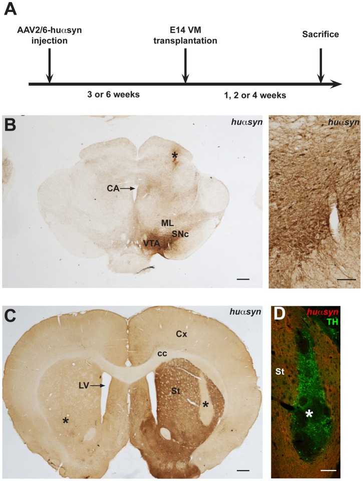
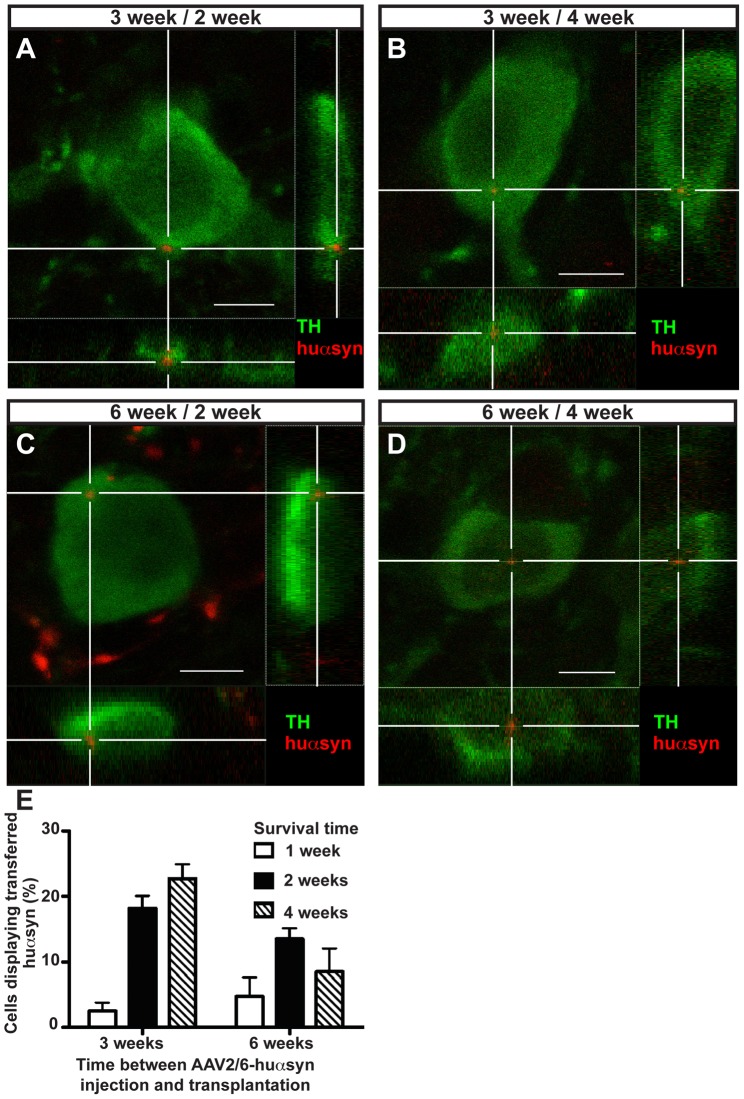
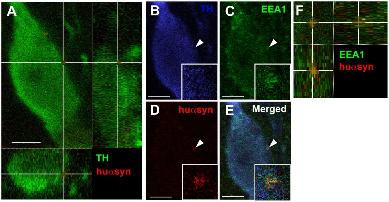
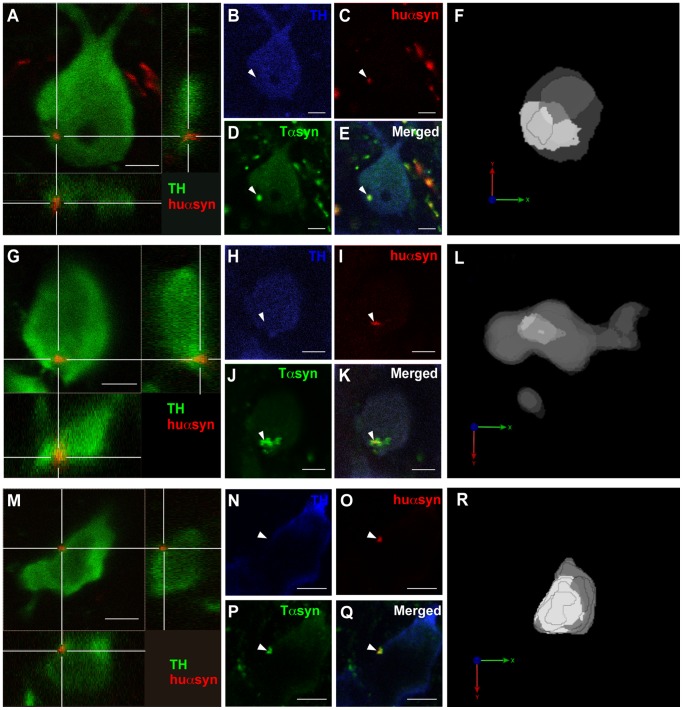
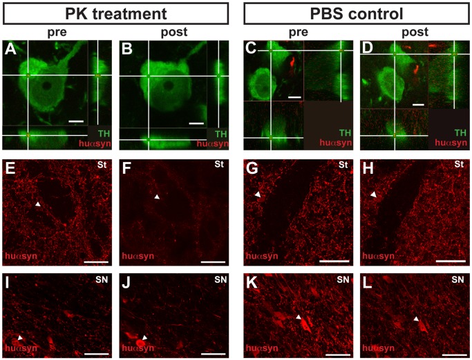
Several people with Parkinson's disease have been treated with intrastriatal grafts of fetal dopaminergic neurons. Following autopsy, 10-22 years after surgery, some of the grafted neurons contained Lewy bodies similar to those observed in the host brain. Numerous studies have attempted to explain these findings in cell and animal models. In cell culture, α-synuclein has been found to transfer from one cell to another, via mechanisms that include exosomal transport and endocytosis, and in certain cases seed aggregation in the recipient cell. In animal models, transfer of α-synuclein from host brain cells to grafted neurons has been shown, but the reported frequency of the event has been relatively low and little is known about the underlying mechanisms as well as the fate of the transferred α-synuclein. We now demonstrate frequent transfer of α-synuclein from a rat brain engineered to overexpress human α-synuclein to grafted dopaminergic neurons. Further, we show that this model can be used to explore mechanisms underlying cell-to-cell transfer of α-synuclein. Thus, we present evidence both for the involvement of endocytosis in α-synuclein uptake in vivo, and for seeding of aggregation of endogenous α-synuclein in the recipient neuron by the transferred α-synuclein. Finally, we show that, at least in a subset of the studied cells, the transmitted α-synuclein is sensitive to proteinase K. Our new model system could be used to test compounds that inhibit cell-to-cell transfer of α-synuclein and therefore might retard progression of Parkinson neuropathology.


Conflict of interest statement

Competing Interests: The authors have the following interest: Peter Ekström is an employee of ImaGene-iT, Lund, Sweden. There are no patents, products in development or marketed products to declare. This does not alter the authors’ adherence to all the PLoS ONE policies on sharing data and materials, as detailed online in the guide for authors.

Figures

Figure 1
Figure 1. AAV2/6-huαsyn- and transplantation-based rat model for prion-like propagation of α-synuclein.
(A) Experimental protocol. Rats were injected in the right substantia nigra with AAV2/6-huαsyn. Three or six weeks later, these rats were transplanted with wild-type rat embryonic day 14 ventral mesencephalic tissue bilaterally in the striatum. Finally, the animals were sacrificed one, two or four weeks after grafting. (B, left) Coronal section at the level of the interpeduncular fossa of a rat ten weeks after AAV2/6 injection. Asterisk marks the injection site. (B, right) High magnification view of the left panel (dashed box) demonstrating expression of huαsyn in the cell bodies of the SNc. (C) Coronal section at the level of the gyrus diagonalis of a rat transplanted with VM tissue six weeks after AAV2/6-huαsyn injection and sacrificed four weeks after grafting. The immunohistochemical analysis with antibodies directed against huαsyn shows the overexpression of this protein in the axon terminals of the right striatum. The center of the bilateral grafts is marked with an asterisk. On the right, the graft is clearly located in the area devoid of signal. (D) Adjacent section from the same animal, subjected to double immunofluorescence with antibodies directed against TH (green) and huαsyn (red). The TH-positive cell bodies of the transplanted neurons are easily distinguished from the surrounding huαsyn-positive host tissue. Here again the asterisk is located in the center of the graft. Abbreviations: CA, cerebral aquaduct; cc, corpus callosum; Cx, cortex; E, embryonic day; LV, lateral ventricle; ML, medial lemniscus; St, striatum; SNc, substantia nigra pars compacta; VM, ventral mesencephalon; VTA, ventral tegmental area. The scale bars for panels B (left), C, and D represent 500 µm, while the scale bar for panel B (right) represents 100 µm.
Figure 2
Figure 2. The propagation of human α-synuclein from host tissue to transplanted dopaminergic neurons is a disease stage- and time-dependent process.
(A-D) Confocal three-dimensional reconstructions of grafted TH-positive neurons (green) displaying intracellular puncta of transferred huαsyn (red), which are marked by the white cross. Reconstructed orthogonal projections are presented as viewed in the x-z (bottom) and y-z (right) planes. These cells were detected in rats transplanted either three (A, B) or six (C, D) weeks after AAV2/6-huαsyn injection and sacrificed two (A, C) or four (B, D) weeks after grafting. (E) Quantification of the percentage of transplanted TH-expressing cells that show intracellular puncta of huαsyn in each experimental group. Each bar represents the number of transferred huαsyn-positive cells compared to the total TH-positive cells counted in each group. We randomly sampled more than 20 TH-positive cells per rat, in three to seven rats per group. Scale bars equal 6 µm.
Figure 3
Figure 3. Endocytosis is involved in human α-synuclein intercellular transfer.
(A) Confocal three-dimensional reconstruction showing the intracellular location of a transferred huαsyn punctum (red) within a TH-positive transplanted neuron (green) from the 3 week/4 week group. (B-D) Triple staining with antibodies raised against TH (B, blue), EEA1 (C, green) and huαsyn (D, red) shows the colocalization of this transmitted huαsyn punctum with EEA1 (arrowheads). Panel E is a merged picture of (B), (C), and (D). The insets in (B), (C), (D) and (E) are higher magnifications of the huαsyn- and EEA1-positive punctum. (F) High magnification three-dimensional reconstruction of the huαsyn- (red) and EEA1-positive (green) punctum. Scale bars represent 6 µm.
Figure 4
Figure 4. Transferred human α-synuclein seeds the aggregation of rat α-synuclein in the recipient cell.
(A, G, M) Confocal three-dimensional reconstructions of three transplanted TH-expressing (green) cells positive for huαsyn (red) transfer. Panel A shows a TH-positive cell from the 6 week/4 week group, panel G displays a TH-expressing cell from the 6 week/2 week group and panel M corresponds to a TH-expressing cell from the 3 week/4 week group. (B-E, H-K, N-Q) Triple staining with antibodies directed against TH (B, H, N, blue), huαsyn (C, I, O, red) and both huαsyn and rodent αsyn (Tαsyn, D, J, P, green) shows the larger area stained with the total αsyn antibody compared to the huαsyn signal alone (arrowheads). The E, K and Q panels are merged pictures of (B), (C), and (D); (H), (I) and (J); and (N), (O) and (P), respectively. (F, L, R) Three-dimensional reconstructions with opaque rendering showing the core of huαsyn in light grey whereas the total αsyn is in dark grey. This view underlines the embedding of huαsyn nucleus within a shell of total αsyn signal. The orientation is indicated by the coordinate system with the x axis in green, y axis in red and z axis in blue. Scale bars represent 5 µm.
Figure 5
Figure 5. Transferred human α-synuclein is proteinase-K sensitive.
(A-D) Confocal three-dimensional reconstructions of two TH-positive neurons (green) transplanted in rats from the 3 week/4 week group. (A) and (C) show the intracellular localization of a transferred huαsyn punctum (red) within these neurons. (B) and (D) depict the same neurons as (A) and (C); however, the antibodies were stripped from (A) and (C), and the sections were then treated with PK (B) or PBS (control, D), then re-stained. Whereas the huαsyn dot is still present in the cell treated with PBS (compare D to C), the transferred huαsyn has been dissolved by the PK treatment (compare B to A). The white cross marks the transferred huαsyn punctum (A, C, D) or its localization prior to the PK treatment (B). (E-H) and (I-L) show confocal planes of coronal sections from, respectively, the striatum and the subtantia nigra of rats of the 3 week/4 week group, stained with antibodies directed against huαsyn. (F), (H), (J) and (L) correspond to the same areas as (E), (G), (I) and (K), respectively; however, the antibodies were stripped from (E), (G), (I) and (K), and the sections were then treated with PK (F, J) or PBS (control, H, L) and re-stained. The arrowheads indicate landmarks that have been used to match the areas of interest before and after treatment. Note the apparent attenuation of signal in total area stained after PK treatment (compare F to E and J to I) and the very high similarity between the staining before and after PBS control treatment (compare H to G and L to K). The sections in (A-B), (E-F), (I-J) on one side and (C-D), (G-H) and (K-L) on the other side have been processed simultaneously, in the exact same conditions. Abbreviations: PK, proteinase K; St, striatum; SN, subtantia nigra. Scale bars represent 5 µm (A-D) or 50 µm (E-L).

References

    1. Hawkes CH, Del Tredici K, Braak H. Parkinson’s disease: the dual hit theory revisited. Ann N Y Acad Sci. 2009;1170:615–622. - PubMed
    1. Chu Y, Kordower JH. Lewy body pathology in fetal grafts. Ann N Y Acad Sci. 2010;1184:55–67. - PubMed
    1. Kordower JH, Chu Y, Hauser RA, Freeman TB, Olanow CW. Lewy body-like pathology in long-term embryonic nigral transplants in Parkinson’s disease. Nat Med. 2008;14:504–506. - PubMed
    1. Kurowska Z, Englund E, Widner H, Lindvall O, Li JY, et al. Signs of Degeneration in 12–22-Year Old Grafts of Mesencephalic Dopamine Neurons in Patients with Parkinson’s Disease. Journal of Parkinson’s Disease. 2011;1:83–92. - PubMed
    1. Li JY, Englund E, Holton JL, Soulet D, Hagell P, et al. Lewy bodies in grafted neurons in subjects with Parkinson’s disease suggest host-to-graft disease propagation. Nat Med. 2008;14:501–503. - PubMed

Publication types