Solitary Extramedullary Plasmacytoma of the Apex of Arytenoid: Endoscopic, CT, and Pathologic Findings
- PMID: 22737292
- PMCID: PMC3380110
- DOI: 10.3342/ceo.2012.5.2.107
Solitary Extramedullary Plasmacytoma of the Apex of Arytenoid: Endoscopic, CT, and Pathologic Findings
Abstract
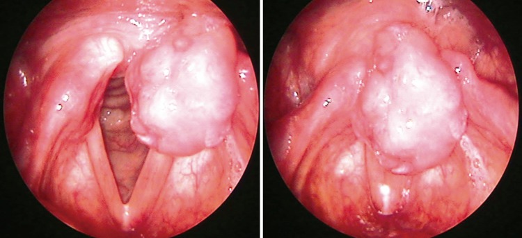
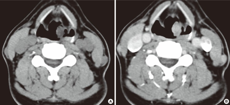
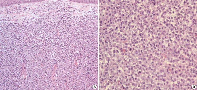
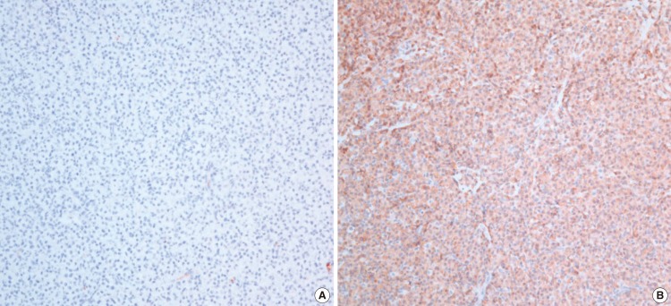
Extramedullary plasmacytoma (EMP) is a rare plasma cell neoplasm that occurs mainly in the soft tissues of head and neck region, with the paranasal sinuses, nasal cavity and nasopharynx being the most common sites. Solitary EMP of the larynx is very rare but increasingly reported recently. Common sites of involvement in larynx in the order of frequency are the epiglottis, ventricles, vocal folds and ventricular folds. We report an extremely rare case of solitary EMP involving in the apex of arytenoids that was successfully treated by only surgical excision. Because solitary EMP of the apex of artytenoids is extremely rare, it should be included in the differential diagnosis for laryngeal mass. Also, solitary, small, pedunculated and localized EMP of the larynx could be completely removed by laryngeal microsurgery.
Keywords: Arytenoid; Extramedullary plasmacytoma; Surgical excision.
Conflict of interest statement
No potential conflict of interest relevant to this article was reported.
Figures
References
-
- Horny HP, Kaiserling E. Involvement of the larynx by hemopoietic neoplasms: an investigation of autopsy cases and review of the literature. Pathol Res Pract. 1995 Mar;191(2):130–138. - PubMed
-
- Kapadia SB, Desai U, Cheng VS. Extramedullary plasmacytoma of the head and neck: a clinicopathologic study of 20 cases. Medicine (Baltimore) 1982 Sep;61(5):317–329. - PubMed
-
- Medini E, Rao Y, Levitt SH. Solitary extramedullary plasmacytoma of the upper respiratory and digestive tracts. Cancer. 1980 Jun;45(11):2893–2896. - PubMed
-
- Kayrouz T, Jose B, Chu AM, Scott RM. Solitary plasmacytoma. J Surg Oncol. 1983 Sep;24(1):46–48. - PubMed
-
- Kost KM. Plasmacytomas of the larynx. J Otolaryngol. 1990 Apr;19(2):141–146. - PubMed
LinkOut - more resources
Full Text Sources
Miscellaneous