. 2011 Sep;13(9):629-33.
doi: 10.5812/kowsar.20741804.2254. Epub 2011 Sep 15.

The healing effect of bioglue in articular cartilage defect of femoral condyle in experimental rabbit model

Affiliations

The healing effect of bioglue in articular cartilage defect of femoral condyle in experimental rabbit model

N Tanideh et al. Iran Red Crescent Med J. 2011 Sep.

Abstract

Background: The full-thickness articular cartilage defects of knee have a poor healing capacity that may progress to osteoarthritis and need a knee replacement. This study determines the healing effect of bioglue in fullthickness articular cartilage defect of femoral condyle in rabbit.

Methods: Forty-eight male rabbits were randomly divided into four equal groups. In group A, 4 mm articular cartilage defects were created in the right and left medial femoral condyles. Then a graft from xiphoid cartilage was transferred into the defect together with a designed bioglue and the knees were closed. In group B, an articular cartilage defect was created identical to group A, but the defect size was 6 mm. In group C, 4 and 6 mm articular cartilage defects were created in the right and left medial femoral condyles respectively. The graft was transferred into the defect and the knees were stitched. In group D, articular cartilage defects were created similar to group C, just filled with bioglue and closed. The rabbits were euthanized and subgroups were defined as A1, B1, C1 and D1 after 30 days and A2, B2, C2 and D2 after 60 days. The cartilages were macroscopically and histologically investigated for any changes.

Results: Microscopic and macroscopic investigations showed that bioglue had a significant healing effect in the femoral condyle.

Conclusion: Addition of bioglue can effectively promote the healing of articular cartilage defects.

Keywords: Articular Cartilage; Bioglue; Defect; Femoral Condyle; Healing; Rabbit.


Conflict of interest statement

Conflict of interest: None declared.

Figures

Fig. 1
Fig. 1
Grossly evaluation of repair articular cartilage of medial femoral condyle in different groups of male rabbits (arrows). A, cartilage graft fixed with designed BioGlue (size 4 mm, time 60 days); B, cartilage graft fixed with designed BioGlue (size 6 mm, 30 days); C, cartilage graft fixed with suture (size 4 mm, time 30 days); D, designed BioGlue filled cartilage defect (size 4 mm, time 30 days).
Fig. 2
Fig. 2
High-power photomicrographs of medial femoral condyle of male rabbit obtained on day 60 with H and E that shows histological changes in relation to healing effects between all groups. A) A2 group, microscopic appearance of reparative tissue showing groups (clusters) of chondrocytes in an extracellular matrix (arrow). The surface is irregular and subchondral area showed granulation tissue formation and remodeling (original magnification×40). B) B2 group, microscopic appearance of reparative tissue showing mixed hyaline/fibrocartilaginous character (arrow). Cartilaginous part revealed predominantly viable individual cells. Tidemark was irregular and interrupted (original magnification×20). C) C2R group, microscopic appearance of reparative tissue showing fibrocartilaginous and fibrous character (arrow). Cartilaginous parts revealed individual disorganized and partially viable cells. (original magnification ×20); D) D2L group, microscopic appearance of reparative tissue showing relatively smooth repaired hyaline cartilage containing columnar arrangement of chondrocytes (arrow; original magnification ×20).
Fig. 3
Fig. 3
High-power photomicrographs of medial femoral condyle obtained on day 60 with toluidene blue that shows histological changes in relation to healing effects between all groups. A) A2 group, histochemical staining of toluidene blue showing defect repaired with hyaline-like cartilage. The subchondral bone and tidemark were well remodeled (original magnification ×40). B) B2 group, histochemical staining of toluidene blue showing densely stained reparative tissue and irregular tidemark (original magnification ×40). C) C2R group, histochemical staining of toluidene blue showing absence of a well formed tidemark (original magnification ×20). D) D2L group, histochemical staining of toluidene blue showing nearly normal cartilage staining. The surface was smooth and the tidemark was well remodeled (original magnification ×40).
Fig. 4
Fig. 4
High-power photomicrographs of medial femoral condyle obtained on 60 days with safranin O that shows histological changes in relation to healing effects between all groups (safranin O ×40). A) A2 group, histochemical staining of safranin O that was more intense in the superficial layer (original magnification×40). B) B2 group, histochemical staining of safranin O with minimal matrix staining (original magnification×40). C) C2R group, histochemical staining of safranin O showing slight staining of extracellular matrix in fibrocartilaginous parts (original magnification ×40). D) D2L group, histochemical staining of safranin O showing moderate staining of extracellular matrix, more intense in the superficial layer (original magnification ×40).
Fig. 5
Fig. 5
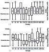
Box and whisker plot of median, mean, and range of changes in surface (a) and matrix (b) of cartilage based on modified ICRS visual histological assessment to evaluate designed BioGlue on articular cartilage graft in male rabbits. A, designed BioGlue and cartilage graft (4 mm); B, designed BioGlue and cartilage graft (6 mm); C, cartilage graft sutured; D, cartilage defect filled with BioGlue; 1, followed after 30 days; 2, followed after 60 days; R, right knee; L, Left knee. The same superscript letters indicates significant statistical difference between comparable groups using Mann-Whitney U test.
Fig. 6
Fig. 6
Box and whisker plot of median, mean, and range of changes in type I collagen staining of the matrix (a) and type II collagen staining of the matrix (b) of cartilage based on modified ICRS visual histological assessment to evaluate designed BioGlue on articular cartilage graft in male rabbits. A, designed BioGlue and cartilage graft (4 mm); B, designed BioGlue and cartilage graft (6 mm); C, cartilage graft sutured; D, cartilage defect filled with BioGlue; 1, followed after 30 days; 2, followed after 60 days; R, right knee; L, Left knee. The same superscript letters indicates significant statistical difference between comparable groups using Mann-Whitney U test.
Fig. 7
Fig. 7
Box and whisker plot of median, mean, and range of changes in cell morphology (a) and matrix staining (b) based on histological grading scale to evaluate designed BioGlue on articular cartilage graft in male rabbits. A, designed BioGlue and cartilage graft (4 mm); B, designed BioGlue and cartilage graft (6 mm); C, cartilage graft sutured; D, cartilage defect filled with BioGlue ; 1, followed after 30 days; 2, followed after 60 days; R, right knee; L, Left knee. The same superscript letters indicates significant statistical difference between comparable groups using Mann-Whitney U test.

Similar articles

Cited by

References

    1. Padua R, Bondì R. Focal articular cartilage defects of the knee: surgical treatment. J Orthop Traumatol. 2004;5:63–5. doi: 10.1007/s10195-004-0043-8. - DOI
    1. Carranza-Bencano A, García-Paino L, Armas Padrón JR, Cayuela Dominguez A. Neochondrogenesis in repair of full-thickness articular cartilage defects using free autogenous periosteal grafts in the rabbit. A follow-up in six months. Osteoarthritis Cartilage. 2000;8:351–8. doi: 10.1053/joca.1999.0309. - DOI - PubMed
    1. Sánchez M, Anitua E, Azofra J, Andía I, Padilla S, Mujika I. Comparison of surgically repaired Achilles tendon tears using plateletrich fibrin matrices. Am J Sports Med. 2007;35:245–51. - PubMed
    1. Beris AE, Lykissas MG, Papageorgiou CD, Georgoulis AD. Advances in articular cartilage repair. Injury. 2005;36:S14–S23. doi: 10.1016/j.injury.2005.10.007. - DOI - PubMed
    1. Gao J, Dennis JE, Solchaga LA, Goldberg VM, Caplan AI. Repair of osteochondral defect with tissue-engineered two-phase composite material of injectable calcium phosphate and hyaluronan sponge. Tissue Eng. 2002;8:827–37. doi: 10.1089/10763270260424187. - DOI - PubMed

LinkOut - more resources