. 2012 Jul;73(7):1035-46.
doi: 10.2460/ajvr.73.7.1035.

Distribution and processing of a disintegrin and metalloproteinase with thrombospondin motifs-4, aggrecan, versican, and hyaluronan in equine digital laminae

Affiliations

Distribution and processing of a disintegrin and metalloproteinase with thrombospondin motifs-4, aggrecan, versican, and hyaluronan in equine digital laminae

Erica Pawlak et al. Am J Vet Res. 2012 Jul.

Abstract

Objective: To determine the expression and distribution of a disintegrin and metalloproteinase with thrombospondin motifs-4 (ADAMTS-4), its substrates aggrecan and versican, and their binding partner hyaluronan in laminae of healthy horses.

Sample: Laminae from the forelimb hooves of 8 healthy horses.

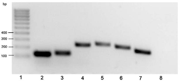
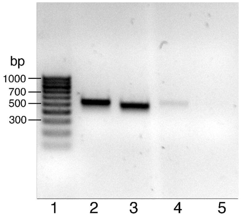
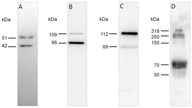
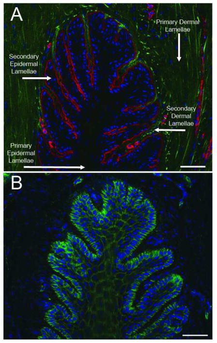
Procedures: Real-time quantitative PCR assay was used for gene expression analysis. Hyaluronidase, chondroitinase, and keratanase digestion of lamina extracts combined with SDS-PAGE and western blotting were used for protein and proteoglycan analysis. Immunofluorescent and immunohistochemical staining of tissue sections were used for protein and hyaluronan localization.

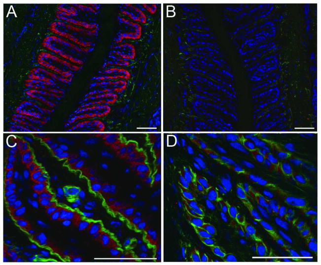
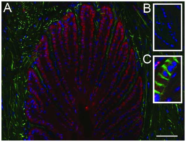
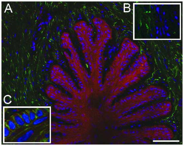
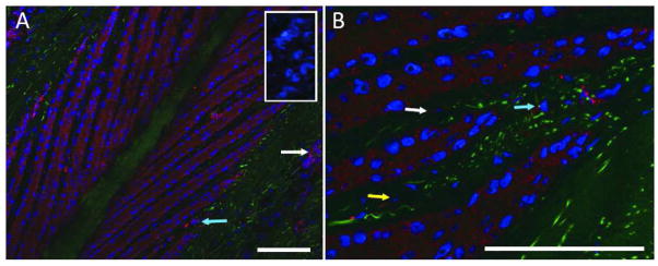
Results: Genes encoding ADAMTS-4, aggrecan, versican, and hyaluronan synthase II were expressed in laminae. The ADAMTS-4 was predominantly evident as a 51-kDa protein bearing a catalytic site neoepitope indicative of active enzyme and in situ activity, which was confirmed by the presence of aggrecan and versican fragments bearing ADAMTS-4 cleavage neoepitopes in laminar protein extracts. Aggrecan, versican, and hyaluronan were localized to basal epithelial cells within the secondary epidermal laminae. The ADAMTS-4 localized to these cells but was also present in some cells in the dermal laminae.

Conclusions and clinical relevance: Within digital laminae, versican exclusively and aggrecan primarily localized within basal epithelial cells and both were constitutively cleaved by ADAMTS-4, which therefore contributed to their turnover. On the basis of known properties of these proteoglycans, it is possible that they can protect the basal epithelial cells of horses from biomechanical and concussive stress.


Figures

Figure 1
Figure 1. Analysis ofRT-qPCR products generated using primer pairs specific for aggrecan, versican, hyaluronan synthase II and ADAMTS4, and cDNA generated using RNA from healthy equine laminae
Lane 1, DNA marker (100 bp ladder); Lane 2 aggrecan; Lane 3 versican; Lane 4 hyaluronan synthase II; Lane 5 ADAMTS-4 N terminal domain; Lane 6 ADAMTS-4 C-terminal domain; Lane 7 GAPDH; Lane 8 blank control.
Figure 2
Figure 2. Analysis of PCR products generated using primer pairs specific for versican isoforms and cDNA generated using RNA from healthy equine laminae
Lane 1, DNA marker (100 bp ladder); Lane 2, V0; Lane 3, V1; Lane 4, V2; Lane 5, V3.
Figure 3
Figure 3. Immunoreactivity of ADAMTS-4 and its substrates in extracts of healthy equine laminae
Western blots of laminar extracts (30 μg protein/lane) were probed using: A) antibody against neoepitope FASLSRFVET exposed on ADAMTS-4 catalytic domain after removal of the regulatory propeptide (NP-40 extract); B) antibody against versican V0/V1 neoeptiope DPEAAE generated by ADAMTS-4 cleavage (NP-40 extract); C) antibody against versican V0/V2 neoeptiope NIVSFE generated by ADAMTS-4 cleavage (NP-40 extract); D) antibody against aggrecan neoepitope ARGSVIL (BC-3) generated by ADAMTS-4 cleavage (guanidine hydrochloride extract digested with hyaluronidase, chondroitinase ABC and karatinase III).
Figure 4
Figure 4. Defining lamellar structure and epidermal/dermal boundaries via localization of laminin and actin
A) Epidermal/dermal lamellar boundary is visualized by immunofluorescent staining against the basement membrane marker laminin in red. Autofluorescent material (putatively collagen) in green, nuclei stained blue; B) Epidermal cellular boundaries visualized with staining against actin in green, nuclei stained blue. The images are representative of samples from 6 animals analyzed. Scale bars 50μm.
Figure 5
Figure 5. Versican uniquely localizes to the basal epithelia of the secondary epidermal lamellae and is not associated with the basement membrane
A) Immunofluorescent staining against versican in red, autofluorescent material in green, nuclei stained blue; B) versican staining blocked by pre-incubation of primary antibody with cognate peptide, autofluorescent material in green, nuclei stained blue; C) versican staining in red, basement membrane visualized by staining against laminin in green, nuclei stained blue; D) versican staining in red, epithelial cell boundaries visualized by staining against actin in green, nuclei stained blue. The images are representative of samples from 6 animal analyzed. Scale bars 50μm.
Figure 6
Figure 6. Aggrecan localizes primarily to the secondary epidermal lamellae
A) Immunofluorescent staining against aggrecan in red, autofluorescent material in green, nuclei stained blue; B) aggrecan staining was blocked by preincubation of the primary antibody with cognate peptide, autofluorescent material in green, nuclei stained blue; C) aggrecan staining in red, epithelial cell boundaries visualized by staining against actin in green, nuclei stained blue. The images are representative of samples from 6 animals analyzed. Scale bars 50μm.
Figure 7
Figure 7. Hyaluronan is present throughout lamellae but enriched in the secondary epidermal lamellae
A) Immunofluorescent staining against hyaluronan in red, autofluorescent material in green, nuclei stained blue; B) hyaluronan staining abrogated by incubation of tissue section with epitope-digesting enzyme (Chondroitinase ABC pH 6.8), autofluorescent material in green, nuclei stained blue; C) hyaluronan staining in red, epithelial cell boundaries visualized by staining against actin in green, nuclei stained blue. The images are representative of samples from 6 animals analyzed. Scale bars 50μm.
Figure 8
Figure 8. ADAMTS-4 is present in epithelial cells of the secondary epidermal lamellae
A) Immunofluorescent staining against ADAMTS-4 “NTPEDSDPDHFD” epitope in red, autofluorescent material in green, nuclei stained blue. Staining blocked by cognate peptide in inset. B) Higher magnification image, ADAMTS-4 visualized in red, autofluorescent material in green, nuclei in blue. Images representative of samples from 6 animals analyzed. Scale bars 50μm. White arrows indicate vascular endothelia, blue arrows indicate mononuclear cells of the dermal lamellae, yellow arrow indicates a dermal fibroblast.

Similar articles

Cited by

References

    1. Pollitt CC. The Anatomy and Physiology of the Suspensory Apparatus of the Distal Phalanx. Veterinary Clinics of North America: Equine Practice. 2010;26:29–49. Advances in Laminitis, Part I. - PubMed
    1. French KR, Pollitt CC. Equine laminitis: loss of hemidesmosomes in hoof secondary epidermal lamellae correlates to dose in an oligofructose induction model: an ultrastructural study. Equine Vet J. 2004;36:230–235. - PubMed
    1. Black SJ. Extracellular matrix, leukocyte migration and laminitis. Vet Immunol Immunopathol. 2009;129:161–163. - PubMed
    1. Alonso L, Fuchs E. Stem cells of the skin epithelium. Proc Natl Acad Sci U S A. 2003;100 (Suppl 1):11830–11835. - PMC - PubMed
    1. Budras KD, Hullinger RL, Sack WO. Light and electron microscopy of keratinization in the laminar epidermis of the equine hoof with reference to laminitis. Am J Vet Res. 1989;50:1150–1160. - PubMed

Publication types

MeSH terms

LinkOut - more resources