. 2012 Jul 24;51(29):5748-62.
doi: 10.1021/bi300409m. Epub 2012 Jul 10.

Aspartate-histidine interaction in the retinal schiff base counterion of the light-driven proton pump of Exiguobacterium sibiricum

Affiliations

Aspartate-histidine interaction in the retinal schiff base counterion of the light-driven proton pump of Exiguobacterium sibiricum

S P Balashov et al. Biochemistry. .

Abstract

One of the distinctive features of eubacterial retinal-based proton pumps, proteorhodopsins, xanthorhodopsin, and others, is hydrogen bonding of the key aspartate residue, the counterion to the retinal Schiff base, to a histidine. We describe properties of the recently found eubacterium proton pump from Exiguobacterium sibiricum (named ESR) expressed in Escherichia coli, especially features that depend on Asp-His interaction, the protonation state of the key aspartate, Asp85, and its ability to accept a proton from the Schiff base during the photocycle. Proton pumping by liposomes and E. coli cells containing ESR occurs in a broad pH range above pH 4.5. Large light-induced pH changes indicate that ESR is a potent proton pump. Replacement of His57 with methionine or asparagine strongly affects the pH-dependent properties of ESR. In the H57M mutant, a dramatic decrease in the quantum yield of chromophore fluorescence emission and a 45 nm blue shift of the absorption maximum with an increase in the pH from 5 to 8 indicate deprotonation of the counterion with a pK(a) of 6.3, which is also the pK(a) at which the M intermediate is observed in the photocycle of the protein solubilized in detergent [dodecyl maltoside (DDM)]. This is in contrast with the case for the wild-type protein, for which the same experiments show that the major fraction of Asp85 is deprotonated at pH >3 and that it protonates only at low pH, with a pK(a) of 2.3. The M intermediate in the wild-type photocycle accumulates only at high pH, with an apparent pK(a) of 9, via deprotonation of a residue interacting with Asp85, presumably His57. In liposomes reconstituted with ESR, the pK(a) values for M formation and spectral shifts are 2-3 pH units lower than in DDM. The distinctively different pH dependencies of the protonation of Asp85 and the accumulation of the M intermediate in the wild-type protein versus the H57M mutant indicate that there is strong Asp-His interaction, which substantially lowers the pK(a) of Asp85 by stabilizing its deprotonated state.


Figures

Figure 1
Figure 1
pH dependence of the retinal chromophore absorption band of ESR, and the effect of the D85N mutation. A. 1, ESR wild type, pH 5; 2, ESR wild type, pH 10.5; 3, D85N mutant, pH 5. B. pH dependence of absorption maximum of ESR. All spectra are measured in 0.2% DDM, 0.1 M NaCl. C. Difference spectra of pH induced absorption changes in ESR: 1, pH7 minus pH 5; 2, pH 10 minus pH 7.
Figure 2
Figure 2
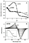
pH dependent transitions in the absorption band of the H57M and R82Q mutants in comparison with the wild type. A. Absorption maximum vs. pH in: 1, H57M; 2, R82Q, 3, wild type. B. Difference spectra upon increasing the pH from 4.5 to 7.4 from deprotonation of the counterion with pKa 6.3 (see inset) in the H57M mutant (the spectra “pHi - pH 4.5” were obtained at the following values of pHi: 4.9, 5.2, 5.5, 5.7, 5.9, 6.2, 6.5, 6.8, 7.1, 7.4).
Figure 3
Figure 3
Fluorescence emission, excitation and absorption spectra of wild type ESR and the D85N and H57M mutants as a tool to estimate the fraction of the pigment with protonated counterion. A. Retinal chromophore fluorescence band measured under 530 nm excitation: 1, D85N mutant of ESR, pH 5; 2, wild type, pH 5; 3, wild type, pH 7. Concentration of retinal proteins was 8 WM. B and C. Comparison of excitation (1) and absorption (2) spectra of D85N (B) and the wild type (C). D. Fluorescence excitation spectra for the 720 nm emission in wild type ESR at: 1, pH 5; 2, pH 6; 3, pH 7; 4, pH 8.8. E. pH dependence of the fluorescence excitation spectrum at 610 nm, proportional to the fraction of protonated counterion: 1, wild type ESR; 2, H57M. The amplitude of the spectrum at 610 nm in H57M at pH 4.5 was taken as 1. F. Fluorescence excitation spectra for the 720 nm emission for the H57M mutant at several pH values, 1 through 6, pH 4.5, 6.1, 6.7, 7.0, 7.5, and 8.8, respectively.
Figure 4
Figure 4
Absorption changes induced by 532 nm laser flash in ESR at pH 7.3 (A, B) and pH 9.1 (C, D) at: A. 1 to 5, spectra taken at 0.2, 1, 2, 5 and 20 Ws after the flash, respectively. B. 1 to 4, spectra taken at 10, 20, 50 and 100 ms after the flash, respectively. C. 1 and 2, spectra taken at 100 Ws and 2 ms, respectively; 3, difference between spectra taken at 2 ms and 100 Ws. D. 1 to 5, spectra taken at 5, 10, 20, 50 and 100 ms after the flash, respectively.
Figure 5
Figure 5
Kinetics of light-induced absorption changes of wild type ESR at 410, 510, 550 and 590 nm at several pH. The bars and numbers on top of the panels represent the kinetic components that were determined from the global fit of absorption changes at the four wavelengths.
Figure 6
Figure 6
The pH dependence of yield of the M intermediate (normalized): 1, wild type ESR; 2, H57M.
Figure 7
Figure 7
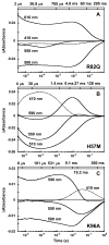
Kinetics of light-induced absorption changes in: A. R82Q mutant at pH 9. B. H57M mutant at pH 7.6. C. K96A mutant at pH 7.6.
Figure 8
Figure 8
Kinetics of light-induced proton release and uptake after flash photoexcitation, followed by absorption changes of pyranine at 456 nm. Changes in the chromophore absorption were subtracted as explained in the text.1, wild type ESR, pH 7.2; 2, H57M, pH 7.5. The measured wild type ESR trace was multiplied by 3.6 to scale with that of H57M. Dashed lines are fits described in the text. Conditions: 0.2 % DDM, 100 mM NaCl.
Figure 9
Figure 9
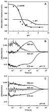
ESR incorporated in liposomes. A. pH dependence of the absorption maximum in: 1, the wild type, fit with pKa ca. 6.5; 2, H57M mutant, fit with pKa ca. 4.5. B and C. Photocycle kinetics of the wild type ESR at selected wavelengths (420, 510 and 590 nm) at pH 7.5 (B) and at pH 4.8 (C), i.e., above and below the corresponding pKa ca. 6.5 of the transition revealed by the absorption maximum shift.
Figure 10
Figure 10
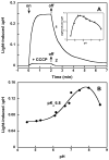
Light-induced proton pumping by ESR. A. 1, time course of light-induced pH changes in suspension of E. coli cells with ESR expressed and reconstituted with all-trans retinal. 2, after addition of 5 10−5 M CCCP. pH 5.5. Inset, pH dependence of light-induced pH changes in E. coli cells with ESR. B. pH dependence of light-induced pH changes in a suspension of proteoliposomes containing ESR.

References

    1. Vishnivetskaya T, Kathariou S, McGrath J, Gilichinsky D, Tiedje JM. Low-temperature recovery strategies for the isolation of bacteria from ancient permafrost sediments. Extremophiles. 2000;4:165–173. - PubMed
    1. Rodrigues DF, Goris J, Vishnivetskaya T, Gilichinsky D, Thomashow MF, Tiedje JM. Characterization of Exiguobacterium isolates from the Siberian permafrost. Description of Exiguobacterium sibiricum sp nov. Extremophiles. 2006;10:285–294. - PubMed
    1. Petrovskaya LE, Lukashev EP, Chupin VV, Sychev SV, Lyukmanova EN, Kryukova EA, Ziganshin RH, Spirina EV, Rivkina EM, Khatypov RA, Erokhina LG, Gilichinsky DA, Shuvalov VA, Kirpichnikov MP. Predicted bacteriorhodopsin from Exiguobacterium sibiricum is a functional proton pump. FEBS Lett. 2010;584:4193–4196. - PubMed
    1. Oesterhelt D, Stoeckenius W. Functions of a new photoreceptor membrane. Proc Natl Acad Sci USA. 1973;70:2853–2857. - PMC - PubMed
    1. Ovchinnikov YA, Abdulaev NG, Feigina MY, Kiselev AV, Lobanov NA. The structural basis of the functioning of bacteriorhodopsin: An overview. FEBS Lett. 1979;100:219–224. - PubMed

Publication types

MeSH terms