. 2011 Sep 27;1(1):2.
doi: 10.1186/2045-5380-1-2.

Medial prefrontal cortex serotonin 1A and 2A receptor binding interacts to predict threat-related amygdala reactivity

Affiliations

Medial prefrontal cortex serotonin 1A and 2A receptor binding interacts to predict threat-related amygdala reactivity

Patrick M Fisher et al. Biol Mood Anxiety Disord. .

Abstract

Background: The amygdala and medial prefrontal cortex (mPFC) comprise a key corticolimbic circuit that helps shape individual differences in sensitivity to threat and the related risk for psychopathology. Although serotonin (5-HT) is known to be a key modulator of this circuit, the specific receptors mediating this modulation are unclear. The colocalization of 5-HT1A and 5-HT2A receptors on mPFC glutamatergic neurons suggests that their functional interactions may mediate 5-HT effects on this circuit through top-down regulation of amygdala reactivity. Using a multimodal neuroimaging strategy in 39 healthy volunteers, we determined whether threat-related amygdala reactivity, assessed with blood oxygen level-dependent functional magnetic resonance imaging, was significantly predicted by the interaction between mPFC 5-HT1A and 5-HT2A receptor levels, assessed by positron emission tomography.

Results: 5-HT1A binding in the mPFC significantly moderated an inverse correlation between mPFC 5-HT2A binding and threat-related amygdala reactivity. Specifically, mPFC 5-HT2A binding was significantly inversely correlated with amygdala reactivity only when mPFC 5-HT1A binding was relatively low.

Conclusions: Our findings provide evidence that 5-HT1A and 5-HT2A receptors interact to shape serotonergic modulation of a functional circuit between the amygdala and mPFC. The effect of the interaction between mPFC 5-HT1A and 5-HT2A binding and amygdala reactivity is consistent with the colocalization of these receptors on glutamatergic neurons in the mPFC.


Figures

Figure 1
Figure 1
Amygdala reactivity to perceptual processing of fearful and angry facial expressions. Statistical parametric map representing bilateral amygdala clusters exhibiting a significant response to task (faces > shapes; right amygdala: (24, -6, -11), z = 6.28, k = 145 voxels (P < 0.05, corrected); left amygdala: (-18, -7, -15), z = 5.77, k = 146 voxels (P < 0.05, corrected). Color bar indicates t-scores.
Figure 2
Figure 2
Association between amygdala reactivity and 5-HT1A BPND and 5-HT2A BPND. (A,B) Plot of non-significant correlation between left and right amygdala reactivity and pgPFC 5-HT1A BPND. (C) Plot of non-significant correlation between left amygdala reactivity and pgPFC 5-HT2A BPND. (D) Plot of significant inverse correlation between right amygdala reactivity and pgPFC 5-HT2A BPND. 5-HT = serotonin; BPND = binding potential, non-displaceable; pgPFC = pregenual prefrontal cortex; sgPFC = subgenual prefrontal cortex.
Figure 3
Figure 3
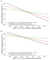
5-HT1A BPND significantly moderated the correlation between 5-HT2A BPND and right amygdala reactivity. (A) pgPFC 5-HT1A BPND moderated the correlation between pgPFC 5-HT2A BPND and right amygdala reactivity. Lines indicate simple slope between pgPFC 5-HT2A BPND and right amygdala reactivity at three arbitrarily chosen pgPFC 5-HT1A BPND values: low (1 SD below mean (-1 SD), solid black line), mean (equivalent to mean, red dotted line) and high (1 SD above mean (+1 SD), green dotted line). (B) sgPFC 5-HT1A BPND significantly moderated the association between sgPFC 5-HT2A BPND and right amygdala reactivity. Lines indicate simple slope between sgPFC 5-HT2A BPND and right amygdala reactivity at three arbitrarily chosen sgPFC 5-HT1A BPND values: low (-1 SD, solid black line), mean (red dotted line) and high (+1 SD, green dotted line). *Indicates simple slope, P < 0.05; 5-HT = serotonin; a.u. = arbitrary units; BPND = binding potential, non-displaceable; pgPFC = pregenual prefrontal cortex; sgPFC = subgenual prefrontal cortex.
Figure 4
Figure 4
Schematic illustrating mPFC projection neurons that act to regulate amygdala response to threat-related stimuli. 5-HT1A and 5-HT2A in mPFC are positioned to modulate this circuitry by biasing excitability of these mPFC neurons, thereby affecting the capacity to regulate amygdala reactivity. 5-HT = serotonin; mPFC = medial prefrontal cortex; CeL = lateral central nucleus of the amygdala; CeM = medial central nucleus of the amygdala; ITC = intercalated cells.
Figure 5
Figure 5
Sagittal image of single-subject magnetic resonance image with pgPFC (top) and sgPFC (bottom) ROIs outlined. Despite its appearance, pgPFC and sgPFC ROIs are drawn on consecutive transaxial slices. pgPFC = pregenual prefrontal cortex; ROIs = regions of interest; sgPFC = subgenual prefrontal cortex.

Similar articles

Cited by

References

    1. Hariri AR, Drabant EM, Weinberger DR. Imaging genetics: perspectives from studies of genetically driven variation in serotonin function and corticolimbic affective processing. Biological Psychiatry. 2006;59(10):888. doi: 10.1016/j.biopsych.2005.11.005. - DOI - PubMed
    1. Pezawas L, Meyer-Lindenberg A, Drabant EM, Verchinski BA, Munoz KE, Kolachana BS, Egan MF, Mattay VS, Hariri AR, Weinberger DR. 5-HTTLPR polymorphism impacts human cingulate-amygdala interactions: a genetic susceptibility mechanism for depression. Nat Neurosci. 2005;8(6):828–34. doi: 10.1038/nn1463. - DOI - PubMed
    1. Phelps EA, Delgado MR, Nearing KI, LeDoux JE. Extinction Learning in Humans: Role of the Amygdala and vmPFC. Neuron. 2004;43(6):897. doi: 10.1016/j.neuron.2004.08.042. - DOI - PubMed
    1. Quirk GJ, Mueller D. Neural mechanisms of extinction learning and retrieval. Neuropsychopharmacology. 2008;33(1):56–72. doi: 10.1038/sj.npp.1301555. - DOI - PMC - PubMed
    1. Likhtik E, Pelletier JG, Paz R, Pare D. Prefrontal control of the amygdala. J Neurosci. 2005;25(32):7429–37. doi: 10.1523/JNEUROSCI.2314-05.2005. - DOI - PMC - PubMed

LinkOut - more resources