. 2012 Aug 10;29(12):2192-208.
doi: 10.1089/neu.2011.2303.

Severe brief pressure-controlled hemorrhagic shock after traumatic brain injury exacerbates functional deficits and long-term neuropathological damage in mice

Affiliations

Severe brief pressure-controlled hemorrhagic shock after traumatic brain injury exacerbates functional deficits and long-term neuropathological damage in mice

Joseph N Hemerka et al. J Neurotrauma. .

Abstract

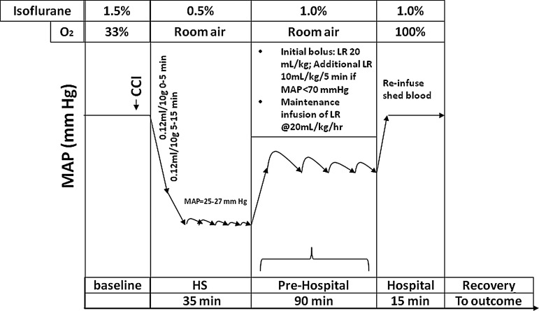
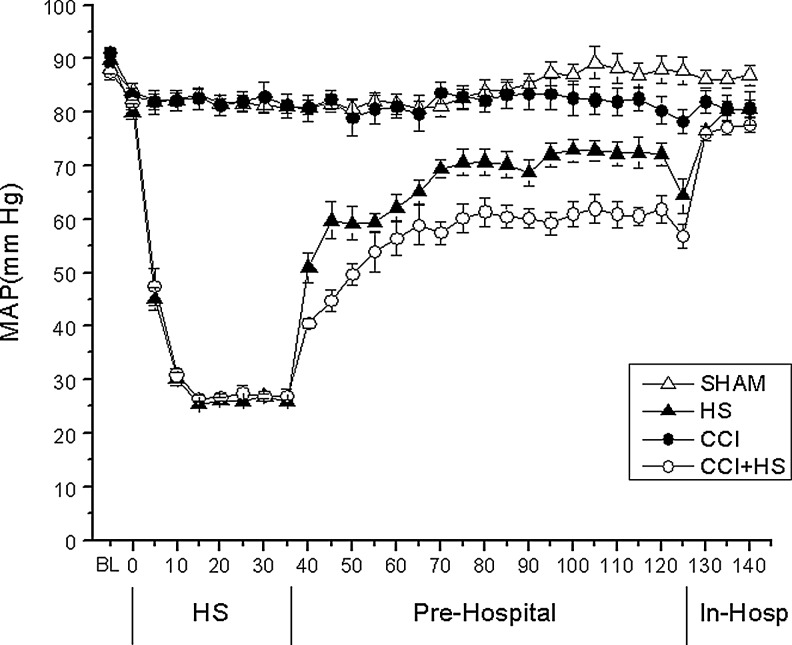
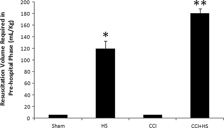
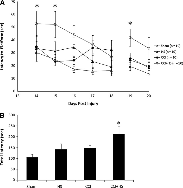
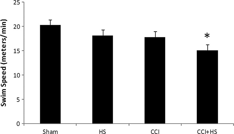
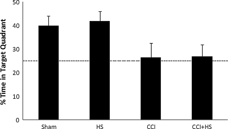
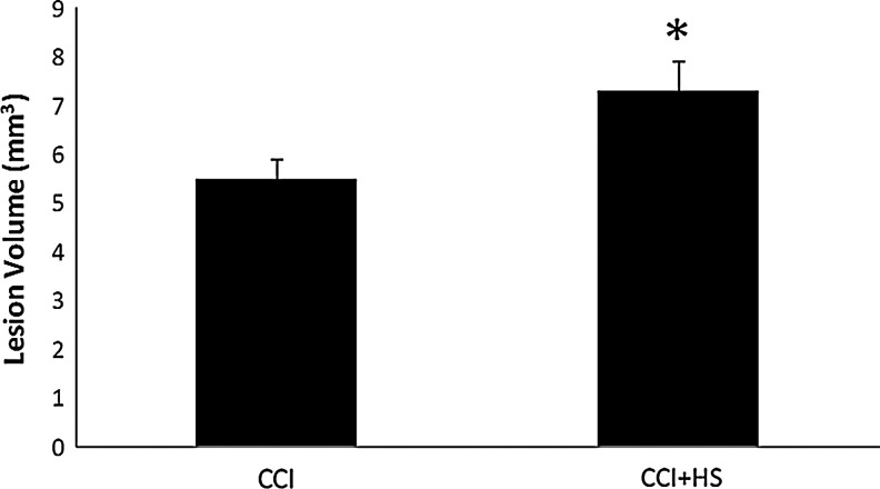
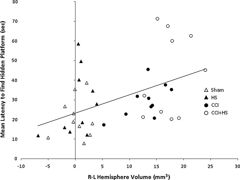
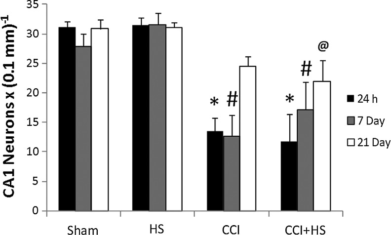
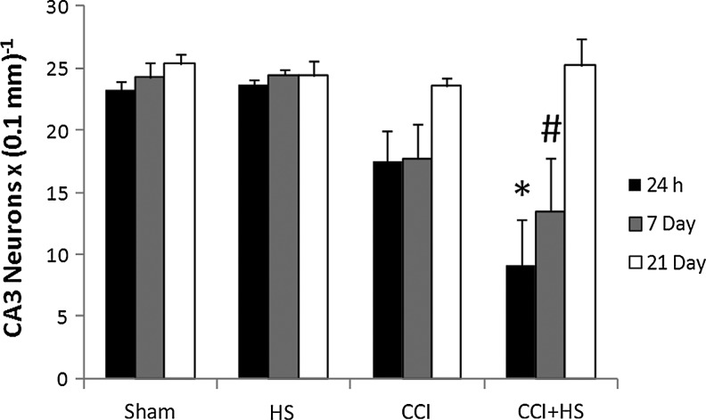
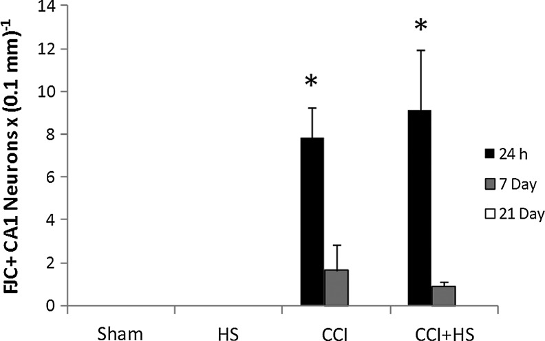
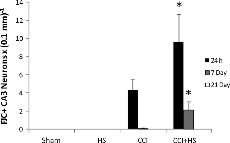
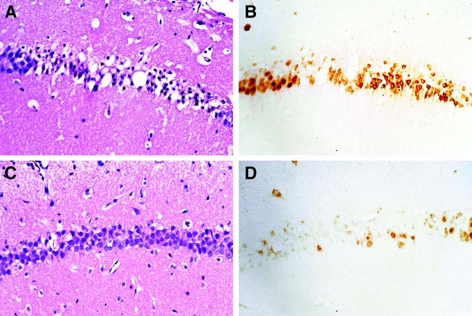
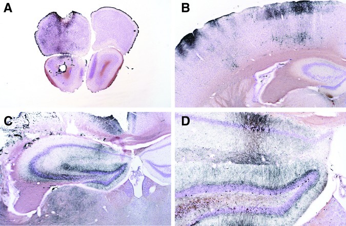
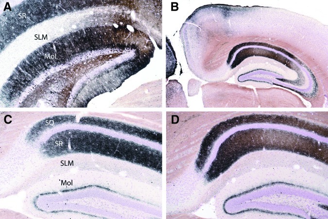
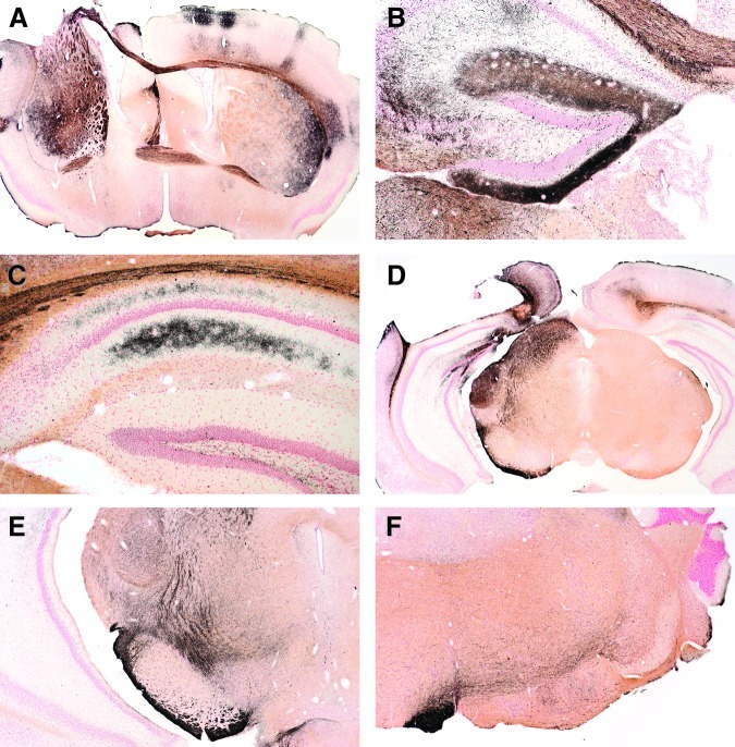
Hypotension after traumatic brain injury (TBI) worsens outcome. We published the first report of TBI plus hemorrhagic shock (HS) in mice using a volume-controlled approach and noted increased neuronal death. To rigorously control blood pressure during HS, a pressure-controlled HS model is required. Our hypothesis was that a brief, severe period of pressure-controlled HS after TBI in mice will exacerbate functional deficits and neuropathology versus TBI or HS alone. C57BL6 male mice were randomized into four groups (n=10/group): sham, HS, controlled cortical impact (CCI), and CCI+HS. We used a pressure-controlled shock phase (mean arterial pressure [MAP]=25-27 mm Hg for 35 min) and its treatment after mild to moderate CCI including, a 90 min pre-hospital phase, during which lactated Ringer's solution was given to maintain MAP >70 mm Hg, and a hospital phase, when the shed blood was re-infused. On days 14-20, the mice were evaluated in the Morris water maze (MWM, hidden platform paradigm). On day 21, the lesion and hemispheric volumes were quantified. Neuropathology and hippocampal neuron counts (hematoxylin and eosin [H&E], Fluoro-Jade B, and NeuN) were evaluated in the mice (n=60) at 24 h, 7 days, or 21 days (n=5/group/time point). HS reduced MAP during the shock phase in the HS and CCI+HS groups (p<0.05). Fluid requirements during the pre-hospital phase were greatest in the CCI+HS group (p<0.05), and were increased in HS versus sham and CCI animals (p<0.05). MWM latency was increased on days 14 and 15 after CCI+HS (p<0.05). Swim speed and visible platform latency were impaired in the CCI+HS group (p<0.05). CCI+HS animals had increased contusion volume versus the CCI group (p<0.05). Hemispheric volume loss was increased 33.3% in the CCI+HS versus CCI group (p<0.05). CA1 cell loss was seen in CCI+HS and CCI animals at 24 h and 7 days (p<0.05). CA3 cell loss was seen after CCI+HS (p<0.05 at 24 h and 7 days). CA1 cell loss at 21 days was seen only in CCI+HS animals (p<0.05). Brief, severe, pressure-controlled HS after CCI produces robust functional deficits and exacerbates neuropathology versus CCI or HS alone.


Figures

FIG. 1.
FIG. 1.
Schematic diagram of the protocol used to study the effect of severe pressure-controlled hemorrhagic shock (HS) on functional and neuropathological outcomes after controlled cortical impact (CCI) in mice. Controls included sham, HS only, and CCI-only groups. A clinically-relevant three-phase model was used that included a 35-min shock phase, during which time the mean arterial pressure (MAP) target was 25–27 mm Hg—a severe HS level. After the shock phase, mice in the HS and CCI+HS groups were resuscitated during a pre-hospital phase, simulating field resuscitation for 90 min using lactated Ringer's (LR) solution. An initial bolus of 20 mL/kg was followed by boluses every 5 min to maintain MAP >70 mm Hg. During the pre-hospital phase, maintenance LR was also infused at 4 mL/kg/h. After the pre-hospital phase, a hospital phase followed, which included reinfusion of the shed blood, simulating transfusion in the trauma bay or emergency department.
FIG. 2.
FIG. 2.
Mean arterial pressure (MAP, mm Hg) versus time (min) in mice subjected to sham, hemorrhagic shock (HS), controlled cortical impact (CCI) traumatic brain injury, or combined CCI+HS. As outlined in Figure 1, a three-phase model was used that included a shock phase, a pre-hospital resuscitation phase, and a hospital phase (In-Hosp). The HS and CCI+HS groups showed the anticipated marked reduction in MAP during the 35-min shock phase (p<0.05 for group×time interaction), since only these two groups were exposed to hemorrhage. In addition, recovery of MAP was slowest in the CCI+HS group during the pre-hospital phase (p<0.05 for group×time interaction).
FIG. 3.
FIG. 3.
Heart rate (HR, beats per minute) versus time (min) in mice subjected to sham, hemorrhagic shock (HS), controlled cortical impact (CCI) traumatic brain injury, or combined CCI+HS. As outlined in Figure 1, a three-phase model was used that included a shock phase, a pre-hospital resuscitation phase, and a hospital phase (In-Hosp). Hemorrhage appeared to produce a divergent HR response in the HS and CCI+HS groups, with a depressor response during the first 25 min of the shock phase in the HS-alone group, but tachycardia in the combined CCI+HS group (p<0.05 for the effect of both time and group×time interaction).
FIG. 4.
FIG. 4.
Fluid requirements during the pre-hospital resuscitation phase to maintain mean arterial pressure (MAP) >70 mm Hg were greatest in the CCI+HS group (**p<0.05 versus all other groups), and were also increased in the HS group (*p<0.05 versus sham and CCI only). Fluid administered to the sham and CCI-only groups represented a maintenance infusion of lactated Ringer's solution at 4 mL/kg/h, thus totaling 6 mL/kg in each of these groups over the 60-min pre-hospital phase (HS, hemorrhagic shock; CCI, controlled cortical impact).
FIG. 5.
FIG. 5.
(A) Graph depicting latency to find the platform on each day of testing in the Morris water maze (MWM) in the sham, hemorrhagic shock (HS) only, controlled cortical impact (CCI) only, and CCI+HS groups. The CCI-only insult was at a mild to moderate injury level that did not produce a significant MWM deficit during hidden platform testing. Also, HS alone also did not produce a MWM deficit versus sham animals. In contrast, the CCI+HS group showed an increased latency on days 14–18 (*p<0.05 versus all other groups), indicating that the secondary HS insult worsened functional outcome after CCI. Latency to find the platform was also increased on the first day of visible platform testing after CCI+HS (*p<0.05 versus all other groups), indicating that the MWM deficit in the acquisition phase of testing in the CCI+HS group cannot necessarily be attributed to spatial memory deficits. (B) Graph of total latency in the hidden platform paradigm (days 14–18) confirmed the deleterious effect of CCI+HS on MWM performance (p<0.05 versus all other groups).
FIG. 6.
FIG. 6.
Swim speed in the Morris water maze (MWM), assessed on days 19–20 for mice subjected to sham, hemorrhagic shock (HS) only, controlled cortical impact (CCI) only, and CCI+HS. Swim speed was significantly reduced only in the CCI+HS group (*p<0.05 versus sham animals by one-way analysis of variance and Student-Newman-Keuls testing).
FIG. 7.
FIG. 7.
Percent of time spent in the target quadrant during a probe trial for mice subjected to sham, hemorrhagic shock (HS) only, controlled cortical impact (CCI) only, and CCI+HS. The percent of time spent in the target quadrant was lower in mice in the CCI and CCI+HS groups than either sham or HS alone. Mice in both the CCI and CCI+HS groups exhibited maximally impaired performance on this task, given that their observed percent of time spent in the target quadrant was similar to random chance (p=0.049 for overall one-way analysis of variance effect).
FIG. 8.
FIG. 8.
Graph depicting contusion volume in mice at 21 days after either controlled cortical impact (CCI), or CCI plus hemorrhagic shock (HS; CCI+HS). Mice in the sham and HS-only groups did not have visible lesions to quantify. Secondary HS after CCI significantly increased contusion volume, by over 33% (*p<0.05 for the CCI+HS group versus the CCI-only group).
FIG. 9.
FIG. 9.
Hemispheric tissue loss represented as right minus left (R-L) hemispheric volume in mice at 21 days after sham, hemorrhagic shock (HS) only, controlled cortical impact (CCI) only, and CCI+HS. In the CCI and CCI+HS groups, the injury was delivered to the left hemisphere. HS alone did not lead to a R-L hemispheric volume difference versus sham animals. CCI alone led to a R-L difference of 12.99 mm3, which represents ∼10% loss of volume (*p<0.05 versus sham or HS only) in the left hemisphere. Combined CCI+HS further expanded tissue loss, to over 15% of the left hemisphere (**p<0.05 versus all other groups), again indicating deleterious neuropathological consequences of a secondary HS insult on CCI.
FIG. 10.
FIG. 10.
Correlation between tissue loss in the damaged left hemisphere, as quantified by the right-left (R-L) difference in hemispheric volume at 21 days after injury, and mean latency to find the hidden platform in the Morris water maze on days 14–18 after injury, in mice subjected to sham, hemorrhagic shock (HS) only, controlled cortical impact (CCI) only, and combined CCI+HS. A significant correlation between tissue loss and behavioral outcome was observed (Pearson's correlation coefficient n=0.434, p=0.005).
FIG. 11.
FIG. 11.
Time course of neuronal survival as assessed in hematoxylin and eosin-stained coronal brain sections in the CA1 hippocampus in the sham, hemorrhagic shock (HS), controlled cortical impact (CCI), and CCI plus hemorrhagic shock (CCI+HS) groups. CA1 survival was reduced at 24 h and 7 days after CCI and CCI+HS versus their respective sham and HS groups. In contrast, CA1 neuron counts were reduced at 21 days after injury only in the CCI+HS group (*p<0.05 versus the respective sham and HS groups at 24 h after the insult; #p<0.05 versus the respective sham and HS groups at 7 days after the insult; @p<0.05 versus the respective sham and HS groups at 21 days after the insult).
FIG. 12.
FIG. 12.
Time course of neuronal survival as assessed in hematoxylin and eosin-stained coronal brain sections in the CA3 hippocampus in the sham, hemorrhagic shock (HS), controlled cortical impact (CCI), and CCI plus hemorrhagic shock (CCI+HS) groups. CA3 survival was significantly reduced at 24 h and 7 days only in the CCI+HS group (*p<0.05 versus the respective sham, HS, and CCI groups at 24 h after the insult; #p<0.05 versus the respective sham and HS groups at 7 days after the insult).
FIG. 13.
FIG. 13.
Time course of neuronal death as assessed in Fluoro-Jade C (FJC)-stained coronal brain sections in the CA1 hippocampus in the sham, hemorrhagic shock (HS), controlled cortical impact (CCI), and CCI plus hemorrhagic shock (CCI+HS) groups. CA1 cell death was significantly increased at 24 h after injury in the CCI and CCI+HS groups versus the sham and HS groups. FJC positivity was not seen in the sham or HS groups at any time point, or in the CCI and CCI+HS groups at 21 days (*p<0.05 versus the respective sham and HS groups at 24 h after the insult).
FIG. 14.
FIG. 14.
Time course of neuronal death as assessed in Fluoro-Jade C (FJC)-stained coronal brain sections in the CA3 hippocampus in the sham, hemorrhagic shock (HS), controlled cortical impact (CCI), and CCI plus hemorrhagic shock (CCI+HS) groups. CA3 cell death was significantly increased at 24 h and 7 days only in the CCI+HS group. FJC positivity was not seen in the sham or HS groups at any time point, or in the CCI and CCI+HS groups at 21 days (*p<0.05 versus the respective sham, HS, and CCI groups at 24 h or 7 days after the insult).
FIG. 15.
FIG. 15.
These panels show hematoxylin and eosin (H&E)- and NeuN-stained sections from mice necropsied at 24 h (A and B), and at 7 days (C and D). A and B are from the same 24-h controlled cortical impact (CCI)-group mouse, are of the same magnification, and represent the exact same region of the CA1 sector of the hippocampus. The H&E-stained section (A) is characterized by extensive eosinophilic neuron degeneration. Although most of the degenerating pyramidal neurons still stain with NeuN, the staining pattern is marked by a transition from nuclear staining, as seen in the left-hand margin of panel B (which is normal), to that of cytoplasmic staining. In contrast, at 7 days (C), the H&E-stained section shows many normal-appearing neurons in the CA1 sector of this CCI-group mouse. However, the bulk of these neurons do not stain (or show only weak nuclear staining) with NeuN (D).
FIG. 16.
FIG. 16.
Hematoxylin and eosin (H&E)- and glial fibrillary acidic protein (GFAP)-stained sections of hippocampi from mice necropsied at 7 days and 21 days post-trauma. All four images are at the same magnification. Panel A is an H&E-stained section from a controlled cortical impact (CCI) mouse necropsied at 7 days. At this magnification, little evidence of degeneration is evident. Panel B is of a GFAP-stained section of hippocampus from a sham mouse, whereas panels C and D show GFAP-stained hippocampal sections from a CCI-group mouse necropsied at 21 days (C), and of a CCI+HS-group mouse necropsied at 7 days. These panels demonstrate the consistent astrocyte response seen at 7 days and 21 days after CCI. At higher magnifications, many reactive astrocytes were also evident within the hippocampus and underlying thalamus of the 7-day and 21-day CCI and CCI+HS rats.
FIG. 17.
FIG. 17.
Representative micrographs of brain sections from mice 24 h after injury. In all micrographs, the left side of the brain (the injured side) is on the left. All sections were stained with cupric silver, so cells and processes staining black are degenerative. Reflecting the sensitivity of the cupric silver stain, panel A shows subtle degeneration in the frontal lobe after temperature probe placement in striatum in a controlled cortical impact plus hemorrhagic shock (CCI+HS) mouse. This injury pattern was seen in all groups. Panel B shows neuron degeneration in the parietal cortex of one of two HS-only mice at 24 h. This pattern of cortical damage, which extended from the frontal to the occipital cortex in this mouse, was limited to the left side of the brain, suggesting that HS can exacerbate brain injury resulting from even a subtle insult such as temperature probe placement. Panels C and D show low- and medium-power magnifications of the same hippocampal section of a 24-h CCI mouse. Many neurons are stained in both the pyramidal sectors (particularly CA1 and CA3), and the dentate gyrus and the underlying dorsal thalamus. Staining is not evident in the contralateral hippocampus at 24 h (partially shown in C).
FIG. 18.
FIG. 18.
Representative micrographs of hippocampal sections from mice 72 h after injury (all sections stained with cupric silver). Panel A shows degenerating neurons within the dentate gyrus, and extensive staining of neuronal processes in the dentate molecular layer (Mol), and within the stratum radiatum (SR) of the pyramidal layer. The stratum lacunosum molecular (SLM) is not affected. Panels B, C, and D are micrographs of the contralateral (non-injured) side of the brain (B and D are low- and medium-power micrographs from the same controlled cortical impact plus hemorrhagic shock [CCI+HS] mouse; C is from a CCI-only mouse). At 72 h the contralateral hippocampi were characterized by extensive neuronal process degeneration within the stratum oriens (SO) and stratum radiatum (SR) of the pyramidal layer, as well as granular staining within the molecular layer (Mol) of the dentate.
FIG. 19.
FIG. 19.
Cupric silver-stained sections from controlled cortical impact (CCI) and CCI plus hemorrhagic shock (CCI+HS) group mice at 14 days. In contrast to the 72-h time point, brains from 14-day mice showed staining (i.e., degeneration) in a variety of nerve fiber tracts. Panel A (CCI+HS) shows staining of the anterior commissure, corpus callosum, and striatum bilaterally. Panel B (CCI) shows persistence of staining in the molecular layer of the dentate and the corpus callosum, the Schaffer collaterals of the hippocampus, and the underlying thalamus (on the injured side). Panel C (CCI+HS) shows staining of processes in the strata oriens and radiatum of the contralateral hippocampus and overlying corpus callosum. Panel D (CCI) shows sub-contusion staining of the superior colliculus, medial geniculate, and cerebral peduncle, whereas E (CCI+HS) shows staining of the cerebral peduncle and fiber tracts associated with the pars compacta of the substantia nigra (nigrostriatal pathway). Panel F, from a more caudal midbrain level of a CCI+HS mouse, shows staining of the pyramidal tract on the left side (rostral to its decussation within the medulla), and staining of associated crossing fibers.

References

    1. Abrahamson E.E. Ikonomovic M.D. Dixon C.E. DeKosky S.T. Simvastatin therapy prevents brain trauma-induced increases in beta-amyloid peptide levels. Ann. Neurol. 2009;66:407–414. - PubMed
    1. Ahmad M. Rose M.E. Vagni V. Griffith R.P. Dixon C.E. Kochanek P.M. Hickey R.W. Graham S.H. Genetic disruption of cyclooxygenase-2 does not improve histological or behavioral outcome after traumatic brain injury in mice. J. Neurosci. Res. 2008;86:3605–3612. - PMC - PubMed
    1. Bulger E.M. May S. Brasel K.J. Schreiber M. Kerby J.D. Tisherman S.A. Newgard C. Slutsky A. Coimbra R. Emerson S. Minei J.P. Bardarson B. Kudenchuk P. Baker A. Christenson J. Idris A. Davis D. Fabian T.C. Aufderheide T.P. Callaway C. Williams C. Banek J. Vaillancourt C. van Heest R. Sopko G. Hata J.S. Hoyt D.B. Out-of-hospital hypertonic resuscitation following severe traumatic brain injury: a randomized controlled trial. JAMA. 2010;304:1455–1464. ROC Investigators. - PMC - PubMed
    1. Carrillo P. Takasu A. Safar P. Tisherman S. Stezoski S.W. Stolz G. Dixon C.E. Radovsky A. Prolonged severe hemorrhagic shock and resuscitation in rats does not cause subtle brain damage. J. Trauma. 1998;45:239–248. - PubMed
    1. Chaiwat O. Sharma D. Udomphorn Y. Armstead W.M. Vavilala M.S. Cerebral hemodynamic predictors of poor 6-month Glasgow Outcome Score in severe pediatric traumatic brain injury. J. Neurotrauma. 2009;26:657–663. - PMC - PubMed

Publication types

MeSH terms