Case Reports
. 2012 Oct 1;96(1):53-63.
doi: 10.1093/cvr/cvs211. Epub 2012 Jun 27.

Dominant-negative effect of SCN5A N-terminal mutations through the interaction of Na(v)1.5 α-subunits

Affiliations
Case Reports

Dominant-negative effect of SCN5A N-terminal mutations through the interaction of Na(v)1.5 α-subunits

Jérôme Clatot et al. Cardiovasc Res. .

Abstract

Aims: Brugada syndrome (BrS) is an autosomal-inherited cardiac arrhythmia characterized by an ST-segment elevation in the right precordial leads of the electrocardiogram and an increased risk of syncope and sudden death. SCN5A, encoding the cardiac sodium channel Na(v)1.5, is the main gene involved in BrS. Despite the fact that several mutations have been reported in the N-terminus of Na(v)1.5, the functional role of this region remains unknown. We aimed to characterize two BrS N-terminal mutations, R104W and R121W, a construct where this region was deleted, ΔNter, and a construct where only this region was present, Nter.

Methods and results: Patch-clamp recordings in HEK293 cells demonstrated that R104W, R121W, and ΔNter abolished the sodium current I(Na). Moreover, R104W and R121W mutations exerted a strong dominant-negative effect on wild-type (WT) channels. Immunocytochemistry of rat neonatal cardiomyocytes revealed that both mutants were mostly retained in the endoplasmic reticulum and that their co-expression with WT channels led to WT channel retention. Furthermore, co-immunoprecipitation experiments showed that Na(v)1.5-subunits were interacting with each other, even when mutated, deciphering the mutation dominant-negative effect. Both mutants were mostly degraded by the ubiquitin-proteasome system, while ΔNter was addressed to the membrane, and Nter expression induced a two-fold increase in I(Na). In addition, the co-expression of N-terminal mutants with the gating-defective but trafficking-competent R878C-Na(v)1.5 mutant gave rise to a small I(Na).

Conclusion: This study reports for the first time the critical role of the Na(v)1.5 N-terminal region in channel function and the dominant-negative effect of trafficking-defective channels occurring through α-subunit interaction.


Figures

Figure 1
Figure 1
Identification of the R104W SCN5A mutation in a patient with BrS. (A) Pedigree of the family. The proband (II.1), who carries the mutation, is indicated by an arrow. Squares represent males, circles females, filled symbols affected subjects, and open symbols healthy subjects. (B) ECG recordings of precordial leads (V1–V6) of the proband and his father at rest, showing typical type1 BrS pattern. (C) Schematic representation of the hNav1.5 α-subunit. Stars represent the location of mutations characterized in this study. (D) Sequences of Nav1.5 N-terminus across species and among voltage-dependent sodium channels (E). Conserved amino acids are in boxes. Residues R104 and R121 are indicated by stars. Sequence accession numbers are given in the Supplementary material online.
Figure 2
Figure 2
Electrophysiological characterization of Nav1.5 channels. In (A–D) panels, the transfected plasmids carry the N-terminal-GFP-tagged Nav1.5. In (E and F) panels, the GFP is fused to the C-terminus of Nav1.5. Numbers of cells and statistical analysis are indicated in Table 1. (A) Representative Na+ current traces of Nav1.5 WT, R104W, and WT+R104W channels. Solid lines indicate the zero current level. (B) Current density–voltage relationships in cells co-transfected with WT, R104W, WT + R104W, WT + R121W, or WT + R878C channels. Only R878C did not exert a dominant-negative suppression on WT INa. (C) Activation curves of WT + mutants were shifted to more positive potentials (D) Current density–voltage relationships in Nav1.5-stable HEK cells transfected with GFP alone as a control, R104W or R121W mutant channels. Cells were perfused with a 25 mM-Na+ solution to avoid saturating currents. R104W and R121W significantly decreased INa when compared with cells transfected with GFP alone (GFP: −59.7 ± 5.8 pA/pF, n = 17; R104W: −25.5 ± 3.4 pA/pF, n = 11, P = 0.03; R121W: −32.8 ± 10.1 pA/pF, n = 7, P = 0.03). (E) Current density–voltage relationships in cells transfected either with WT, ΔNter, or WT + ΔNter. Deletion of the Nav1.5 N-terminus did not exert a dominant-negative effect on WT channels. (F) Peak current density at −20 mV of WT channels alone (n = 10) or co-expressed with the N-terminal fragment, Nter (n = 9; ***P<0.001).
Figure 3
Figure 3
R104W and R121W are mostly retained in the ER in RNC. Three-dimensional deconvolution images of RNC co-transfected with GFP-Nav1.5 (green) and CD4-KKXX (red). Nuclei are stained with DAPI (blue). (AC) Nav1.5 WT, (DF) R104W, and (GI) R121W. Note that in the merged image (C) Nav1.5 WT is mostly expressed at the plasma membrane, as opposed to CD4-KKXX, which is retained in the ER. In contrast, N-terminal mutant merged images (F and I) show numerous internal yellow dots, indicating that Nav1.5 mutants are mostly retained in the ER, similarly to CD4-KKXX. Scale bar: 10 μm.
Figure 4
Figure 4
R104W impairs WT channel trafficking in RNC. Three-dimensional deconvolution images of RNCs co-transfected with GFP-Nav1.5 (green) and Flag-Nav1.5 (red). (AC) GFP-Nav1.5-WT + Flag-Nav1.5-WT channels, (DF) GFP-Nav1.5-R104W + Flag-Nav1.5-WT channels. The co-expression of both WT channel constructs exhibits clear membrane staining, whereas the co-expression of WT and R104W mutant shows more intracellular labelling of both channels. Scale bar: 10 μm.
Figure 5
Figure 5
Co-immunoprecipitation of Nav1.5 α-subunits. Co-immunoprecipitation of Nav1.5 α-subunits tagged with either HA or GFP was performed in HEK293 cells. HA-WT, GFP-WT, HA-WT + GFP-WT, or HA-WT + GFP-R104W were transfected as indicated above the lanes. To assess interaction between Nav1.5 α-subunits, the total cell lysates were immunoprecipitated with anti-HA antibody crosslinked to beads. The blots were hybridized with an anti-HA antibody (top gels: blot Ab: HA) or an anti-GFP antibody (bottom gels: blot Ab: GFP). The left side corresponds to the total cell lysates of transfected cells before IP. The right side (IP with HA Ab) corresponds to the elution fractions from beads. The results demonstrated an interaction between Nav1.5 α-subunits, both for WT + WT and WT + mutant (boxed area). These are representative blots from a total of three identical experiments.
Figure 6
Figure 6
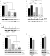
Biochemical analysis. In panels (A, B, and D), cells were transfected with GFP-Nav1.5 and in panel (E), with Nav1.5-GFP. NS indicates not significant, **P = 0.004 and ***P≤ 0.001. Data are represented as arbitrary units. Glyceraldehyde 3-phosphate dehydrogenase (GAPDH) was used as total protein loading control. (A) Western blots of total cell lysates show that N-terminal mutant protein quantities are significantly lower than WT (B) Incubation of transfected cells with MG132 prevented degradation of N-terminal mutants (C) Representative western blot of total protein extracted from cells co-transfected with GFP-Nav1.5 WT and the non-tagged Nav1.5 WT or R104W constructs. The anti-Nav1.5 antibody revealed both channels (tagged and non-tagged) and showed a decrease in the total Nav1.5 channel after cotransfection with the mutant. The anti-GFP antibody revealed only the GFP-Nav1.5-WT channels and showed no degradation of GFP-Nav1.5-WT in the presence of the R104W mutant. (D) Cell surface biotinylation experiments. Transferrin receptor (TfnR) was used as a biotinylated protein loading control. Note that the surface membrane expression of N-terminal mutants was markedly reduced compared with WT and R878C. (E) Western blot of total cell lysates and biotinylation of cells transfected with ΔNter-GFP. ΔNter was slightly degraded compared with WT, while normally expressed at the membrane.
Figure 7
Figure 7
N-terminal mutants are complemented by R878C. (A) Representative INa traces of R878C, R104W, and R121W alone or co-expressed with R878C. Cells were bathed with a 135 mM Na+ solution because of small INa. (B) I/V relationships of R878C, R878C + R104W, and R878C + R121W channels. R878C was able to restore a small INa when co-expressed with R104W and R121W mutants (2 and 8.5% of WT INa density). (C) Activation curves of WT, R121W + WT and R121W + R878C channels. Note that the activation was shifted by 7.4 and 11.7 mV compared with WT.

Comment in

References

    1. Antzelevitch C, Brugada P, Borggrefe M, Brugada J, Brugada R, Corrado D, et al. Brugada syndrome: report of the second consensus conference: endorsed by the heart rhythm society and the european heart rhythm association. Circulation. 2005;111:659–670. - PubMed
    1. Kapplinger JD, Tester DJ, Alders M, Benito B, Berthet M, Brugada J, et al. An international compendium of mutations in the SCN5A-encoded cardiac sodium channel in patients referred for Brugada syndrome genetic testing. Heart Rhythm. 2010;7:33–46. - PMC - PubMed
    1. Wilde AA, Brugada R. Phenotypical manifestations of mutations in the genes encoding subunits of the cardiac sodium channel. Circ Res. 2011;108:884–897. - PubMed
    1. Abriel H. Cardiac sodium channel Na(v)1.5 and interacting proteins: Physiology and pathophysiology. J Mol Cell Cardiol. 2010;48:2–11. - PubMed
    1. Keller DI, Rougier JS, Kucera JP, Benammar N, Fressart V, Guicheney P, et al. Brugada syndrome and fever: Genetic and molecular characterization of patients carrying SCN5A mutations. Cardiovasc Res. 2005;67:510–519. - PubMed

Publication types

Substances