. 2013 Jan;6(1):69-82.
doi: 10.1038/mi.2012.49. Epub 2012 Jun 27.

IL-22 from conventional NK cells is epithelial regenerative and inflammation protective during influenza infection

Affiliations

IL-22 from conventional NK cells is epithelial regenerative and inflammation protective during influenza infection

P Kumar et al. Mucosal Immunol. 2013 Jan.

Abstract

Influenza infection primarily targets the upper respiratory system, leading to a severe destruction of the epithelial cell layer. The role of immune cells in the regeneration of tracheal and bronchial epithelial cells is not well defined. Here, we investigated the production of pro-constructive cytokine, Interleukin-22 (IL-22), in the bronchoalveolar lavage (BAL), trachea, lung tissue, and spleen during influenza infection. We found that conventional natural killer (NK) cells (NCR1(+)NK1.1(+)CD127(-)RORγt(-)) were the predominant IL-22-producers in the BAL, trachea, and lung tissues. Tracheal epithelial cells constitutively expressed high levels of IL-22R and underwent active proliferation in response to IL-22 in the wild-type mice. Infection of IL-22(-/-) mice with influenza virus resulted in a severe impairment in the regeneration of tracheal epithelial cells. In addition, IL-22(-/-) mice continued to lose body weight even after 10 days post infection without any recovery. Tracheal epithelial cell proliferation was significantly reduced in IL-22(-/-) mice during influenza infection. Adoptive transfer of IL-22-sufficient but not IL-22-deficient NK cells into IL-22(-/-) mice restored the tracheal/bronchial epithelial cell regeneration and conferred protection against inflammation. Our findings strongly suggest that conventional NK cells have evolved to both kill virus-infected cells and also to provide vital cytokines for tissue regeneration.


Conflict of interest statement

Conflict of interest: The authors claim no financial conflict of interest.

Figures

Figure 1
Figure 1. Tracheal epithelial cell death correlates with influenza viral titer
(A) Tracheal sections from PR8-infected mice were stained with anti-E-Cadherin (E-Cad, epithelial cells, blue), Annexin-V (Anxn-V, apoptotic cells, red) and are shown with Differential Interference Contrast (DIC) images. Data shown are one set of representative panels from a group of five mice infected with PR8 for each DPI and from a total of three different experiments. (B) Levels of PR8 viral titers were quantified using a novel infrared dye-based assay. MDCK cells were incubated with BAL fluids from PR8-infected mice for 16 h. Nucleoprotein (NP) of influenza virus was detected in the infected MDCK cells using monoclonal anti-NP antibody. BAL fluids from five infected mice for each DPI were collected and analyzed, and data from one representative of five independent experiments are shown. (C) Influenza viral titers were quantified through a IR-dye based method using a standard curve. Data shown are averages of PFU from duplicate wells and a representative of five independent experiments.
Figure 2
Figure 2. NCR1+ cells predominantly generate IL-22 in the lung during influenza infections
(A) Single cell suspensions from indicated organs of infected mice were stained with anti-CD3, anti-CD4, anti-CD8, anti-γδTCR, anti-NCR1, and anti-IL-22 antibodies and analyzed through flow cytometry to define the cell populations that are positive for intracellular IL-22. Dot plots of IL-22+ cells among CD3+CD4+ T, CD3+CD8+ T, CD3+γδTCR+ T and CD3NCR1+ cells are shown. Data shown in (A) are one representative of six mice analyzed on DPI 7. (B) Absolute numbers and percentages of total and IL-22+ cell types based on CD3+CD4+, CD3+CD8+, CD3+γδTCR+ T, or NCR1+ gating in various organs on DPI 7. Data shown in (B) are averages and standard deviations of six mice each and one of three independent experiments. Asterisks in (B) denote: *=p<0.05.
Figure 3
Figure 3. IL-22-producing NCR1+ cells in the influenza-infected lungs are conventional NK cells
Phenotypes of IL-22-generating NCR1+ cells and their absolute numbers are shown. Flow cytometry analyses of IL-22+ cells from (A) lung tissue or (B) spleen from mice at different DPI are shown. Single cell suspensions were stained for CD3, NCR1, NK1.1, CD127 and intracellular IL-22. CD3NCR1+ cells were gated and analyzed for IL-22/Isotype and NK1.1 or IL-22/Isotype and CD127. Individual dot plots (A,B) or average absolute numbers (C,D) of IL-22 producing NCR1+ cells are shown. Data shown are one representative (A,B) and average absolute numbers with standard deviations (C,D,) of five mice for each DPI. Data shown in A and B are from one representative of three independent experiments. (E) Absolute numbers of total and (F) IL-22+NCR1+NK1.1+ cells on indicated DPI in the trachea of infected mice. Cell numbers were calculated and shown per 10,000 total lymphocytes. Data shown in E, F are the averages with standard deviations of six mice each from three independent experiments. Asterisks in (C–F) denote: *=p<0.01 and **= p<0.001.
Figure 4
Figure 4. Lack of IL-22 significantly reduces the proliferation of tracheal epithelial cells
(A) Tracheal sections from infected mice on different DPI were stained with anti-IL-22R, anti-Ki-67 or isotype antibodies and analyzed using confocal microscopy. Data shown are one representative of a minimum of three independent experiments with three mice each DPI. (B) Flow cytometric analyses of IL-22R expression by tracheal epithelial cells or of (C) Ki-67 in influenza-infected WT and IL-22−/− mice on DPI 0 and 7. Data presented averages and standard deviations of percent IL-22R or Ki-67 positive epithelial cells (E-Cadherin+) were calculated from three of WT and IL-22−/− mice each.
Figure 5
Figure 5. Adoptive transfer and detection of IL-22-producing NK cells in IL-22−/− mice
(A) Sorting schema for CD3NK1.1+NCR1+ NK cells. CD45.1+ lung-derived lymphocytes from B6.SJL mice (H-2b) were stained with anti-CD3, anti-NK1.1 and anti-NCR1 antibodies and the CD3NK1.1+NCR1+ NK cells were sorted and cultured with IL-2 for 8–12 days before they were adoptively transferred intravenously into IL-22−/− mice (H-2b, CD45.2+) on DPI 4 of influenza infection. (B) BAL fluid and (C) spleens of the host IL-22−/− mice were tested for donor-derived IL-22-generating NK cells. Lungs from the host mice were analyzed through confocal microcopy for the presence of CD45.1+ IL-22-producing NK cells after (D) one or (E) three days of adoptive transfer. (F,G) Quantification of IL-22-producing NK cells in (D) and (E). IL-22+CD45.1+ or IL-22+CD45.2+ NK cells were counted in 10 independent fields per combination and are shown as averages with standard deviations.
Figure 6
Figure 6. Adoptively transferred IL-22 sufficient but not IL-22 deficient NK cells promote epithelial cell regeneration in IL-22−/− mice
(A,B) Weight loss after influenza infection was monitored in WT and in IL-22−/− mice with or without the adoptive transfer of NK cells. Weight loss in individual mice is represented by lines and the average values are shown in red circles. Data presented were from a total of 17 WT and 16 IL-22−/− with no adoptive transfer and 5–9 mice with adoptive transfer. Arrows indicate the time of adoptive transfer of NK cells into IL-22−/− mice. (C, D) Integrity of tracheal epithelial cell layers after adoptive transfer of IL-22-sufficient or deficient NK cells into IL-22−/− mice. (C) Hematoxylin and Eosin staining of trachea from WT and IL-22−/− mice on indicated DPI that were adoptively transferred with IL-22 sufficient or IL-22 deficient NK cells. Exploded views are shown for details. (D) Epithelial cell damage per cartilage length was calculated from 3–6 mice for each DPI and presented with p values and NS denotes not significant. ‘n’ denotes the number of cartilages analyzed for each group.
Figure 7
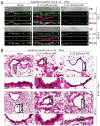
Figure 7. Adoptive transfer of IL-22 sufficient NK cells augments epithelial cell proliferation in IL-22−/− mice
(A) Tracheal sections from IL-22−/− mice with or without the adoptive transfer of NK cells were immunostained with anti-IL-22R and anti-Ki-67. Data shown are representatives of confocal images from indicated mice. Tracheae were collected after indicated DPI. White scale bars in each confocal image represent five microns. (B) Lungs from the IL-22−/− mice with or without adoptive transfer of IL-22 sufficient or IL-22 deficient NK cells were sectioned and stained with Hematoxylin and Eosin to analyze the regeneration of bronchial epithelial cells on indicated DPI. Photomicrographs of selected areas of apical lobes of the lungs show the levels of epithelial cell regeneration in the lumen side of the bronchia. Boxes denote the area selected and magnified. The black bar in the top left panel represents a scale of 100 micron. Data presented were from a total of at least three mice for each DPI from one of three independent experiments.
Figure 8
Figure 8. Lack of IL-22 increases lung inflammation
(A) Severity of inflammation after adoptive transfer of IL-22-sufficient or IL-22 deficient NK cells as visualized by collagen deposition (Mason’s trichrome staining, blue) in the lung tissue. (B) Quantification of the degree of inflammation in the lung tissues on indicated DPI. Levels of collagen deposition were determined by a double-blind assay with a scale of 1–10, with 10 being severe. Data shown were collected from a total of 3–6 mice per DPI and were representatives of a minimum of three independent experiments. Asterisks in (B) denote: **=p<0.001 and NS= not significant.

References

    1. Mandelboim O, et al. Recognition of haemagglutinins on virus-infected cells by NKp46 activates lysis by human NK cells. Nature. 2001;409:1055–1060. - PubMed
    1. Achdout H, et al. Enhanced recognition of human NK receptors after influenza virus infection. J Immunol. 2003;171:915–923. - PubMed
    1. Gill JR, et al. Pulmonary pathologic findings of fatal 2009 pandemic influenza A/H1N1 viral infections. Arch Pathol Lab Med. 2010;134:235–243. - PMC - PubMed
    1. Capelozzi VL, et al. Pathological and ultrastructural analysis of surgical lung biopsies in patients with swine-origin influenza type A/H1N1 and acute respiratory failure. Clinics (Sao Paulo) 2010;65:1229–1237. - PMC - PubMed
    1. Matrosovich MN, Matrosovich TY, Gray T, Roberts NA, Klenk HD. Human and avian influenza viruses target different cell types in cultures of human airway epithelium. Proc Natl Acad Sci U S A. 2004;101:4620–4624. - PMC - PubMed

Publication types

MeSH terms

Substances