. 2012;49(5):405-16.
doi: 10.1159/000338758. Epub 2012 Jun 26.

Expression of pannexin isoforms in the systemic murine arterial network

Affiliations

Expression of pannexin isoforms in the systemic murine arterial network

Alexander W Lohman et al. J Vasc Res. 2012.

Abstract

Aims: Pannexins (Panx) form ATP release channels and it has been proposed that they play an important role in the regulation of vascular tone. However, distribution of Panx across the arterial vasculature is not documented.

Methods: We tested antibodies against Panx1, Panx2 and Panx3 on human embryonic kidney cells (which do not endogenously express Panx proteins) transfected with plasmids encoding each Panx isoform and Panx1(-/-) mice. Each of the Panx antibodies was found to be specific and was tested on isolated arteries using immunocytochemistry.

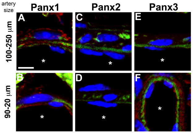
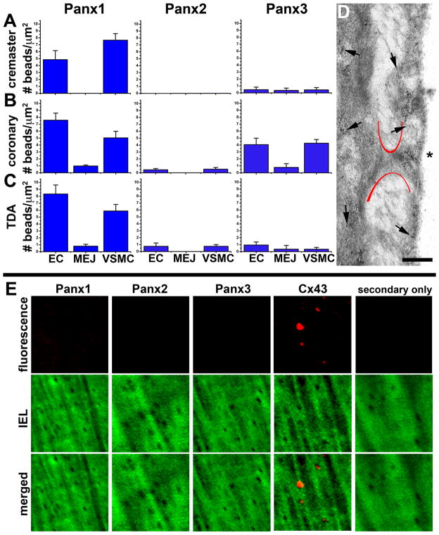
Results: We demonstrated that Panx1 is the primary isoform detected in the arterial network. In large arteries, Panx1 is primarily in endothelial cells, whereas in small arteries and arterioles it localizes primarily to the smooth muscle cells. Panx1 was the predominant isoform expressed in coronary arteries, except in arteries less than 100 µm where Panx3 became detectable. Only Panx3 was expressed in the juxtaglomerular apparatus and cortical arterioles. The pulmonary artery and alveoli had expression of all 3 Panx isoforms. No Panx isoforms were detected at the myoendothelial junctions.

Conclusion: We conclude that the specific localized expression of Panx channels throughout the vasculature points towards an important role for these channels in regulating the release of ATP throughout the arterial network.


Figures

Figure 1
Figure 1. Specificity of pannexin antibodies in cultured cells
HEK293T cells were tested for endogenous pannexin expression by mRNA via RT-PCR (A) and by protein via Western blot (B). In A, mRNA expression is normalized to the housekeeping gene β2 microglobulin (B2M). The HEK cells were transfected with Panx1, Panx2 or Panx3 plasmids, stained using anti-Panx1 CT antibody, anti-Panx1 EL antibody, anti-Panx2 antibody or anti-Panx3 antibody and detected with anti-rabbit IRDye 800CW (LiCOR) for Western blots (B) or anti-rabbit Alexa 594 for immunofluorescence (C–E). In addition, for immunofluorescence the antibody corresponding to the transfected pannexin isoform was incubated with its respective blocking peptide for negative controls. In C–E, blue are DAPI stained nuclei and red is the expression of each pannexin; scale bar is 10 μm in unstained images and 5 μm in stained images.
Figure 2
Figure 2. Specificity of Panx1 antibodies in arteries
The anti-Panx1 antibodies were tested on mouse TDA from C57Bl/6 (A) and Panx1−/− mice (B). Blue are DAPI stained nuclei, green is autofluorescence of internal elastic lamina and red is pannexin staining. Scale bar in each image is 20 μm and asterisks indicate the lumen of the artery.
Figure 3
Figure 3. Pannexin expression in the murine systemic arterial network
In A–H, each Panx antibody was tested on arteries of progressively decreasing size starting with the aorta (A), then carotid artery (B), femoral artery (C), renal artery (D), TDA (E), abdominal artery (F), an arteriole from the spinotrapezius muscle (G) and finally a cremasteric arteriole (H). Blue are DAPI stained nuclei, green is autofluorescence of internal elastic lamina and red is pannexin staining. Scale bar in each image is 10 μm and is representative for the row of staining; asterisks indicate the lumen of the artery.
Figure 4
Figure 4. Pannexin expression in the coronary arteries
Coronary arteries that were between 100–250 μm in diameter (A, C, E) or 20–90 μm in diameter (B, D, F) were assessed for expression of Panx1 (A–B), Panx2 (C–D) or Panx3 (E–F). In each image, blue is DAPI stained nuclei, green is autofluorescence of the internal elastic lamina and red is pannexin staining. Scale bar is 5 μm and representative for all images; asterisks indicate the artery lumen.
Figure 5
Figure 5. Minimal pannexin expression at myoendothelial junctions
In A–C, the number of gold beads after staining with Panx1, Panx2 or Panx3 per μm2 in endothelial cells (EC), MEJ and vascular smooth muscle (VSMC) was quantified in cremaster arterioles (20–40 μm; A), coronary arteries (50–100 μm; B), and TDAs (200 μm; C). In D, representative immuno-TEM from coronary arterioles is shown with gold beads representing Panx1. Asterisks indicate the artery lumen and arrows indicate representative location of gold beads. Scale bar is 0.5 μm. In E, holes in the internal elastic lamina from TDAs, corresponding to possible MEJs, were examined for protein expression. Green is autofluorescence of the internal elastic lamina and red is the protein of interest. Only when Cx43 antibody was used punctate fluorescence could be detected in the holes. Each image is 15 μm × 15 μm.
Figure 6
Figure 6. Pannexin expression in the kidney
In A–D, the glomerulus of the kidney was stained for Panx1 (A), Panx2 (B) and Panx3 (C), with Panx3 peptide competition shown in D. In E–H, arterioles of the cortical kidney were stained for Panx1 (E), Panx2 (F) and Panx3 (G), with Panx3 peptide competition shown in H. In all images, pannexins are in red and blue represents DAPI stained nuclei. Scale bar in A is 20 μm and representative for all images.
Figure 7
Figure 7. Pannexin expression in the pulmonary artery and lung alveoli
The pulmonary artery (lumen diameter approximately 400 μm) was stained for each of the pannexin isoforms in A–D. In C, Panx2 peptide competition is shown. The asterisks indicate the luminal side of the arterial wall. In E–H, the distal lung alveoli were also stained with each of the pannexin isoforms, with G showing Panx2 peptide competition. In all images, green is autofluorescence of matrix proteins and red blood cells (E–H), red is the pannexin isoform of interest, and blue represents DAPI stained nuclei. Scale bars in A and E are both 20 μm.

References

    1. Panchin YV. Evolution of gap junction proteins--the pannexin alternative. The Journal of experimental biology. 2005;208:1415–1419. - PubMed
    1. Baranova A, Ivanov D, Petrash N, Pestova A, Skoblov M, Kelmanson I, Shagin D, Nazarenko S, Geraymovych E, Litvin O, Tiunova A, Born TL, Usman N, Staroverov D, Lukyanov S, Panchin Y. The mammalian pannexin family is homologous to the invertebrate innexin gap junction proteins. Genomics. 2004;83:706–716. - PubMed
    1. Bao L, Locovei S, Dahl G. Pannexin membrane channels are mechanosensitive conduits for atp. FEBS Lett. 2004;572:65–68. - PubMed
    1. Ray A, Zoidl G, Weickert S, Wahle P, Dermietzel R. Site-specific and developmental expression of pannexin1 in the mouse nervous system. Eur J Neurosci. 2005;21:3277–3290. - PubMed
    1. Santiago MF, Veliskova J, Patel NK, Lutz SE, Caille D, Charollais A, Meda P, Scemes E. Targeting pannexin1 improves seizure outcome. PloS one. 2011;6:e25178. - PMC - PubMed

Publication types