

Review
doi: 10.1038/ejhg.2012.121.
Epub 2012 Jun 27.
Inherited ichthyoses/generalized Mendelian disorders of cornification
Affiliations
- PMID: 22739337
- PMCID: PMC3548255
- DOI: 10.1038/ejhg.2012.121
Item in Clipboard
Review
Inherited ichthyoses/generalized Mendelian disorders of cornification
Eur J Hum Genet.
2013 Feb.
Abstract
Inherited ichthyoses, defined as the generalized form of Mendelian disorders of cornification, are characterized by visible scaling and/or hyperkeratosis of most or all of the skin. This etiologically and phenotypically heterogenous group of conditions is caused by mutations in various different genes important for keratinocyte differentiation and epidermal barrier function. Diagnosing a specific entity is a particular challenge for the nonspecialist presented with the common clinical scaling. For the clinician, this review outlines an algorithmic approach for utilizing diagnostic clues to narrow down the differential diagnosis and to guide further testing and treatment options.
Figures
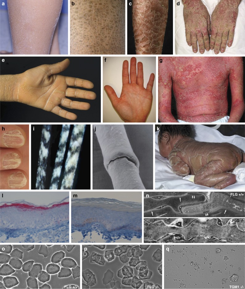
Variety of phenotypical and histological features and other dermatologic findings in different MeDOC. (a) Fine scaling phenotype in IV (FLG). (b) Moderate scaling phenotype in recessive X-linked ichthyosis (STS). (c) Large, brown scaling phenotype in ARCI (TGM1). (d) Hyperkeratosis in KI (KRT10). (e) Palmar keratoderma in LK (LOR). (f) Palmar hyperlinearity in IV (FLG). (g) Generalized fine scaling phenotype with concomitant severe erythema in NS (SPINK5). (h) Onychoschisis of fingernails in TTD, (for mutations see Table 1). (i) Tiger-tail pattern of hair under polarizing microscopy in TTD. (j) Bamboo hair (trichorrhexis invaginata) under electron microscopy in NS. (k) Collodion membrane in ARCI (TGM1). (l) Immunohistochemistry. Normal FLG staining in the granulous layer of a healthy control person (FLG +/+) and (m) absence of FLG staining in the granulous layer of a homozygous IV subject (FLG −/−). (n) Normal lamellar bilayers (arrows), corneodesmosomes (double-arrows) and corneodesmosome-derived lacunae (asterisks) in a FLG +/+ control (upper part). Foci of nonlamellar, electron-dense material (asterisks) but normal appearing corneodesmosomes (double-arrows) in a FLG −/− subject (lower part). Ruthenium tetroxide postfixation. Scale bars 0.25 μm. (o) Phase microscopy of corneocytes. Whereas corneocytes of controls appear normal after pretreatment in the ‘SDS heating test' (see text), (p) in IV subjects a subgroup of corneocytes displays ragged fragile outlines and nuclear remnants. (q) In patients with ARCI (TGM1), corneocytes are completely destructed by detergent/heat treatment as a consequence of instability of cornified envelopes due to lacking of TGM1.
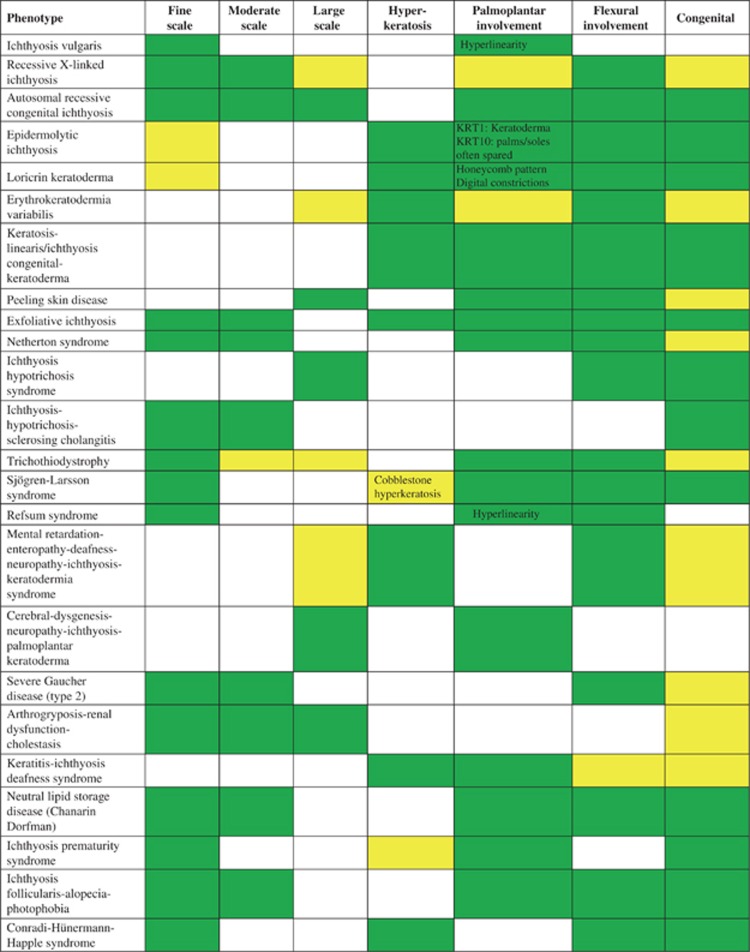
Scaling- and hyperkeratosis phenotype characteristics in selected examples of ichthyosis/MeDOC; for further phenotype details c.f. Oji et al. present  ; variable
  ; not present
  .
.
; variable ; not present .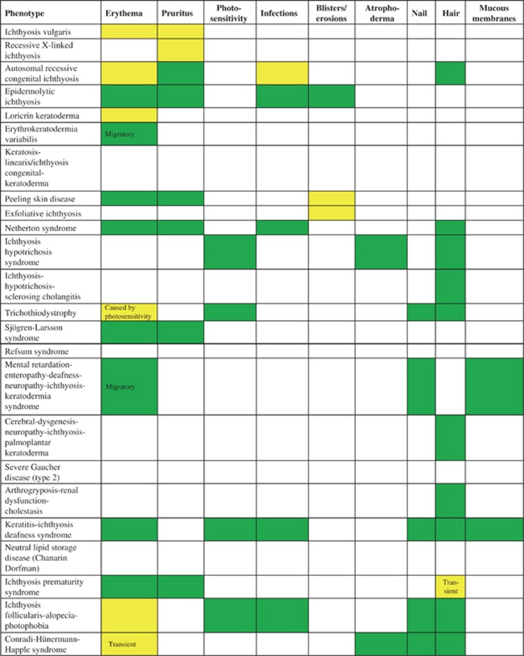
Other dermatologic features in selected examples of ichthyosis/MeDOC; for further phenotype details c.f. Oji et al. present  ; variable
  ; not present
  .
.
; variable ; not present .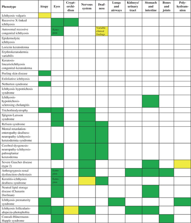
Organ involvement in selected examples of ichthyosis/MeDOC; for further phenotype details c.f. Oji et al. present  ; variable
  ; not present
  .
.
; variable ; not present .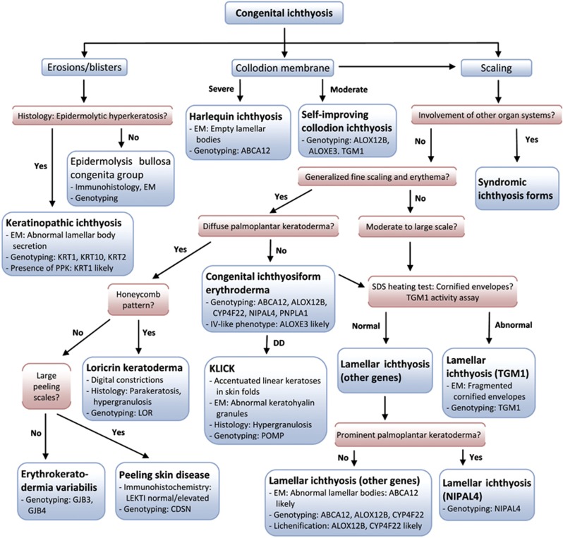
Algorithm for the diagnosis of congenital ichthyosis/MeDOC. EM, electron microscopy; KLICK, keratosis linearis-ichthyosis congenita-keratoderma; SDS, sodium dodecyl sulfate.
Comment in
-
Congenital disorders of glycosylation: other causes of ichthyosis.Eur J Hum Genet. 2014 Apr;22(4):444. doi: 10.1038/ejhg.2013.168. Epub 2013 Jul 31. Eur J Hum Genet. 2014. PMID: 23900269 Free PMC article. No abstract available.
References
-
- Styperek AR, Rice ZP, Kamalpour L, et al. Annual direct and indirect health costs of the congenital ichthyoses. Pediatr Dermatol. 2010;27:325–336. - PubMed
-
- Oji V, Tadini G, Akiyama M, et al. Revised nomenclature and classification of inherited ichthyoses. results of the First Ichthyosis Consensus Conference in Soreze 2009. J Am Acad Dermatol. 2010;63:607–641. - PubMed
-
- Oji V, Traupe H. Ichthyoses: differential diagnosis and molecular genetics. Eur J Dermatol. 2006;16:349–359. - PubMed
-
- Jonca N, Leclerc EA, Caubet C, Simon M, Guerrin M, Serre G. Corneodesmosomes and corneodesmosin: from the stratum corneum cohesion to the pathophysiology of genodermatoses. Eur J Dermatol. 2011;21 (Suppl 2:35–42. - PubMed
Publication types
MeSH terms
LinkOut - more resources
Full Text Sources
Other Literature Sources
