. 2012 Jun 28;3(6):e331.
doi: 10.1038/cddis.2012.71.

Cannabidiol protects oligodendrocyte progenitor cells from inflammation-induced apoptosis by attenuating endoplasmic reticulum stress

Affiliations

Cannabidiol protects oligodendrocyte progenitor cells from inflammation-induced apoptosis by attenuating endoplasmic reticulum stress

M Mecha et al. Cell Death Dis. .

Abstract

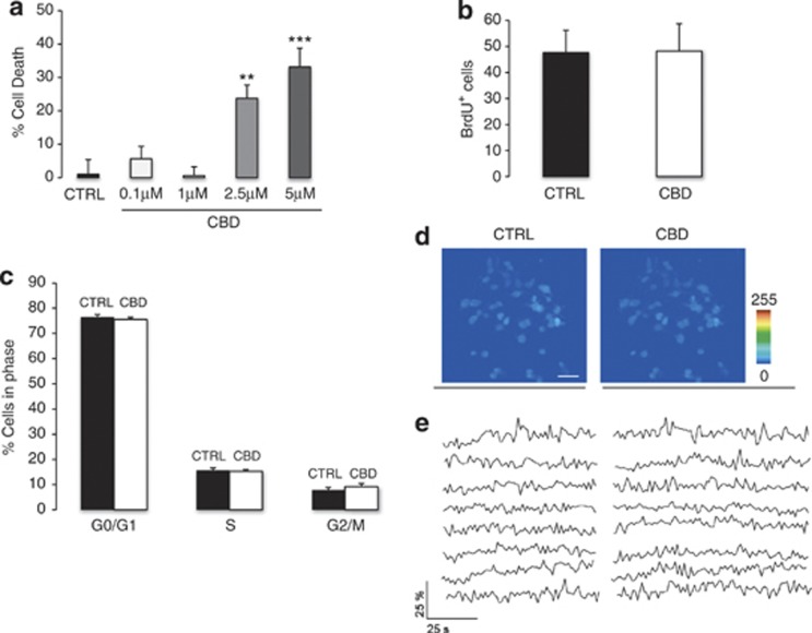
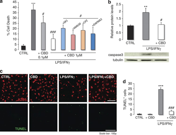
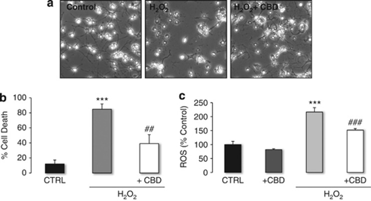
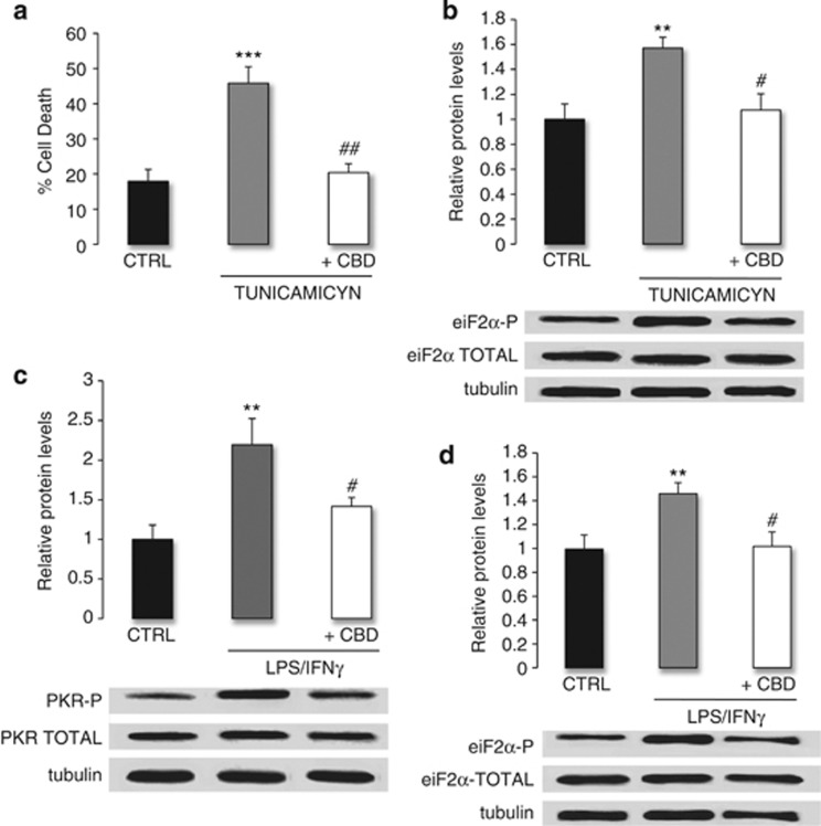
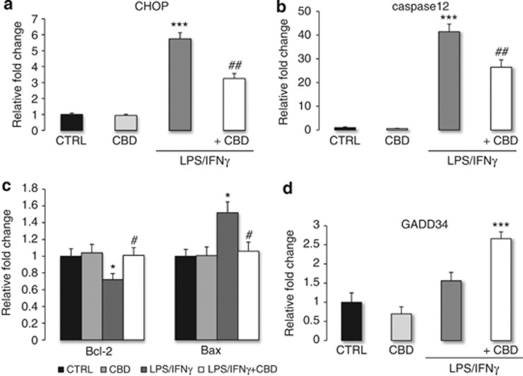
Cannabidiol (CBD) is the most abundant cannabinoid in Cannabis sativa that has no psychoactive properties. CBD has been approved to treat inflammation, pain and spasticity associated with multiple sclerosis (MS), of which demyelination and oligodendrocyte loss are hallmarks. Thus, we investigated the protective effects of CBD against the damage to oligodendrocyte progenitor cells (OPCs) mediated by the immune system. Doses of 1 μM CBD protect OPCs from oxidative stress by decreasing the production of reactive oxygen species. CBD also protects OPCs from apoptosis induced by LPS/IFNγ through the decrease of caspase 3 induction via mechanisms that do not involve CB1, CB2, TRPV1 or PPARγ receptors. Tunicamycin-induced OPC death was attenuated by CBD, suggesting a role of endoplasmic reticulum (ER) stress in the mode of action of CBD. This protection against ER stress-induced apoptosis was associated with reduced phosphorylation of eiF2α, one of the initiators of the ER stress pathway. Indeed, CBD diminished the phosphorylation of PKR and eiF2α induced by LPS/IFNγ. The pro-survival effects of CBD in OPCs were accompanied by decreases in the expression of ER apoptotic effectors (CHOP, Bax and caspase 12), and increased expression of the anti-apoptotic Bcl-2. These findings suggest that attenuation of the ER stress pathway is involved in the 'oligoprotective' effects of CBD during inflammation.


Figures

Figure 1
Figure 1
Effects of CBD on OPC cytotoxicity. (a) Although CBD did not induce cell death at low concentrations (0.1 and 1 μM), cytotoxic effects were observed at 2.5 and 5 μM. OPCs were exposed to CBD and cell death was quantified 48 h later using the LDH method. Data represent the mean±S.E.M. of n=3 cultures analyzed in triplicate. Statistical significance was determined by a one-way ANOVA: **P⩽0.01 and ***P⩽0.001 versus untreated cells. (b) CBD did not augment OPC proliferation evident as an increase in BrdU incorporation. OPCs were incubated with BrdU (10 μM) for 24 h and at least 10 000 cells were then quantified by immunocytochemistry. (c) Flow cytometry analysis revealed the CBD does not affect the cell cycle progression of OPCs. Freshly isolated OPCs were incubated for 24 h in the presence or absence of CBD (1 μM), and the cell cycle of a minimum of 10 000 cells was analyzed on a FACSAria flow cytometer following PI staining. (d, e) CBD did not alter intracellular Ca2+ levels under basal conditions. OPCs were loaded with the Ca2+-sensitive dye Fluo-4 and the fluorescence signal was monitored on a scanning confocal microscope under basal conditions and after adding CBD (1 μM: n=5 coverslips). (d) Representative pseudocolor images showing Ca2+ levels in cultured OPCs under basal conditions and in the presence of CBD (1 μM). Scale bar=50 μm. (e) Fluorescence traces showing the Ca2+ time course in individual OPCs (n=8) from the experiment shown in (d), both under basal conditions and in the presence of CBD.
Figure 2
Figure 2
CBD protects against inflammatory damage by diminishing apoptosis and decreases the number of TUNEL+ OPCs through a mechanism that does not involve CB1, CB2, TRPV1 or PPARγ receptors. (a) LPS/IFNγ-induced cytotoxicity in OPCs was attenuated by CBD, a dose of 1 μM proving to be the most effective). Administration of CB1, CB2, TRPV1 or PPARγ antagonists (SR1, AM639, CPZ: 1 μM and GW9662: 50 nM) 30 min before the stimulus had no effect, indicating that none of these receptors are implicated in the protective effects of CBD. The data represent the mean±S.E.M. of n=3 independent cultures analyzed in triplicate, and the statistical analysis was performed using Kruskal–Wallis ANOVA followed by Mann–Whitney U test: ***P⩽0.001 versus untreated cells, #P⩽0.05 and ###P⩽0.001 versus cells exposed to LPS/IFNγ alone. (b) Cleaved caspase3 western blot shows that LPS/IFNγ treatment induces the activation of the apoptotic pathway, whereas the co-treatment with CBD reduces this induction. OPCs were incubated with LPS/IFNγ in presence or absence of CBD (1 μM). Total protein extracts were prepared 24 h later and cleaved caspase 3 (19 KDa) was assessed in western blots probed with specific antibodies. The data represent the mean±S.E.M. optical density normalized to tubulin from three independent cultures analyzed in triplicate, and statistical analysis was performed using one-way ANOVA followed by the Bonferroni post-hoc test: **P⩽0.01 compared with non-treated cells, #P⩽0.05 compared with LPS/IFNγ group. (c and d) A2B5 and TUNEL staining show that LPS/IFNγ decreased OPC number by inducing apoptosis, an effect that was reversed by CBD as previously confirmed by caspase 3 measurement. OPCs were exposed for 24 h to the cytotoxic stimulus, in the presence or absence of CBD (1 μM), and 6000 OPCs cells were counted. Data represent the mean±S.E.M., and the statistical significance was determined using the Kruskal–Wallis ANOVA followed by Mann–Whitney U test: ***P⩽0.001 versus untreated cells, and ###P⩽0.001 versus cells exposed to LPS/IFNγ alone
Figure 3
Figure 3
Oxidative stress induced by H2O2 induces OPC detachment and death, an effect that was attenuated by CBD by decreasing ROS production. (a) Phase contrast images ( × 10) show that treatment with H2O2 induced the swelling and detachment of OPCs, an effect prevented by CBD treatment. (b) CBD protects OPCs from oxidative stress. OPCs were treated with H2O2 in the presence or absence of CBD (1 μM) and cell death was quantified 18 h later using the LDH method. The data represent the mean±S.E.M. of four independent cultures analyzed in triplicate, and the statistical significance was determined using one-way ANOVA followed by the Bonferroni post-hoc test: ***P⩽0.001 versus untreated cells, ##P⩽0.01 versus cells exposed to H2O2 alone. (c) CBD decreased ROS production under conditions of oxidative stress. OPCs were loaded with DCF-DA for 30 min and stimulated with H2O2 in the presence or absence of CBD (1 μM). Fluorescence was measured 2 h later at 485/530 nm in a microplate reader and the data represent the means±S.E.M. of n=3 independent cultures analyzed in triplicate. Statistical significance was determined using Kruskal–Wallis ANOVA followed by Mann–Whitney U test: ***P⩽0.001 versus untreated cells, ###P⩽0.001 versus cells exposed to H2O2 alone
Figure 4
Figure 4
OPC death is mediated by ER stress, an effect that is attenuated by CBD through decreased PKR and eiF2α phosphorylation in conditions of inflammation. (a) CBD attenuated tunicamycin-induced OPC death. OPCs were incubated with tunicamycin (1 μg/ml) in the presence or absence of CBD (1 μM), and cell death was quantified 24 h later by the LDH method. The data represent the mean±S.E.M. of three independent cultures analyzed in triplicate, and the statistical significance was determined using Kruskal–Wallis ANOVA followed by Mann–Whitney U test: ***P⩽0.001 versus untreated cells, ##P⩽0.01 versus cells exposed to tunicamycin alone. (b) Tunicamycin treatment induced the eiF2α phosphorylation, an effect that was attenuated by CBD. OPCs were incubated with tunicamycin (1 μg/ml) in the presence or absence of CBD (1 μM). Total protein extracts were prepared 5 min later and the phosphorylated (38 kDa) and total (38 kDa) eiF2α was assessed in western blots probed with specific antibodies. The data represent the mean±S.E.M. optical density normalized to tubulin from four independent cultures analyzed in triplicate, and the statistical significance was determined using Kruskal–Wallis ANOVA followed by Mann–Whitney U test: **P⩽0.01 versus untreated cells, #P⩽0.05 versus cells exposed to tunicamycin alone. (c and d) Inflammation-induced PKR and eiF2α phosphorylation, an effect that was attenuated by CBD. OPCs were treated with LPS/IFNγ in the presence or absence of CBD (1 μM). Total protein extracts were prepared 5 min later and PKR (phosphorylated, 68 kDa; total, 68 kDa) and eiF2α (phosphorylated, 38 kDa; total, 38 kDa) was assessed in western blots probed with specific antibodies. The data represent the mean±S.E.M. optical density normalized to tubulin from five cultures, and the statistical significance was determined using Kruskal–Wallis ANOVA followed by Mann–Whitney U test: **P⩽0.01 versus untreated cells, #P⩽0.05 versus cells exposed to LPS/IFNγ alone
Figure 5
Figure 5
Inflammation activates the ER apoptotic pathway in OPCs, an effect that was attenuated by CBD. (a) CBD attenuated the inflammation-induced increase in CHOP expression. (b) The upregulation of caspase 12 expression in conditions of inflammation was attenuated by CBD. (c) CBD restores the Bcl-2/Bax balance in OPCs treated with LPS/IFNγ. (d) CBD increases the expression of the negative feedback regulator GADD34 under conditions of inflammation. In all cases, OPCs were incubated with LPS/IFNγ in the presence or absence of CBD (1 μM). The mRNA expression of each gene was measured 24 h later by quantitative RT-PCR and normalized to the expression of the 18S gene. The data represent the mean±S.E.M. of three independent cultures analyzed in triplicate, and the statistical significance was determined using Kruskal–Wallis ANOVA followed by Mann–Whitney U test: *P<0.05, ***P⩽0.001 versus untreated cells; and #P⩽0.05, ##P⩽0.01 versus cells exposed to LPS/IFNγ alone

Similar articles

Cited by

References

    1. Izzo AA, Borrelli F, Capasso R, Di Marzo V, Mechoulam R. Non-psychotropic plant cannabinoids: new therapeutic opportunities from an ancient herb. Trends Pharmacol Sci. 2009;30:515–527. - PubMed
    1. Sastre-Garriga J, Vila C, Clissold S, Montalban X. THC and CBD oromucosal spray (Sativex(R)) in the management of spasticity associated with multiple sclerosis. Expert Rev Neurother. 11:627–637. - PubMed
    1. Kozela E, Lev N, Kaushansky N, Eilam R, Rimmerman N, Levy R, et al. Cannabidiol inhibits pathogenic T cells, decreases spinal microglial activation and ameliorates multiple sclerosis-like disease in C57BL/6 mice. Br J Pharmacol. 163:1507–1519. - PMC - PubMed
    1. Levine JM, Reynolds R, Fawcett JW. The oligodendrocyte precursor cell in health and disease. Trends Neurosci. 2001;24:39–47. - PubMed
    1. Bradl M, Lassmann H. Oligodendrocytes: biology and pathology. Acta Neuropathol. 2010;119:37–53. - PMC - PubMed

Publication types