. 2012 Jun 28;3(6):e336.
doi: 10.1038/cddis.2012.78.

BRCA1 negatively regulates IGF-1 expression through an estrogen-responsive element-like site

Affiliations

BRCA1 negatively regulates IGF-1 expression through an estrogen-responsive element-like site

H J Kang et al. Cell Death Dis. .

Abstract

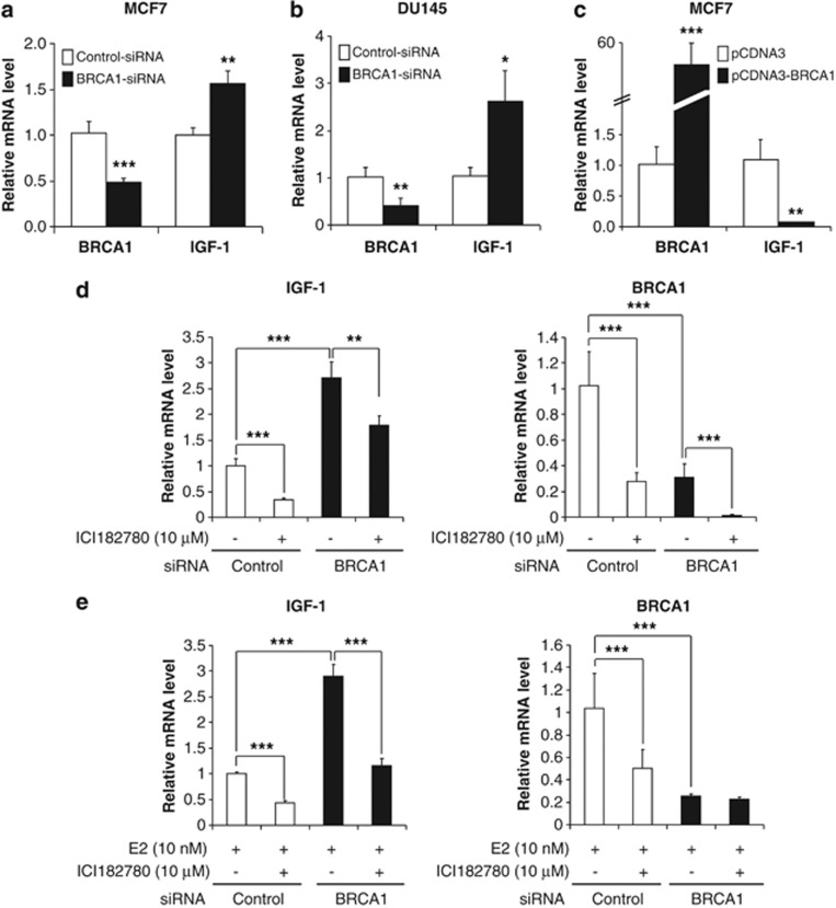
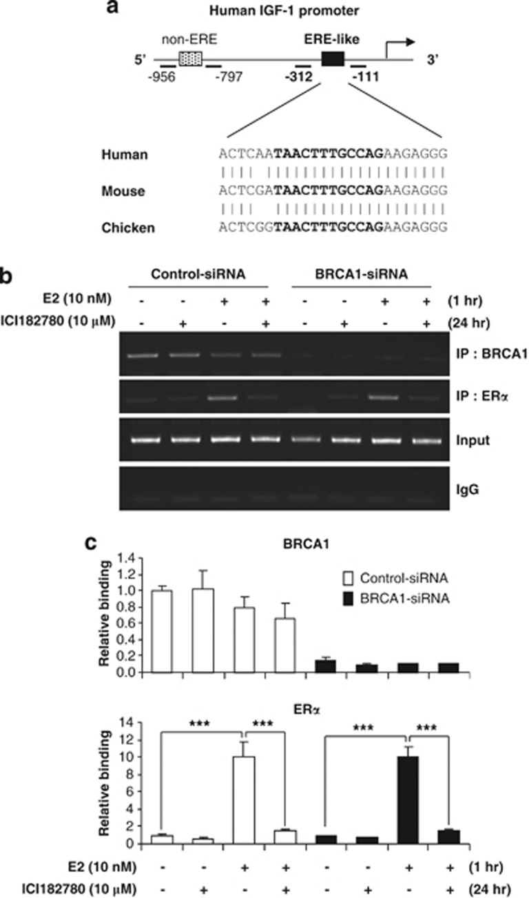
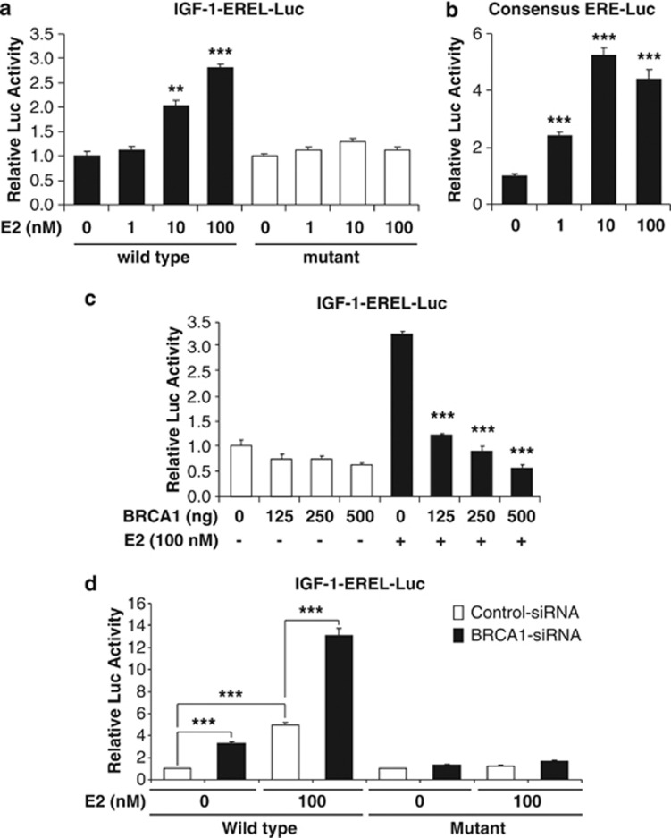
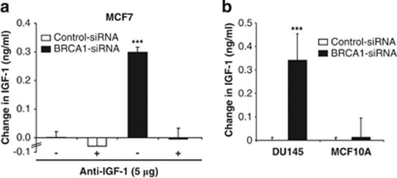
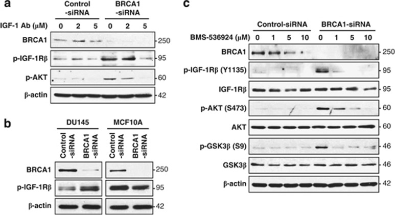
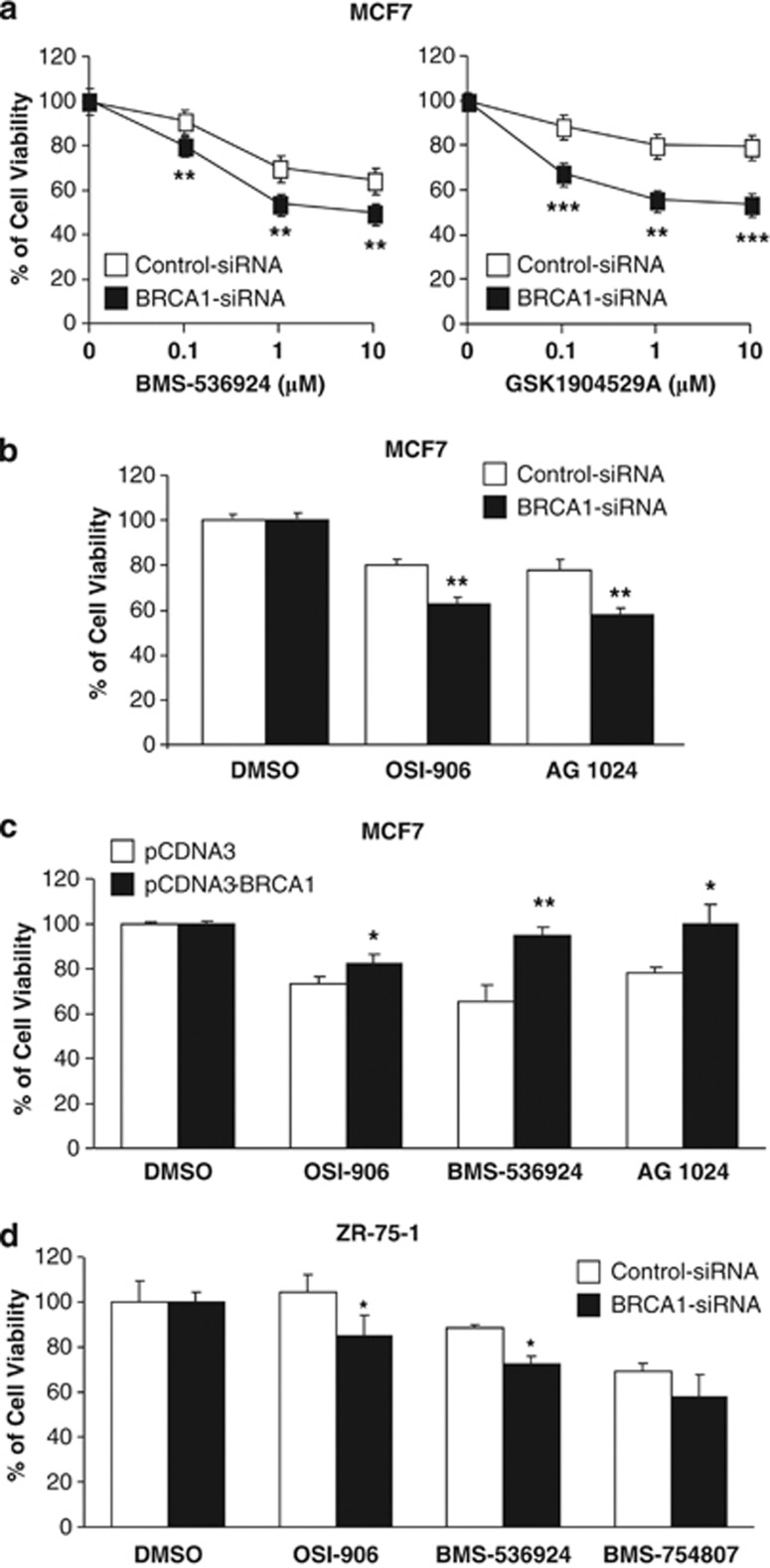
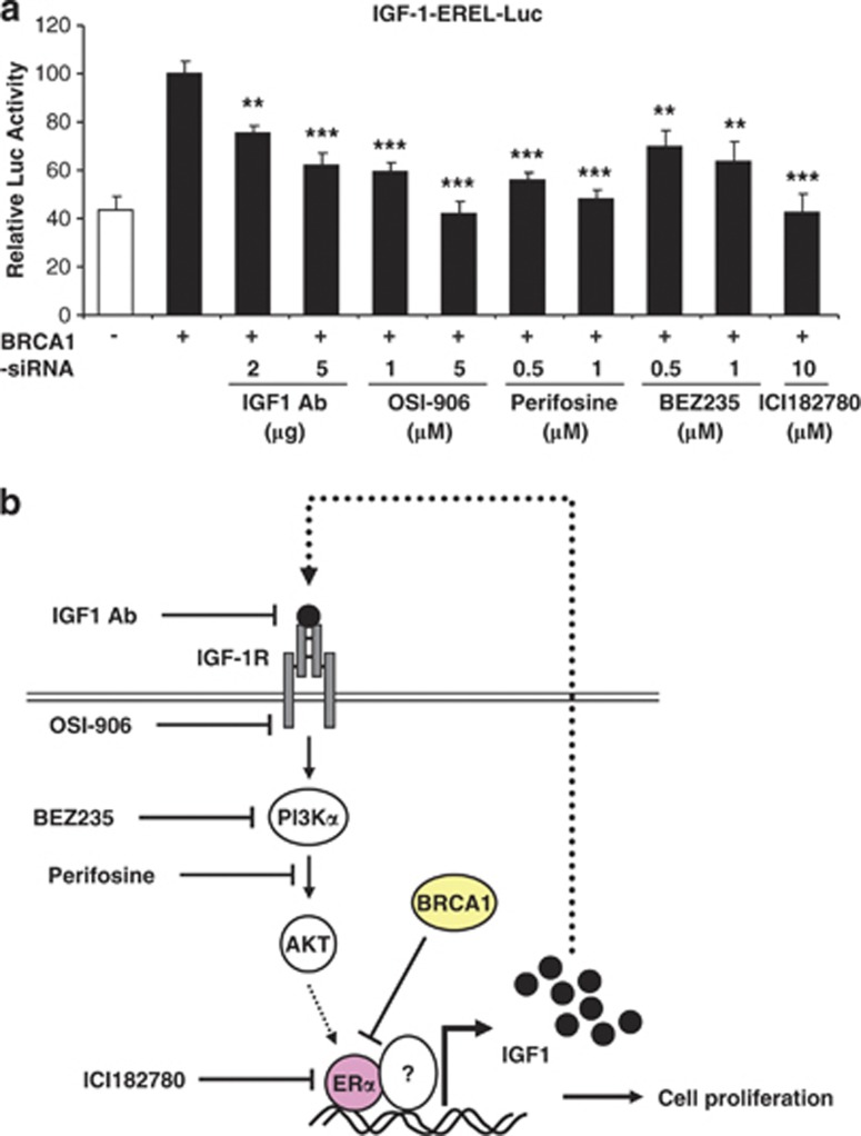
The insulin-like growth factor-1 receptor (IGF-1R) signaling pathway is critical for both normal mammary gland development and malignant transformation. It has been reported that the IGF-1 stimulates breast cancer cell proliferation and is upregulated in tumors with BRCA1/2 mutations. We report here that IGF-1 is negatively regulated by BRCA1 at the transcriptional level in human breast cancer cells. BRCA1 knockdown (BRCA1-KD) induces the expression of IGF-1 mRNA in MCF7 cells in an estrogen receptor α (ERα)-dependent manner. We found that both BRCA1 and ERα bind to the endogenous IGF-1 promoter region containing an estrogen-responsive element-like (EREL) site. BRCA1-KD does not significantly affect ERα binding on the IGF-1 promoter. Reporter analysis demonstrates that BRCA1 could regulate IGF-1 transcripts via this EREL site. In addition, enzyme-linked immunosorbent assay revealed that de-repression of IGF-1 transcription by BRCA1-KD increases the level of extracellular IGF-1 protein, and secreted IGF-1 seems to increase the phospho-IGF-1Rβ and activate its downstream signaling pathway. Blocking the IGF-1/IGF-1R/phosphoinositide 3-kinase (PI3K)/AKT pathway either by a neutralizing antibody or by small-molecule inhibitors preferentially reduces the proliferation of BRCA1-KD cells. Furthermore, the IGF-1-EREL-Luc reporter assay demonstrates that various inhibitors, which can inhibit the IGF-1R pathway, can suppress this reporter activity. These findings suggest that BRCA1 defectiveness keeps turning on IGF-1/PI3K/AKT signaling, which significantly contributes to increase cell survival and proliferation.


Figures

Figure 1
Figure 1
Negative regulation of IGF-1 mRNA expression by BRCA1. (a and b) Total RNAs extracted from cells transfected with 100 nM of siRNA (control versus BRCA1) for 72 h were used for qRT-PCR assay to quantify the level of BRCA1 and IGF-1 transcripts in MCF7 and DU145 cells. (c) Total RNAs isolated from the cells transfected with 1 μg of DNA expression vector (control versus BRCA1) for 24 h were subjected to qRT-PCR assay analysis as in (a and b). (d) Total RNAs were extracted from MCF7 cells transfected with 100 nM of siRNA (control versus BRCA1) for 48 h followed by 24 h treatment of ICI182780 under normal growth conditions and used for qRT-PCR assay to monitor the levels of BRCA1 and IGF-1 transcripts. (e) MCF7 cells cultured with phenol-red-free DMEM supplemented with charcoal-stripped FBS (CS-FBS) were transfected with 100 nM siRNA (control versus BRCA1) for 3 days. Transfected cells were further treated with 10 μM ICI182780 for 24 h and stimulated by 10 nM of E2 in phenol-red-free DMEM containing CS-FBS. After extraction of total RNAs, qRT-PCR assays were performed as in (d). Representative data from two independent experiments performed in triplicate are shown as mean±S.E.M. *P<0.05; **P<0.01 and ***P<0.001
Figure 2
Figure 2
Binding of endogenous BRCA1 and ERα to the promoter regions of human IGF-1. (a) Schematic diagram of the human IGF-1 promoter showing the location and sequence of the ERE-like (EREL) sequence. (b) Cells pretreated with siRNA (control versus BRCA1) were used for ChIP assay. Endogenous promoter regions associated with BRCA1 and/or ERα were immunoprecipitated with anti-BRCA1 or anti-ERα antibody, respectively. The relative amounts of IGF-1 promoter-specific DNA containing the EREL site in immunoprecipitated complexes were then determined by semiquantitative PCR as described in Materials and Methods. The non-ERE region in IGF-1 promoter was used as negative control. (c) Using the same DNA samples obtained in (b) qRT-PCR was performed. (b and c) Representative data from two independent experiments performed in duplicate are shown as mean±S.E.M. ***P<0.001
Figure 3
Figure 3
Regulation of IGF-1-EREL-Luc reporter activity by BRCA1 in the presence or absence of estradiol (E2). (a) MCF7 cells transfected with IGF-1-EREL-Luc (wild type versus mutant) or (b) consensus ERE-Luc for 24 h were further treated with or without E2 under estrogen-deprived conditions. After 24 h of treatment, cells were harvested and luciferase activity was measured as described in Materials and Methods. (c) Effects of wild-type BRCA1 overexpression on the wild-type IGF-1-EREL-Luc promoter reporter in the absence or presence of E2. Cells transfected with the wild-type IGF-1-EREL-Luc reporter and increasing amounts of BRCA1 expression vector (pcDNA3-BRCA1) overnight were further incubated in the absence or presence of E2 under estrogen-deprived conditions. Then, cells were harvested to measure luciferase activity. (d) Effects of BRCA1-KD on the wild-type IGF-1-EREL-Luc promoter reporter. Cells pretreated with siRNA (control versus BRCA1) for 72 h were transfected with wild-type IGF-1-EREL-Luc for 24 h under E2-stimulated conditions and luciferase activity was measured. (ad) Representative data from two independent experiments performed in triplicate are shown as mean±S.E.M. **P<0.01 and ***P<0.001
Figure 4
Figure 4
Effects of BRCA1 mutants on the wild-type IGF-1-EREL-Luc reporter in MCF7 cells. (Left) Schematic diagrams show the structure of the BRCA1 mutant constructs. (Right) Effects of wild and mutant types of BRCA1 expressing plasmid DNAs on IGF-1-EREL-Luc reporter. Cells transfected with the IGF-1-EREL-Luc reporter and various BRCA1 mutant expression vectors overnight were further incubated in the presence of E2 under estrogen-deprived conditions. Cells were then harvested to measure luciferase activity. Representative data from two independent experiments performed in triplicate are shown as mean±S.E.M.
Figure 5
Figure 5
Effect of BRCA1-KD on secretion of IGF-1 protein. (a) The levels of secreted IGF-1 proteins in MCF7 cells transfected with siRNA (control versus BRCA1) in the absence or presence of neutralizing IGF-1 antibody were measured by ELISA assay as described in Materials and Methods. (b) The secreted IGF-1 proteins were measured by ELISA assay in DU145 or MCF10A cells transfected with siRNA (control versus BRCA1). ***P<0.001
Figure 6
Figure 6
Autocrine activation of the IGF-1R pathway by IGF-1 in BRCA1-KD cells. (a) Cells pretreated with siRNA (control versus BRCA1) were incubated with an anti-IGF-1-neutralizing antibody and their lysates were subjected to western blot analysis with indicated antibodies. (b) DU145 and MCF10A cells treated with siRNA (control versus BRCA1) were subjected to western blot analysis as in (a). (c) Cells pretreated with siRNA were incubated with BMS-536924 and changes of p-IGF-1Rβ, p-AKT, and p-GSK3 were monitored by western blot analysis. β-actin was used as a loading control
Figure 7
Figure 7
Sensitization of cells to IGF-1R inhibitors by BRCA1-KD. (a) MTT assays were performed to measure the viability of MCF7 cells pretreated with siRNA (control versus BRCA1) after an additional 48 h treatment of IGF-1R inhibitors (BMS-536924 or GSK1904529A). (b) Cells pretreated with siRNA as in a were treated with 1 μM of IGF-1R inhibitors (OSI-906 or AG 1024) for 48 h and the viability of cells was measured by MTT assay. (c) Cells transfected with expression vectors (control versus BRCA1) were treated with 1 μM of OSI-906, 10 μM of AG 1024, or 1 μM of BMS-536924 for 72 h and the viability of cells was measured by MTT assay. (d) ZR-75-1 cells, pretreated with siRNA (control versus BRCA1), were further treated with IGF-1R inhibitors (1 μM of OSI-906, 1 μM of BMS-536924, or 1 μM of BMS-754807, respectively) for 48 h and cell viability was measured by MTT assay. (ad) Representative data are shown as mean±S.E.M. from at least three independent experiments performed in triplicate. *P<0.05; **P<0.01; and ***P<0.001
Figure 8
Figure 8
Pharmacological inhibition of IGF-1-EREL-Luc reporter activity in BRCA1-KD MCF7 cells. (a) MCF7 cells, pretreated with wild-type IGF-1-EREL-Luc and BRCA1 siRNA, were further treated with an IGF-1 monoclonal antibody or a small-molecule IGF-1R inhibitor (OSI-906), an AKT translocation inhibitor (Perifosine), a PI3K inhibitor (BEZ235), or an antiestrogen (ICI182780) for 24 h and subjected to the luciferase reporter assay. Representative data from two independent experiments are shown as mean±S.E.M. **P<0.01 and ***P<0.001. (b) Schematic diagram of the IGF-1/IGF-1R pathway regulation by BRCA1

References

    1. Ewing GP, Goff LW. The insulin-like growth factor signaling pathway as a target for treatment of colorectal carcinoma. Clin Colorectal Cancer. 2010;9:219–223. - PubMed
    1. Fagan DH, Yee D. Crosstalk between IGF1R and estrogen receptor signaling in breast cancer. J Mammary Gland Biol Neoplasia. 2008;13:423–429. - PubMed
    1. Pollak M. Insulin and insulin-like growth factor signalling in neoplasia. Nat Rev Cancer. 2008;8:915–928. - PubMed
    1. Riedemann J, Macaulay VM. IGF1R signalling and its inhibition. Endocr Rel Cancer. 2006;13:S33–S43. - PubMed
    1. Law JH, Habibi G, Hu K, Masoudi H, Wang MY, Stratford AL, et al. Phosphorylated insulin-like growth factor-I/insulin receptor is present in all breast cancer subtypes and is related to poor survival. Cancer Res. 2008;68:10238–10246. - PubMed

Publication types