. 2012 Jun 28;3(6):e337.
doi: 10.1038/cddis.2012.79.

Suppression of PP2A is critical for protection of melanoma cells upon endoplasmic reticulum stress

Affiliations

Suppression of PP2A is critical for protection of melanoma cells upon endoplasmic reticulum stress

K H Tay et al. Cell Death Dis. .

Abstract

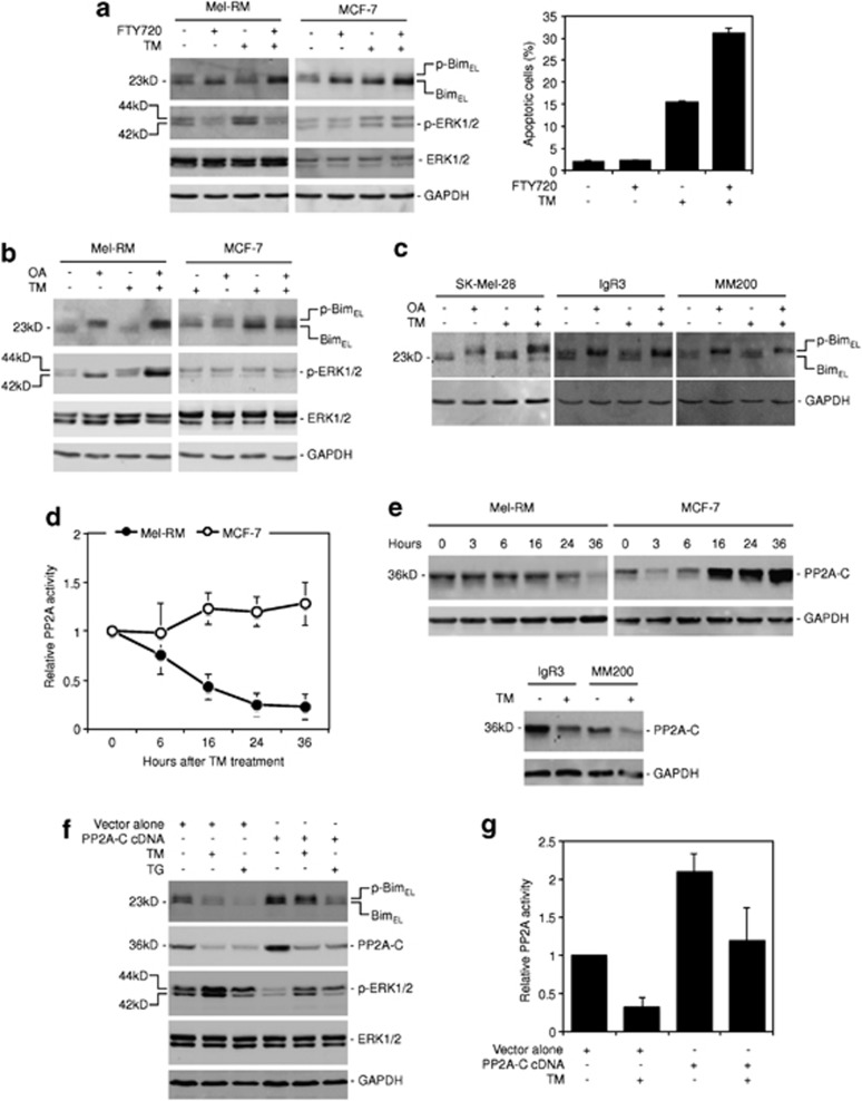
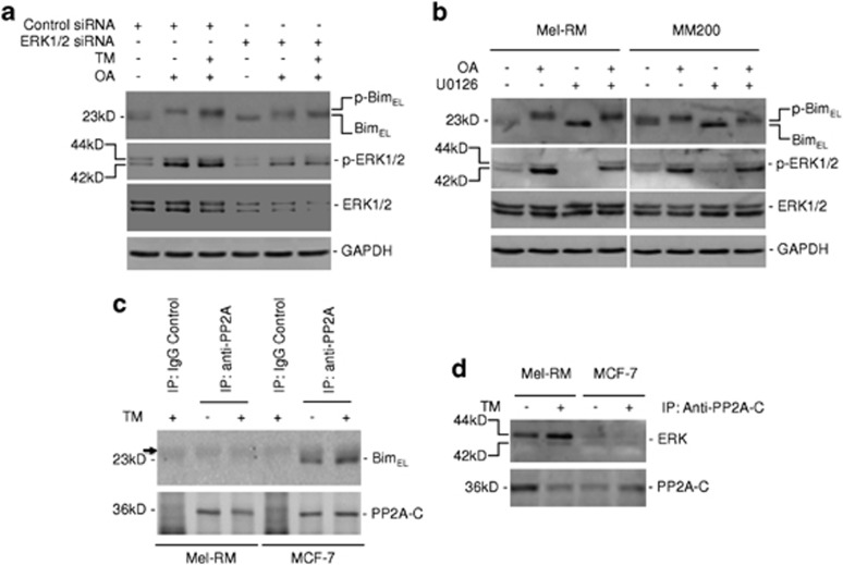
Endoplasmic reticulum (ER) stress triggers apoptosis by activating Bim in diverse types of cells, which involves dephosphorylation of Bim(EL) by protein phosphatase 2A (PP2A). However, melanoma cells are largely resistant to ER stress-induced apoptosis, suggesting that Bim activation is suppressed in melanoma cells undergoing ER stress. We show here that ER stress reduces PP2A activity leading to increased ERK activation and subsequent phosphorylation and proteasomal degradation of Bim(EL). Despite sustained upregulation of Bim at the transcriptional level, the Bim(EL) protein expression was downregulated after an initial increase in melanoma cells subjected to pharmacological ER stress. This was mediated by increased activity of ERK, whereas the phosphatase activity of PP2A was reduced by ER stress in melanoma cells. The increase in ERK activation was, at least in part, due to reduced dephosphorylation by PP2A, which was associated with downregulation of the PP2A catalytic C subunit. Notably, instead of direct dephosphorylation of Bim(EL), PP2A inhibited its phosphorylation indirectly through dephosphorylation of ERK in melanoma cells. Taken together, these results identify downregualtion of PP2A activity as an important protective mechanism of melanoma cells against ER stress-induced apoptosis.


Figures

Figure 1
Figure 1
ER stress does not induce sustained upregulation of BimEL in melanoma cells. (a) TM induces sustained upregulation of BimEL in MCF-7 breast cancer cells but not in Mel-RM melanoma cells. Left: 30μg of total protein of whole-cell lysates from Mel-RM and MCF-7 cells treated with TM (3 μM) for indicated periods were subjected to western blot analysis of Bim and GAPDH (as a loading control). The arrowhead points to a band of unknown origin generated with the antibody against Bim. Right: quantitative expression levels of BimEL as shown the left panel that were normalized to GAPDH. Quantitation of each band was carried out with ImageReader LAS-4000. The data shown are representative of three individual experiments. (b) TM does not induce sustained upregulation of BimEL in IgR3, Sk-Mel-28, and MM200 melanoma cells. Thirty microgram of total protein of whole-cell lysates from IgR3, Sk-Mel-28, and MM200 treated with TM (3 μM) for indicated periods were subjected to western blot analysis of Bim and GAPDH (as a loading control). The arrowhead points to a band of unknown origin generated with the antibody against Bim. The data shown are representative of three individual experiments. (c) Melanoma cells are not sensitive to ER stress-induced apoptosis. Mel-RM and MM200 melanoma cells, and MCF-7 breast cancer cells were treated with TM (3 μM) for 48 h before apoptosis was measured by the PI method using flow cytometry. The data shown are the mean±S.E. of three individual experiments. (d) TG does not induce sustained upregulation of BimEL in melanoma cells. Thirty microgram of total protein of whole-cell lysates from Mel-RM and MM200 melanoma cells treated with TG (1 μM) for indicated periods were subjected to western blot analysis of Bim and GAPDH (as a loading control). The data shown are representative of three individual experiments. (e) Induction of ER stress by TM in Mel-RM and MM200 melanoma cells and MCF-7 breast cancer cells. Mel-RM, MM200, and MCF-7 cells were treated with TM (3 μM) for indicated periods. For examining the expression of GRP78 and CHOP, 30 μg of total protein of whole-cell lysates were subjected to western blot analysis of GRP78, CHOP, and GAPDH (as a loading control). The arrowhead points to a non-specific band generated with the antibody against CHOP. For examining activation of XBP1, RT-PCR products of XBP1 mRNA and β-actin mRNA (as a control) from total RNA extracts were digested with ApaLI for 90 min followed by electrophoresis. The longer fragment derived from the active form of XBP1 mRNA and two shorter bands derived from the inactive form are indicated. The data shown are representative of three individual experiments
Figure 2
Figure 2
ER stress induces increased apoptosis when BimEL is expressed at high levels. (a) Induction of BimEL expression in Mel-RM.Bim cells that carried a lentivirus-based 4-OHT-responsive inducible BimEL expression system. Mel-RM cells and Mel-RM.Bim cells were treated with 4-OHT (10 nM), TM (3 μM), or 4-OHT plus TM for 24 h. Thirty microgram of total protein of whole-cell lysates were then subjected to western blot analysis of Bim and GAPDH (as a loading control). The data shown are representative of three individual western blot analyses. (b) Induction of BimEL sensitizes Mel-RM.Bim to TM-induced apoptosis. Mel-RM cells and Mel-RM.Bim cells that carried a lentivirus-based 4-OHT-responsive inducible BimEL expression system were treated with 4-OHT (10 nM), TM (3 μM), or 4-OHT plus TM for 48 h. Apoptosis was measured by the PI method using flow cytometry. The data shown are the mean±S.E. of three individual experiments. (c) Overexpression of BimEL in IgR3 and MM200 cells transiently transfected with cDNA encoding BimEL. IgR3 and MM200 cells were transfected with vector alone or BimEL cDNA. Twenty-four hours later, cells were treated with TM (3 μM) for a further 24 h. Thirty microgram of total protein of whole-cell lysates were then subjected to western blot analysis of Bim and GAPDH (as a loading control). The data shown are representative of three individual western blot analyses. (d) Overexpression of Bim sensitizes IgR3 and MM200 cells to ER stress-induced apoptosis. IgR3 (left) and MM200 (right) cells were transfected with vector alone or BimEL cDNA. Twenty-four hours later, cells were treated with TM (3 μM) for a further 48 h. Apoptosis was measured by the PI method using flow cytometry. The data shown are the mean±S.E. of three individual experiments
Figure 3
Figure 3
ER stress induces transcriptional upregulation of Bim in melanoma cells. (a) TM induces sustained upregulation of Bim mRNA in melanoma cells. Mel-RM melanoma cells and MCF-7 breast cancer cells were treated with TM (3 μM) for indicated periods before total RNA was isolated and subjected to qPCR analysis for the Bim mRNA expression. The relative abundance of the Bim mRNA in each cell line before treatment was arbitrarily designated as 1. The data shown are the mean±S.E. of three individual experiments. (b) CHOP is required for transcriptional upregulation of Bim by ER stress in melanoma cells. Upper panel: Mel-RM melanoma cells and MCF-7 breast cancer cells were transfected with the control or CHOP siRNA. Twenty-four hours later, cells were treated with TM (3 μM) for a further 6 h. Thirty microgram of total protein of whole-cell lysates were then subjected to western blot analysis of CHOP and GAPDH (as a loading control). The data shown are representative of three individual western blot analyses. Lower panel: Mel-RM melanoma cells and MCF-7 breast cancer cells were transfected with the control or CHOP siRNA. Twenty-four hours later, cells were treated with TM (3 μM) for a further 36 h before total RNA was isolated and subjected to qPCR analysis for the Bim mRNA expression. The relative abundance of the Bim mRNA in each cell line transfected with the control siRNA without treatment was arbitrarily designated as 1. The data shown are the mean±S.E. of three individual experiments. (c) The general transcription inhibitor actinomycin D (Act.D) efficiently blocks transcriptional upregulation of Bim in Mel-RM melanoma cells and MCF-7 breast cancer cells. Mel-RM and MCF-7 cells were treated with Act.D (100 ng/ml) for 1 h before the addition of TM (3 μM) for a further 24 h. Total RNA was then isolated and subjected to qPCR analysis for the Bim mRNA expression. The relative abundance of the Bim mRNA in each cell line before treatment was arbitrarily designated as 1. The data shown are the mean±S.E. of three individual experiments. (d) Overexpression of CHOP induces upregulation of the Bim mRNA and the BimEL protein in melanoma cells. Left panel: Mel-RM.CHOP cells that carried a lentivirus-based 4-OHT-responsive inducible CHOP expression system were treated with 4-OHT (10 nM) for indicated periods. Thirty microgram of total protein of whole-cell lysates were then subjected to western blot analysis of CHOP, Bim, and GAPDH (as a loading control). The data shown are representative of three individual western blot analyses. Right panel: Mel-RM.CHOP cells that carried a lentivirus-based 4-OHT-responsive inducible CHOP expression system were treated with 4-OHT (10 nM) for indicated periods. Total RNA was isolated and subjected to qPCR analysis for the Bim mRNA expression. The relative abundance of the Bim mRNA in cells without 4-OHT treatment was arbitrarily designated as 1. The data shown are the mean±S.E. of three individual experiments. (e) Overexpression of CHOP induces apoptosis in melanoma cells. Mel-RM.CHOP cells that carried a lentivirus-based 4-OHT-responsive inducible CHOP expression system were treated with 4-OHT (10 nM) for 48 h before apoptosis was measured by the PI method using flow cytometry. The data shown are the mean±S.E. of three individual experiments
Figure 4
Figure 4
Downregulation of the BimEL protein in melanoma cells undergoing ER stress is mediated by MEK/ERK signaling. (a) The proteasome inhibitor MG132 reverses downregulation of the BimEL in melanoma cells under ER stress. Mel-RM cells with or without pretreatment with the proteasome inhibitor MG132 (10 μM) for 1 h were treated with TM (3 μM) for 24 h. Thirty microgram of total protein of whole-cell lysates were then subjected to western blot analysis of Bim and GAPDH (as a loading control). The data shown are representative of three individual experiments. (b) ER stress accelerates the turnover rate of BimEL in melanoma cells. Upper panel: Mel-RM cells were treated with the protein synthesis inhibitor cycloheximide (CHX) (10 μg/ml) with or without the addition of TM (3 μM) for indicated periods. Thirty microgram of total protein of whole-cell lysates were then subjected to western blot analysis of Bim and GAPDH (as a loading control). The data shown are representative of three individual experiments. Lower panel: quantitative expression levels of BimEL as shown the upper panel that were normalized to GAPDH. Quantitation of each band was determined using ImageReader LAS-4000. The data shown are representative of three individual experiments. (c) TM phosphorylates BimEL in melanoma cells but dephosphorylates it in MCF-7 cells. Mel-RM and MCF-7 (upper panel) and IgR3, Sk-Mel-28, and MM200 (lower panel) cells were treated TM (3 μM) for indicated periods. Thirty microgram of total protein of whole-cell lysates were then subjected to western blot analysis using an antibody that specifically recognizes BimEL phosphorylated at Ser69. Western blot analysis of GAPDH was included as a loading control. The data shown are representative of three individual experiments. (d) TG induces phosphorylation of BimEL in melanoma cells. Mel-RM and MM200 cells were treated with TG (1 μM) for indicated periods. Thirty microgram of total protein of whole-cell lysates were then subjected to western blot analysis using an antibody that specifically recognizes BimEL phosphorylated at Ser69. Western blot analysis of GAPDH was included as a loading control. The data shown are representative of three individual experiments. (e) TM increases an increase in ubiquitination of BimEL in Mel-RM melanoma cells, but a decrease in MCF-7 breast cancer cells. Whole-cell lysates from Mel-RM melanoma cells and MCF-7 breast cancer cells with or without treatment with TM (3 μM) for 36 h were subjected to immunoprecipitation using an antibody against Bim. Thirty microgram of total protein of the resulting precipitates were subjected to SDS-PAGE and probed with an antibody against ubiquitin and an antibody against Bim. The data shown are representative of three individual experiments. (f) The MEK inhibitor U0126 inhibits phosphorylation of BimEL and increases its expression in melanoma cells undergoing ER stress. Thirty microgram of total protein of whole-cell lysates from Mel-RM cells treated with U0126 (20 μM), TM (3 μM), or U0126 plus TM for 24 h were subjected to western blot analysis of Bim, pERK, ERK, and GAPDH (as a loading control). The data shown are representative of three individual experiments. (g) Knockdown of MEK1 by siRNA inhibits phosphorylation of BimEL and increases its expression in melanoma cells undergoing ER stress. Mel-RM cells were transfected with the control or MEK1 siRNA. Twenty-four hours later, cells were treated with TM (3 μM) for a further 24 h. Thirty microgram of total protein of whole-cell lysates were then subjected to western blot analysis of Bim, MEK1, pERK (pERK), ERK, and GAPDH (as a loading control). The data shown are representative of three individual western blot analyses
Figure 5
Figure 5
Downregulation of the BimEL protein in melanoma cells under ER stress is associated with reduction in PP2A activity that is, at least in part, due to downregulation of the PP2A catalytic C subunit (PP2A-C). (a) Pharmacological activation of PP2A reverses downregulation of BimEL by ER stress in melanoma cells. Left panel: 30 μg of total protein of whole-cell lysates from Mel-RM melanoma cells or MCF-7 breast cancer cells treated with the pharmacological PP2A activator FTY720 (2.5 μM), TM (3 μM), or FTY720 plus TM for 24 h were subjected to western blot analysis of Bim, pERK, ERK, and GAPDH (as a loading control). The data shown are representative of three individual experiments. Right panel: Mel-RM cells were treated with FTY720 (2.5 μM), TM (3 μM), or FTY720 plus TM for 48 h before apoptosis was measured by the PI method using flow cytometry. The data shown are the mean±S.E. of three individual experiments. (b) Inhibition of PP2A further promotes phosphorylation of BimEL and increases phosphorylation of ERK in Mel-RM cells under ER stress. Thirty microgram of total protein of whole-cell lysates from Mel-RM melanoma cells or MCF-7 breast cancer cells treated with the OA (50 nM), TM (3 μM), or OA plus TM for 24 h were subjected to western blot analysis of Bim, pERK, ERK, and GAPDH (as a loading control). The data shown are representative of three individual experiments. (c) Inhibition of PP2A further promotes phosphorylation of BimEL in Sk-Mel-28, IgR3, and MM200 melanoma cells. Thirty microgram of total protein of whole-cell lysates from Sk-Mel-28, IgR3, and MM200 cells treated with the OA (50 nM), TM (3 μM), or OA plus TM for 24 h were subjected to western blot analysis of Bim and GAPDH (as a loading control). The data shown are representative of three individual experiments. (d) ER stress reduces PP2A activity in Mel-RM melanoma cells, but increases PP2A activity in MCF-7 breast cancer cells. Mel-RM cells and MCF-7 cells were treated with TM (3 μM) for indicated periods before the phosphatase activity of PP2A was quantitated using a PP2A-C immunoprecipitation phosphatase assay kit. The PP2A phosphatase activity in cells without treatment was arbitrarily designated as 1. The data shown are the mean±S.E. of three individual experiments. (e) ER stress downregulates PP2A-C in melanoma cells but upregulates its expression in MCF-7 breast cancer cells. Upper panel: Thirty microgram of total protein of whole-cell lysates from Mel-RM melanoma cells or MCF-7 breast cancer cells treated with TM (3 μM) for indicated periods were subjected to western blot analysis of PP2A-C and GAPDH (as a loading control). The data shown are representative of three individual experiments. Lower panel: Thirty microgram of total protein of whole-cell lysates from IgR3 and MM200 melanoma cells treated with TM (3 μM) for 24 h were subjected to western blot analysis of PP2A-C and GAPDH (as a loading control). The data shown are representative of three individual experiments. (f) Overexpression of PP2A-C upregulates BimEL and decrease ERK phosphorylation in melanoma cells. Mel-RM cells were transiently transfected with vector alone or cDNA encoding PP2A-C. Twenty-four hours later, cells were treated with TM (3 μM) or TG (1 μM) for a further 24 h. Thirty microgram of total protein of whole-cell lysates were then subjected to western blot analysis of Bim, PP2A-C, pERK, ERK, and GAPDH (as a loading control). The data shown are representative of three individual western blot analyses. (g) Overexpression of PP2A-C causes increased PP2A activity in melanoma cells with or without undergoing ER stress. Mel-RM cells were transiently transfected with vector alone or cDNA encoding PP2A-C. Twenty-four hours later, cells were treated with TM (3 μM) for a further 24 h. The phosphatase activity of PP2A was then quantitated using a PP2A-C immunoprecipitation phosphatase assay kit. The PP2A phosphatase activity in cells transfected with vector alone without TM treatment was arbitrarily designated as 1. The data shown are the mean±S.E. of three individual experiments
Figure 6
Figure 6
PP2A inhibits BimEL phosphorylation through dephosphorylation of ERK in melanoma cells. (a) Knockdown of ERK1/2 blocks phosphorylation of BimEL in melanoma cells with or without undergoing ER stress. Mel-RM cells were transfected with the control siRNA or ERK1 siRNA plus ERK2 siRNA. Twenty-four hours later, cells were treated with OA (50 nM) for 24 h in the presence or absence of TM (3 μM). Thirty microgram of total protein of whole-cell lysates were then subjected to western blot analysis of Bim, pERK, ERK, and GAPDH (as a loading control). The data shown are representative of three individual western blot analyses. (b) The MEK inhibitor U0126 blocks phosphorylation of BimEL mediated by OA in melanoma cells. Thirty microgram of total protein of whole-cell lysates from Mel-RM and MM200 cells treated with OA (50 nM), U0126 (20 μM), or OA plus U0126 for 24 h were subjected to western blot analysis of Bim, pERK, ERK, and GAPDH (as a loading control). The data shown are representative of three individual experiments. (c) PP2A-C is physically associated with BimEL in MCF-7 breast cancer cells but not in melanoma cells. Whole-cell lysates from Mel-RM melanoma cells with or without treatment with TM (3 μM) for 6 h and those from MCF-7 breast cancer cells with or without treatment with TM (3 μM) for 16 h were subjected to immunoprecipitation with a mouse antibody against PP2A-C. Immunoprecipitation of whole-cell lysates from cells treated with TM with purified mouse IgG was included as a control. Thirty microgram of total protein of the resulting precipitates were subjected to SDS-PAGE and probed with an antibody against Bim and an antibody against PP2A-C. The data shown are representative of three individual experiments. (d) PP2A-C is physically associated with ERK in melanoma cells but not in MCF-7 breast cancer cells. Whole-cell lysates from Mel-RM melanoma cells and MCF-7 breast cancer cells with or without treatment with TM (3 μM) for 6 h were subjected for immunoprecipitation with a mouse antibody against PP2A-C. Thirty microgram of total protein of the resulting precipitates were subjected to SDS-PAGE and probed with an antibody against ERK and an antibody against PP2A-C. The data shown are representative of three individual experiments

Similar articles

Cited by

References

    1. Harding HP, Calfon M, Urano F, Novoa I, Ron D. Transcriptional and translational control in the mammalian unfolded protein response. Annu Rev Cell Dev Biol. 2002;18:575–599. - PubMed
    1. Schroder M, Kaufman RJ. The mammalian unfolded protein response. Annu Rev Biochem. 2005;74:739–789. - PubMed
    1. Boyce M, Yuan J. Cellular response to endoplasmic reticulum stress: a matter of life or death. Cell Death Differ. 2006;13:363–373. - PubMed
    1. Xu C, Bailly-Maitre B, Reed JC. Endoplasmic reticulum stress: cell life and death decisions. J Clin Invest. 2005;115:2656–2664. - PMC - PubMed
    1. Zhang L, Lopez H, George NM, Liu X, Pang X, Luo X. Selective involvement of BH3-only proteins and differential targets of Noxa in diverse apoptotic pathways. Cell Death Differ. 2011;18:864–873. - PMC - PubMed

Publication types

MeSH terms