. 2012 Dec;17(10):1348-61.
doi: 10.1177/1087057112451921. Epub 2012 Jun 26.

Cell-permeable, small-molecule activators of the insulin-degrading enzyme

Affiliations

Cell-permeable, small-molecule activators of the insulin-degrading enzyme

Sayali S Kukday et al. J Biomol Screen. 2012 Dec.

Abstract

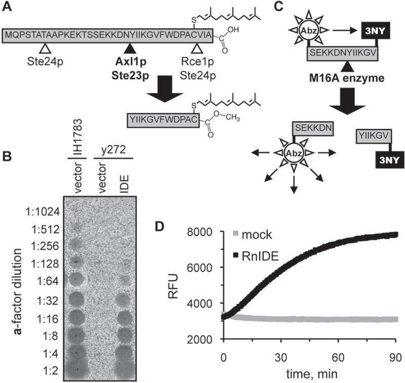
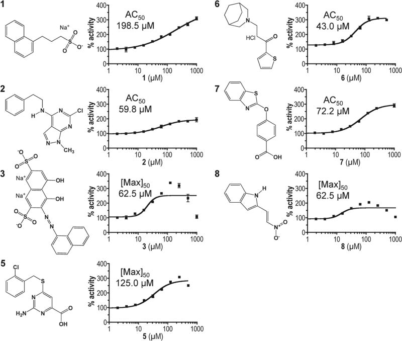
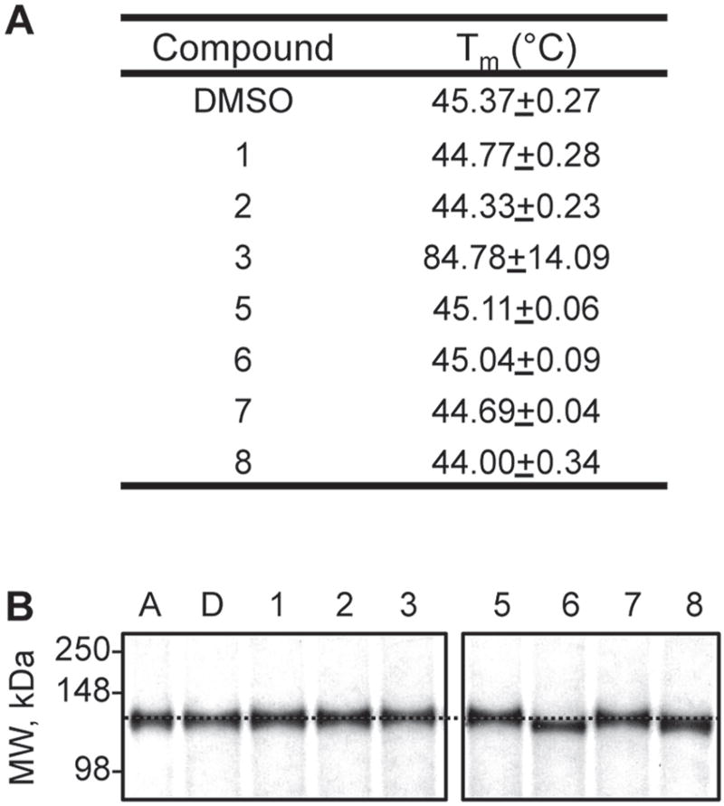
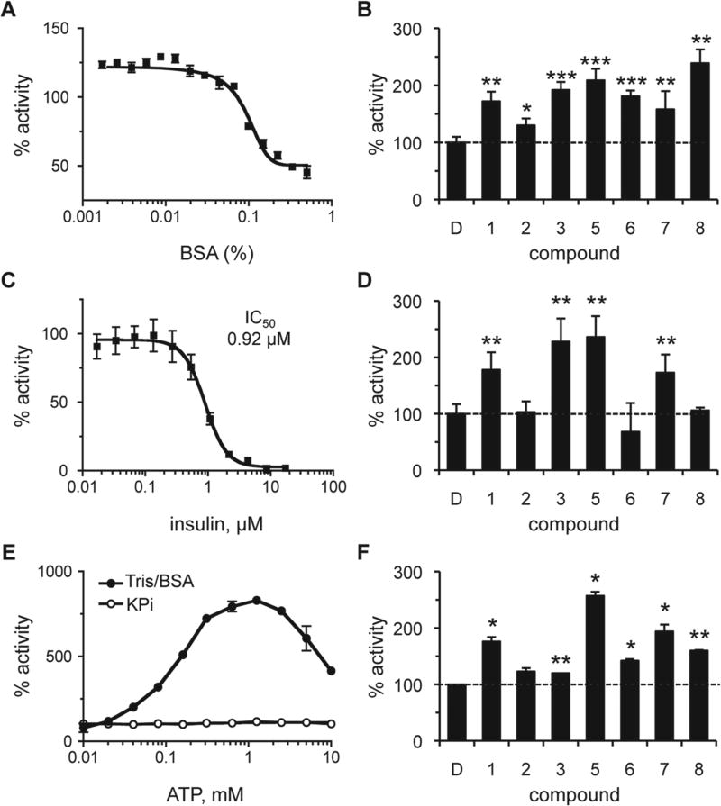
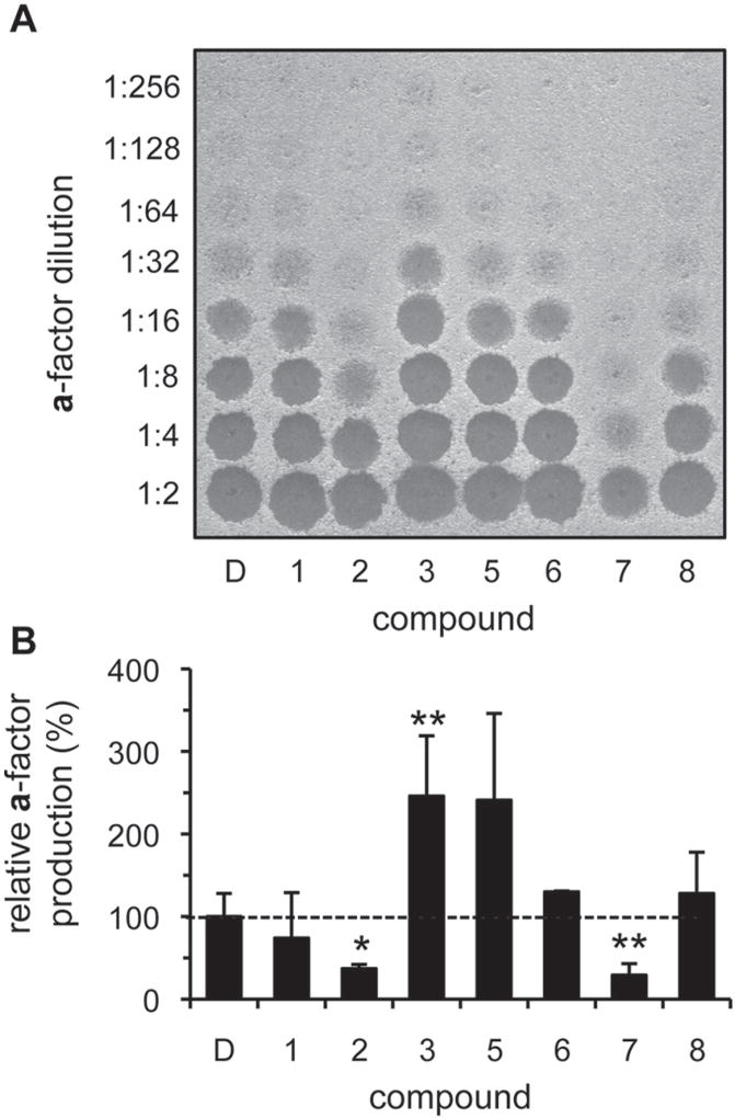
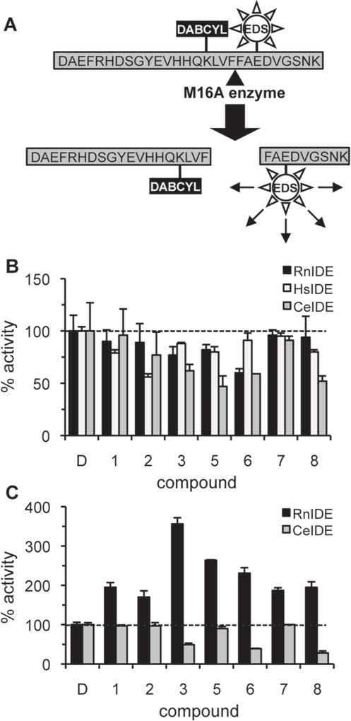
The insulin-degrading enzyme (IDE) cleaves numerous small peptides, including biologically active hormones and disease-related peptides. The propensity of IDE to degrade neurotoxic Aβ peptides marks IDE as a potential therapeutic target for Alzheimer disease. Using a synthetic reporter based on the yeast a-factor mating pheromone precursor, which is cleaved by multiple IDE orthologs, we identified seven small molecules that stimulate rat IDE activity in vitro. Half-maximal activation of IDE by the compounds is observed in vitro in the range of 43 to 198 µM. All compounds decrease the K(m) of IDE. Four compounds activate IDE in the presence of the competing substrate insulin, which disproportionately inhibits IDE activity. Two compounds stimulate rat IDE activity in a cell-based assay, indicating that they are cell permeable. The compounds demonstrate specificity for rat IDE since they do not enhance the activities of IDE orthologs, including human IDE, and they appear specific for a-factor-based reporters since they do not enhance rat IDE-mediated cleavage of Aβ-based reporters. Our results suggest that IDE activators function in the context of specific enzyme-substrate pairs, indicating that the choice of substrate must be considered in addition to target validation in IDE activator screens.


Conflict of interest statement

Declaration of Conflicting Interests

The authors declared no potential conflicts of interest with respect to the research, authorship, and/or publication of this article.

Figures

Figure 1
Figure 1
Reporters of M16A enzyme activity. (A) Production of the yeast a-factor mating pheromone is dependent on the action of several proteases, including the M16A enzymes Axl1p and Ste23p. (B) Rat insulin-degrading enzyme (IDE) can substitute for the yeast M16A enzymes in a-factor production in vivo. Yeast strain y272 (MATa axl1 Δ ste23Δ) was transformed with an IDE-encoding plasmid (pWS491) or an empty vector (pRS316), and resultant strains were evaluated for their ability to produce a-factor. A wild-type MATa strain (IH1783) transformed with an empty vector (pRS316) was evaluated in parallel. The appearance of a clear spot (i.e., zone of reduced growth) within the MATα lawn indicates the presence of a-factor. (C) An internally quenched fluorogenic dodecapeptide modeled on the M16A cleavage site in the yeast a-factor precursor. The NH2-terminal fluorophore is aminobenzoic acid (Abz), and the COOH-terminal quenching group is 3-nitro-tyrosine (3NY). (D) Progress curves demonstrating time-dependent fluorescent output in the presence or absence of recombinant rat IDE. The reactions contained rat IDE (10 µg/ml; 87.7 nM) or enzyme storage buffer (mock). RFU, relative fluorescence units.
Figure 2
Figure 2
Chemical structures and dose-response profiles of insulin-degrading enzyme (IDE) activators. Compounds were identified by their ability to enhance rat IDE-mediated in vitro cleavage of the peptide reporter depicted in Figure 1C. Structures were downloaded from the Developmental Therapeutics Program (DTP) structure database (http://dtp.nci.nih.gov/branches/dscb/diversity_explanation.html). Compound 4 was eliminated due to a lack of measurable effect on IDE kinetic parameters and inconsistent behavior across experiments. Compounds were evaluated for their effectiveness at stimulating rat IDE activity over the indicated dose range using the fluorescence-based IDE activity assay described in Figure 1. A best-fit nonlinear dose-response curve was determined for data points using GraphPad Prism 4.0 and a four-parameter logistic equation (solid line). Where sigmoidal dose-response curves were observed, AC50 values were determined. Where hormetic response curves were observed (i.e., 3, 5, 8), the fitted curves could not be used to determine accurate AC50 values, so the lowest half-maximal activating concentration is reported ([Max]50).
Figure 3
Figure 3
Effect of compounds on the biophysical properties of rat insulin-degrading enzyme (IDE). (A) Thermal melt midpoints (Tm) observed in the presence of IDE activators (100 µM) were determined using a thermal shift assay. Compound-treated rat IDE (0.5 µM) was evaluated across the temperature range 28 to 70 °C. (B) Mobility shifts observed in the presence of IDE activators. The indicated compounds were incubated with 1 mg/mL IDE for 60 min at 37 °C and then analyzed by native polyacrylamide gel electrophoresis (10%). Adenosine triphosphate (A) was used at 3 mM; compounds 1 to 8 were used at 100 µM. D, DMSO. A dashed horizontal line has been drawn across the image at the expected mobility of IDE.
Figure 4
Figure 4
Effect of assay conditions on the properties of rat insulin-degrading enzyme (IDE) activators. The effect of bovine serum albumin (BSA), insulin, and adenosine triphosphate (ATP) on the activity of IDE was determined both in the absence and presence of compounds. Rat IDE was used (87.7 nM) in the assay described in Figure 1. Values are reported as percentages relative to a water or DMSO-treated control as appropriate (n = 4); *p < 0.05, **p < 0.01, and ***p < 0.001 relative to the mock-treated control. For dose-response curves, data points were plotted, and a best-fit nonlinear dose-response curve was determined using GraphPad Prism 4.0 as described in Figure 2. (A) Observed activity of IDE in 0.1 M KPi over a range of BSA (0%–0.5% final). (B) Observed activity in the presence of compounds (100 µM) in 0.1 M KPi/0.01% BSA. The dashed line is a visual reference for 100% activity (also present in panels D and F). (C) Observed activity of IDE in 0.1 M KPi over a range of human insulin (0–17.2 µM). (D) Observed activity in the presence of compounds (100 µM) in 0.1 M KPi containing 0.92 µM (IC50) insulin. (E) The effect of ATP (0–10 mM) on IDE activity was evaluated in 0.1 M KPi or 50 mM Tris (pH 7.5) containing 0.01% BSA (Tris/BSA). (F) Observed activity of IDE in the presence of compounds in Tris/BSA containing 1 mM ATP. Compounds were used at optimal concentrations as derived from dose-response curves in Tris/BSA buffer (S. P. Manandhar and W. K. Schmidt, unpublished observations). Compound 1 was used at 1000 µM; compounds 5, 6, and 7 at 500 µM; compound 2 at 250 µM; and compounds 3 and 8 at 125 µM.
Figure 5
Figure 5
Select compounds enhance in vivo insulin-degrading enzyme (IDE)–dependent production of yeast a-factor. Diluted yeast cultures (1:2000; y272 cotransformed with pWS192 and pWS496) were grown to saturation (72 h) in the presence of activators, and the a-factor produced was recovered and analyzed. Compounds were used at 100 µM, with the exception of compounds 5, 6, and 8 (12.5 µM, 50 µM, and 25 µM, respectively). The raw data (A) were quantified and mean values graphed relative to a DMSO-treated control (B). Each value is normalized to the density of the culture at the time a-factor was collected (n = 4 for all compounds, except 6, for which n = 2); *p < 0.05 and **p < 0.01 relative to the DMSO-treated control. A nearly identical graph is observed in the absence of normalization.
Figure 6
Figure 6
Rat insulin-degrading enzyme (IDE) activators are substrate and species specific. (A) An internally quenched fluorogenic peptide was modeled on Aβ1–28. The quencher DABCYL is conjugated to Lys16 and the fluorophore EDANS (EDS) to Glu22. The peptide has unmodified N- and C-termini. (B) Effect of compounds (100 µM) on rat (RnIDE; 10 µg/mL), human (HsIDE; 100 µg/mL), and worm IDE (CeIDE; 10 µg/mL) mediated cleavage of the Aβ1–28 reporter (50 µM) was evaluated in 0.1 M KPi, pH 7.6. Mean activity values are reported as percentages relative to a DMSO-treated control (n = 3). (C) Effect of compounds (100 µM) on the ability of RnIDE and CeIDE (each at 10 µg/mL) to cleave the a-factor reporter was evaluated as in Figure 1 HsIDE does not recognize the a-factor–based reporter and thus was not evaluated. Mean activity values are reported as percentages relative to the DMSO-treated control (n = 3).

References

    1. Haass C, Selkoe DJ. Soluble Protein Oligomers in Neurodegeneration: Lessons from the Alzheimer’s Amyloid Beta-Peptide. Nat. Rev. Mol. Cell. Biol. 2007;8(2):101–112. - PubMed
    1. Nalivaeva NN, Beckett C, Belyaev ND, Turner AJ. Are Amyloid-Degrading Enzymes Viable Therapeutic Targets in Alzheimer’s Disease? J. Neurochem. 2012;120(Suppl. 1):167–185. - PubMed
    1. Farris W, Mansourian S, Chang Y, Lindsley L, Eckman EA, Frosch MP, Eckman CB, Tanzi RE, Selkoe DJ, Guenette S. Insulin-Degrading Enzyme Regulates the Levels of Insulin, Amyloid Beta-Protein, and the Beta-Amyloid Precursor Protein Intracellular Domain In Vivo. Proc. Natl. Acad. Sci. U. S. A. 2003;100(7):4162–4167. - PMC - PubMed
    1. Leissring MA, Farris W, Chang AY, Walsh DM, Wu X, Sun X, Frosch MP, Selkoe DJ. Enhanced Proteolysis of Beta-Amyloid in APP Transgenic Mice Prevents Plaque Formation, Secondary Pathology, and Premature Death. Neuron. 2003;40(6):1087–1093. - PubMed
    1. Kim M, Hersh LB, Leissring MA, Ingelsson M, Matsui T, Farris W, Lu A, Hyman BT, Selkoe DJ, Bertram L, Tanzi RE. Decreased Catalytic Activity of the Insulin Degrading Enzyme in Chromosome 10-Linked Alzheimer’s Disease Families. J. Biol. Chem. 2007;282(11):7825–7832. - PubMed

Publication types

MeSH terms

LinkOut - more resources