

The limited role of interferon-γ in systemic juvenile idiopathic arthritis cannot be explained by cellular hyporesponsiveness
- PMID: 22740319
- PMCID: PMC3482423
- DOI: 10.1002/art.34604
The limited role of interferon-γ in systemic juvenile idiopathic arthritis cannot be explained by cellular hyporesponsiveness
Abstract
Objective: Systemic juvenile idiopathic arthritis (JIA) is an autoinflammatory syndrome in which the myelomonocytic lineage appears to play a pivotal role. Inflammatory macrophages are driven by interferon-γ (IFNγ), but studies have failed to demonstrate an IFN- induced gene signature in active systemic JIA. This study sought to characterize the status of an IFN-induced signature within affected tissue and to gauge the integrity of IFN signaling pathways within peripheral monocytes from patients with systemic JIA.
Methods: Synovial tissue from 12 patients with active systemic JIA and 9 with active extended oligoarticular JIA was assessed by real-time polymerase chain reaction to quantify IFN-induced chemokine gene expression. Peripheral monocytes from 3 patients with inactive systemic JIA receiving anti-interleukin-1β (anti-IL-1β) therapy, 5 patients with active systemic JIA, and 8 healthy controls were incubated with or without IFNγ to gauge changes in gene expression and to measure phosphorylated STAT-1 (pSTAT-1) levels.
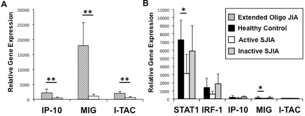
Results: IFN-induced chemokine gene expression in synovium was constrained in active systemic JIA compared to the known IFN-mediated extended oligoarticular subtype. In unstimulated peripheral monocytes, IFN-induced gene expression was similar between the groups, except that lower levels of STAT1, MIG, and PIAS were observed in patients with active disease, while higher levels of PIAS1 were observed in patients with inactive disease. Basal pSTAT-1 levels in monocytes tended to be higher in systemic JIA patients compared to healthy controls, with the highest levels seen in those with inactive disease. Upon stimulation of monocytes, the fold increase in gene expression was roughly equal between groups, except for a greater increase in STAT1 in patients with inactive systemic JIA compared to controls, and a greater increase in IRF1 in those with active compared to inactive disease. Upon stimulation, the fold increase in pSTAT-1 was highest in monocytes from patients with inactive systemic JIA.
Conclusion: Monocytes in patients with active systemic JIA retain the ability to respond to IFNγ, suggesting that the lack of an IFN-induced gene signature in patients with active disease reflects a limited in vivo exposure to IFNγ. In patients with inactive systemic JIA who received treatment with anti-IL-1β, hyperresponsiveness to IFNγ was observed.
Copyright © 2012 by the American College of Rheumatology.
Figures
 ) Healthy controls; (
 ) Extended oligoarticular JIA (
 ) Active SJIA patients; (
 ) Inactive SJIA patients. Results shown as mean + SD. JIA = juvenile idiopathic arthritis; SJIA = systemic JIA; IFN-γ = interferon-γ; STAT1 = signal transducer and activator of transcription 1; IRF-1 = interferon regulatory factor 1; IP-10 = interferon gamma-induced protein 10; MIG = monokine induced by interferon-γ; I-TAC = Interferon-inducible T-cell alpha chemoattractant. * = p < 0.05, ** = p < 0.01.
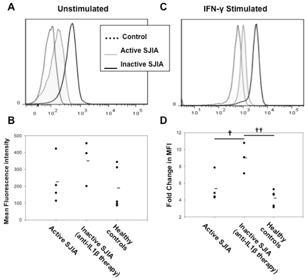
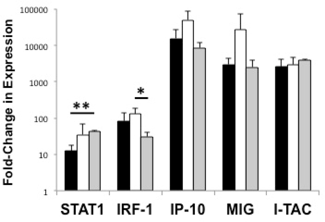 ) Healthy controls; () Active SJIA patients; () Inactive SJIA patients. Results shown as mean + SD. SJIA = systemic JIA; IFN-γ = interferon-γ; STAT1 = signal transducer and activator of transcription 1; IRF-1 = interferon regulatory factor 1; IP-10 = interferon gamma-induced protein 10; MIG = monokine induced by interferon-γ; I-TAC = Interferon-inducible T-cell alpha chemoattractant. * = p < 0.05, ** = p < 0.01.
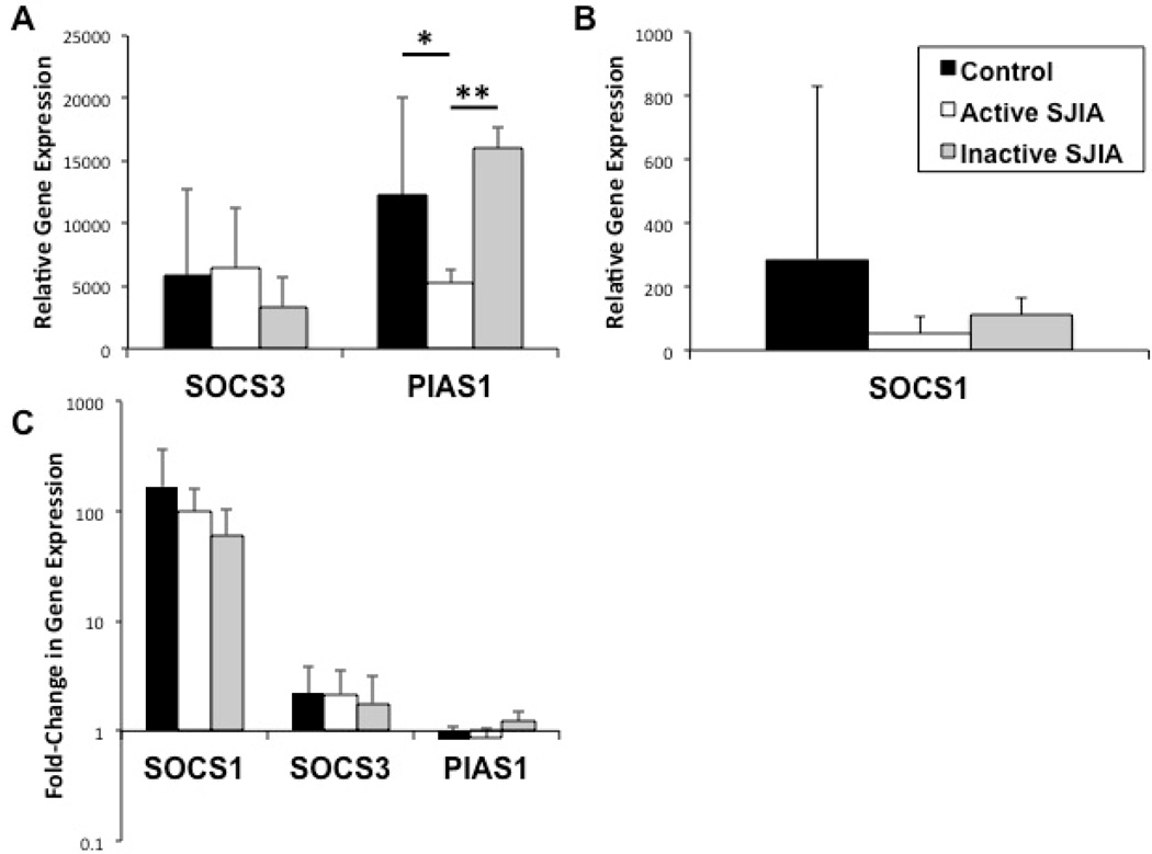 ) Healthy controls; () Active SJIA patients; () Inactive SJIA patients. Results shown as mean + SD. IFN-γ = interferon-γ; GAPDH = glyceraldehyde 3-phosphate dehydrogenase; SOCS1/3 = suppressor of cytokine signaling 1/3; PIAS1 = protein inhibitor of activated STAT1; IL-1β = interleukin-1β. * = p < 0.05, ** = p < 0.01.
Similar articles
-
Interferon signals and monocytic sensitization of the interferon-γ signaling pathway in the peripheral blood of patients with rheumatoid arthritis.Arthritis Rheum. 2012 Feb;64(2):400-8. doi: 10.1002/art.33347. Arthritis Rheum. 2012. PMID: 21953607
-
Proinflammatory Cytokine Environments Can Drive Interleukin-17 Overexpression by γ/δ T Cells in Systemic Juvenile Idiopathic Arthritis.Arthritis Rheumatol. 2017 Jul;69(7):1480-1494. doi: 10.1002/art.40099. Epub 2017 Jun 12. Arthritis Rheumatol. 2017. PMID: 28296284
-
Identification of enhanced IFN-γ signaling in polyarticular juvenile idiopathic arthritis with mass cytometry.JCI Insight. 2018 Aug 9;3(15):e121544. doi: 10.1172/jci.insight.121544. eCollection 2018 Aug 9. JCI Insight. 2018. PMID: 30089725 Free PMC article.
-
Systemic JIA: new developments in the understanding of the pathophysiology and therapy.Best Pract Res Clin Rheumatol. 2009 Oct;23(5):655-64. doi: 10.1016/j.berh.2009.08.003. Best Pract Res Clin Rheumatol. 2009. PMID: 19853830 Free PMC article. Review.
-
Therapeutic Potential of Interferon-γ and Its Antagonists in Autoinflammation: Lessons from Murine Models of Systemic Juvenile Idiopathic Arthritis and Macrophage Activation Syndrome.Pharmaceuticals (Basel). 2015 Nov 25;8(4):793-815. doi: 10.3390/ph8040793. Pharmaceuticals (Basel). 2015. PMID: 26610523 Free PMC article. Review.
Cited by
-
The cytokine storms of COVID-19, H1N1 influenza, CRS and MAS compared. Can one sized treatment fit all?Cytokine. 2021 Aug;144:155593. doi: 10.1016/j.cyto.2021.155593. Epub 2021 May 26. Cytokine. 2021. PMID: 34074585 Free PMC article. Review.
-
Interleukin-18: Biological properties and role in disease pathogenesis.Immunol Rev. 2018 Jan;281(1):138-153. doi: 10.1111/imr.12616. Immunol Rev. 2018. PMID: 29247988 Free PMC article. Review.
-
Neutrophil Homeostasis and Emergency Granulopoiesis: The Example of Systemic Juvenile Idiopathic Arthritis.Front Immunol. 2021 Dec 13;12:766620. doi: 10.3389/fimmu.2021.766620. eCollection 2021. Front Immunol. 2021. PMID: 34966386 Free PMC article. Review.
-
Interferon-γ mediates anemia but is dispensable for fulminant toll-like receptor 9-induced macrophage activation syndrome and hemophagocytosis in mice.Arthritis Rheum. 2013 Jul;65(7):1764-75. doi: 10.1002/art.37958. Arthritis Rheum. 2013. PMID: 23553372 Free PMC article.
-
Macrophage activation syndrome and cytokine-directed therapies.Best Pract Res Clin Rheumatol. 2014 Apr;28(2):277-92. doi: 10.1016/j.berh.2014.03.002. Best Pract Res Clin Rheumatol. 2014. PMID: 24974063 Free PMC article. Review.
References
-
- Schneider R, Laxer R. Systemic-onset juvenile idiopathic rheumatoid arthritis [review] Baillieres Clin Rheumatol. 1998;12:245–271. - PubMed
-
- Fall N, Barnes M, Thornton S, Luyrink L, Olson J, Ilowite NT, et al. Gene expression profiling of peripheral blood from patients with new-onset systemic juvenile idiopathic arthritis reveals molecular heterogeneity that may predict macrophage activation syndrome. Arthritis Rheum. 2007;56:3793–3804. - PubMed
-
- Stéphan JL, Zeller J, Hubert P, Herbelin C, Dayer JM, Prieur AM. Macrophage activation syndrome and rheumatic disease in childhood: a report of four new cases. Clin Exp Rheumatol. 1993;11:451–456. - PubMed
Publication types
MeSH terms
Substances
Grants and funding
LinkOut - more resources
Full Text Sources
Other Literature Sources
Medical
Research Materials
Miscellaneous
