. 2012 Sep;86(18):9696-707.
doi: 10.1128/JVI.01012-12. Epub 2012 Jun 27.

Negative elongation factor-mediated suppression of RNA polymerase II elongation of Kaposi's sarcoma-associated herpesvirus lytic gene expression

Affiliations

Negative elongation factor-mediated suppression of RNA polymerase II elongation of Kaposi's sarcoma-associated herpesvirus lytic gene expression

Zsolt Toth et al. J Virol. 2012 Sep.

Abstract

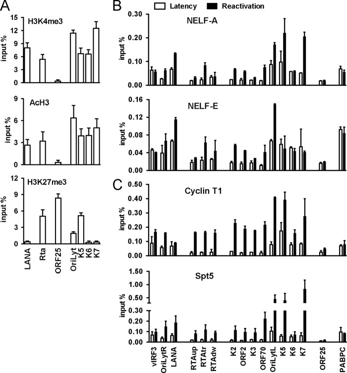
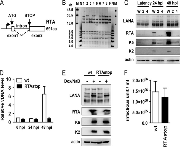
Genome-wide chromatin immunoprecipitation assays indicate that the promoter-proximal pausing of RNA polymerase II (RNAPII) is an important postinitiation step for gene regulation. During latent infection, the majority of Kaposi's sarcoma-associated herpesvirus (KSHV) genes is silenced via repressive histone marks on their promoters. Despite the absence of their expression during latency, however, several lytic promoters are enriched with activating histone marks, suggesting that mechanisms other than heterochromatin-mediated suppression contribute to preventing lytic gene expression. Here, we show that the RNAPII-mediated transcription of the KSHV OriLytL, K5, K6, and K7 (OriLytL-K7) lytic genes is paused at the elongation step during latency. Specifically, the RNAPII-mediated transcription is stalled by the host's negative elongation factor (NELF) at the promoter regions of OriLytL-K7 lytic genes during latency, leading to the hyperphosphorylation of the serine 5 residue and the hypophosphorylation of the serine 2 of the C-terminal domain of the RNAPII large subunit, a hallmark of stalled RNAPII. Consequently, depletion of NELF expression induced transition of stalled RNAPII into a productive transcription elongation at the promoter-proximal regions of OriLytL-K7 lytic genes, leading to their RTA-independent expression. Using an RTA-deficient recombinant KSHV, we also showed that expression of the K5, K6, and K7 lytic genes was highly inducible upon external stimuli compared to other lytic genes that lack RNAPII on their promoters during latency. These results indicate that the transcription elongation of KSHV OriLytL-K7 lytic genes is inhibited by NELF during latency, but can also be promptly reactivated in an RTA-independent manner upon external stimuli.


Figures

Fig 1
Fig 1
Binding of RNA polymerase II on the KSHV genome. (A) Genome-wide mapping of RNAPII on the KSHV genome during latency and reactivation using the ChIP-chip assay. H-224 RNAPII antibody was used for chromatin immunoprecipitation (ChIP) with the lysates of uninduced (latency) and reactivated (by 8 h of stimulation with 1 μg/ml doxycycline) TRExBCBL1-Rta cells. Missing probes in specific genomic regions that contain highly repetitive sequences are shown below the graph of the KSHV genome (**). The alternating dark and light blue squares represent viral ORFs. (B) Schematic representation of the promoters of OriLytL-K7/PAN genes showing their known regulatory elements and the relative positions of RNAPII during latency. (C) RNAPII ChIPs. ChIP DNA was measured by real-time qPCRs using primers for the latent promoters (La) and the selected early or late (L) promoters. PABPC was used as a cellular control, representing a constitutively active cellular promoter. RTAup, RTApr, and RTAdw represent the upstream promoter region of RTA, the transcription start site of RTA, and the downstream portion of the RTA promoter within the gene body, respectively. (D) ChIP analysis of the serine 5 phosphorylated RNAPII (pS5 RNAPII) and (E) the serine 2 phosphorylated RNAPII (pS2 RNAPII) in latent and reactivated TRExBCBL1-Rta cells. (F and G) ChIP assays for the total RNAPII (F) and the serine 5 phosphorylated RNAPII (pS5 RNAPII) (G) using uninduced KSHV-infected BC1 and VG1 cells.
Fig 2
Fig 2
Dynamic association of transcription elongation factors with viral promoters during latency and reactivation. ChIP experiments were done as described in the legend to Fig. 1. (A) The enrichment of activating (H3K4me3, AcH3) and repressive (H3K27me3) histone modifications on the promoters of the LANA latent gene, the RTA immediate-early (IE) gene, the OriLytL, K5, K6, and K7 early (E) genes, and the ORF25 late (L) gene during latency. (B) Recruitment of NELF-A and NELF-E on viral promoters during latency and upon lytic reactivation of KSHV. (B) Binding of cyclin T1 and Spt5 to viral promoters.
Fig 3
Fig 3
Depletion of NELF genes induces the expression of viral lytic genes that possess RNAPII on their promoters during latency. (A) NELF gene depletion. BCBL-1 cells were transduced by lentiviruses carrying either scrambled shRNA (shcontrol) or NELF-A- and NELF-E-specific shRNAs (shNELF). At 3 days postinfection, whole-cell lysates were used for immunoblotting with the indicated antibodies. (B and C) RT-PCR (B) and immunoblotting (C) analysis of viral gene expression. Total RNAs were purified from mock-treated, shcontrol lentivirus-infected or shNELF lentivirus-infected BCBL-1 cells. TERxBCBL1-Rta cells induced with doxycycline for 24 h (TREX24) were included as controls. Semiquantitative RT-PCR analyses (B) and immunoblotting (C) were performed for the indicated viral genes. Glyceraldehyde-3-phosphate dehydrogenase (GAPDH) and JunB (B) as well as actin (C) were included as cellular controls. (D) Immunofluorescent detection of K5 (green) and RTA (red) expression in lentiviral shcontrol or lentiviral shNELF-transduced BCBL-1 cells. The nucleus was counterstained with DAPI. (E) Immunoblotting analysis of viral protein expression. At 3 days postinfection with shcontrol or shNELF lentivirus, 293T-rKSHV.219 cell lysates were used for immunoblotting analysis. TREX24 cell lysates were used as controls for RTA expression. (F) RFP expression in lentiviral shcontrol- or lentiviral shNELF-transduced 293T-rKSHV.219 cells. The RFP expression of rKSHV.219 virus is controlled by the RTA-inducible PAN lytic promoter, whereas GFP is constitutively expressed from the EF-1 cellular promoter.
Fig 4
Fig 4
NELF-mediated RNAPII stalling at the OriLyt-K7 lyitc promoters. ChIP assays were performed with lentiviral shcontrol- or lentiviral shNELF-infected BCBL-1 cells using antibodies of total RNAPII (N20), serine 5 phosphorylated RNAPII (pS5 RNAPII), serine 2 phosphorylated RNAPII (pS2 RNAPII), and the transcription elongation factor Spt5.
Fig 5
Fig 5
Construction of an RTA-deficient KSHV. (A) Schematic diagram of the RTA gene. The frameshift/Stop mutation was introduced into the second exon of RTA. (B) NheI restriction enzyme digestion of BAC16RTAstop mutant bacmids (lanes 1 to 8) and wild-type BAC16 (lane 9). Lanes M and N indicate the midrange PFGE DNA marker and the 1-kb DNA marker, respectively. (C) Expression of viral proteins in 293TBAC16 (W) and 293TBAC16-RTAstop (carrying either BAC16RTAstop bacmid clone 2 or 4) cell lines. Lysates of uninduced cells (Latency) or cells induced with 3 mM NaB for 24 or 48 h were used for the immunoblotting assay with antibodies to the indicated proteins. (D) Viral genomic DNA copy number. Genomic DNA was prepared from 293TBAC16 (wt) and 293TBAC16RTAstop (clone 4, RTAstop) either mock treated (0 hpi) or treated with 3 mM NaB (24 and 48 hpi). The levels of viral genomic DNA were determined by real-time qPCR and were calculated relative to the mock-treated samples (0 hpi). (E) Immunoblot analysis of viral protein expression. Lysates of iSLKBAC16 (wt) or iSLKBAC16RTAstop (clone 4) cells either mock treated (−) or treated with 1 μg/ml of doxycycline and 3 mM NaB (+) were used for immunoblotting assays with antibodies to the indicated proteins. (F) Comparison of infectious virus production. iSLKBAC16 (wt) and iSLKBAC16RTAstop (clone 4) cells were induced with 1 μg/ml of doxycycline and 3 mM NaB for 3 days. Virus titers were determined by infecting 293T cells and quantifying the numbers of GFP-expressing cells by flow cytometry at 24 h postinfection.
Fig 6
Fig 6
RTA-independent KSHV lytic gene expression. (A) Viral protein expression. At 72 h postinfection with shcontrol or shNELF lentivirus, the immunoblotting assay was performed using lysates from infected 293TBAC16RTAstop (clone 4) cells with the indicated antibodies. (B) Immunofluorescence analysis of the KSHV lytic protein K5 in lentivirus shcontrol- or shNELF-transduced 293TBAC16 and 293TBAC16RTAstop (clone 4) cells. DAPI staining shows the nuclei. (C) Real-time qPCR analysis of KSHV viral gene expression. At 8, 24, and 48 h poststimulation of 293TBAC16 or 293TBAC16RTAstop (clone 4) cells with 3 mM NaB, KSHV gene expression was determined by real-time qPCR. The induction levels of viral gene expression were calculated relative to that of mock-treated cells.
Fig 7
Fig 7
Immunofluorescence analysis of lytic protein expression in BCBL-1 cells. (A) Immunofluorescence analysis of K5 and RTA expression. Mock-treated (latent cycle) or NaB-treated BCBL-1 (lytic cycle) cells were permeabilized and costained with anti-K5 mouse monoclonal and anti-RTA rabbit polyclonal antibodies, followed by staining with FITC-coupled anti-mouse and TRITC-coupled anti-rabbit secondary antibodies, respectively. (B) Immunofluorescence analysis of ORF59 and RTA expression. Mock-treated or NaB-treated BCBL-1 cells were permeabilized and costained with anti-ORF59 mouse monoclonal and anti-RTA rabbit polyclonal antibody, followed by staining with FITC-coupled anti-mouse and TRITC-coupled anti-rabbit secondary antibody. (C) Ratio of BCBL-1 cells expressing lytic protein alone (viral protein indicated below the graph), RTA alone, or both RTA and lytic protein. For details of the percent calculation, see Materials and Methods.

References

    1. Adelman K, et al. 2009. Immediate mediators of the inflammatory response are poised for gene activation through RNA polymerase II stalling. Proc. Natl. Acad. Sci. U. S. A. 106:18207–18212 - PMC - PubMed
    1. Aida M, et al. 2006. Transcriptional pausing caused by NELF plays a dual role in regulating immediate-early expression of the junB gene. Mol. Cell. Biol. 26:6094–6104 - PMC - PubMed
    1. Barboric M, Nissen RM, Kanazawa S, Jabrane-Ferrat N, Peterlin BM. 2001. NF-kappaB binds P-TEFb to stimulate transcriptional elongation by RNA polymerase II. Mol. Cell 8:327–337 - PubMed
    1. Bernstein BE, et al. 2006. A bivalent chromatin structure marks key developmental genes in embryonic stem cells. Cell 125:315–326 - PubMed
    1. Brookes E, Pombo A. 2009. Modifications of RNA polymerase II are pivotal in regulating gene expression states. EMBO Rep. 10:1213–1219 - PMC - PubMed

Publication types

MeSH terms