. 2012 Sep;86(18):9617-28.
doi: 10.1128/JVI.00813-12. Epub 2012 Jun 27.

Functional analysis of the Autographa californica multiple nucleopolyhedrovirus GP64 terminal fusion loops and interactions with membranes

Affiliations

Functional analysis of the Autographa californica multiple nucleopolyhedrovirus GP64 terminal fusion loops and interactions with membranes

Sicong Dong et al. J Virol. 2012 Sep.

Abstract

The Autographa californica multiple nucleopolyhedrovirus (AcMNPV) glycoprotein GP64 is the major envelope protein of the budded virus (BV). GP64 is a class III fusion protein that mediates BV attachment to the cell surface and low-pH-triggered membrane fusion between the BV envelope and the endosome membrane during entry. Class III fusion proteins contain terminal looped structures that are believed to interact with membranes. To examine the functions of 3 loops found at the apex of the GP64 postfusion structure, we generated 2-alanine substitutions that scanned the two so-called fusion loops (loop 1 and loop 2) plus an adjacent loop structure (loop 3) that is closely attached to loop 2 and is also found at the apex of the GP64 postfusion structure. We identified essential residues from Y75 to T86 (loop 1) and N149 to H156 (loop 2) that are required for fusion activity, but no essential residues in loop 3. Further analysis revealed that critical fusion loop residues fall within two groups that are associated with either membrane merger (hemifusion) or fusion pore expansion. We next examined the interactions of soluble GP64 proteins and BV with membranes composed of various phospholipids. BV interacted directly with small unilamellar vesicles (SUVs) comprised of phospholipids phosphatidylcholine and phosphatidic acid (PC/PA) or phosphatidylcholine and phosphatidylserine (PC/PS) under neutral and acidic pH. We also examined the interactions of soluble GP64 constructs containing substitutions of the most hydrophobic residues within each of the two fusion loops. We found that a 2-residue substitution in either single loop (loop 1 [positions 81 and 82] or loop 2 [positions 153 and 154]) was not sufficient to substantially reduce the GP64-liposome interaction, but the same substitutions in both fusion loops severely reduced the GP64-liposome association at neutral pH. These results suggest that critical hydrophobic residues in both fusion loops may be involved in the interaction of GP64 with host cellular membranes and direct GP64-membrane interactions may represent a receptor-binding step prior to a low-pH-triggered conformational change.


Figures
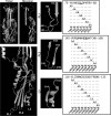
Fig 1
Fig 1
The GP64 postfusion structures of the homotrimer and monomer (Protein Data Bank [PDB] accession no. 3DUZ) are shown as ribbon structures in the top left panels. A close-up view of the loops from the lower portion of the monomer is shown in the bottom left panel. Fusion loop positions (FL1 and FL2) as well as loop 3 (L3) are indicated. Higher-resolution views of the 3 loops that were targeted for analysis by 2-alanine scanning are shown in the central panels, and several amino acid positions are indicated for reference. The sequence of each loop region is shown on the right, and specific substitutions are indicated below the sequence. The construct name is listed below each specific 2-Ala substitution. Each construct is named according to the position of the first substituted amino acid. Underlined amino acids represent the most highly hydrophobic residues at the apex of loops 1 and 2, and substitutions of those residues were examined in the experiment shown in Fig. 5. Structure diagrams were generated with PyMOL.
Fig 2
Fig 2
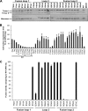
Analysis of expression levels and fusion activities of GP64 constructs containing loop substitutions. (A) Relative expression levels of GP64 constructs in Sf9 cells. The top and bottom panels show Western blots of cell lysates from Sf9 cells transiently expressing GP64 constructs (36 h p.t.) with 2 amino acid substitutions and detected using anti-GP64 MAb AcV5. The construct names and GP64 regions are indicated above the lanes. The top panels show GP64 constructs prepared and electrophoresed under nonreducing conditions, and the bottom panels show constructs were processed under reducing conditions. (B) Relative cell surface levels of GP64 constructs were examined by cELISA with MAb AcV5 at 36 h p.t. Sf9 cells were transfected with 2 μg of each plasmid expressing the indicated GP64 construct. The leftmost 10 columns show the results for a reference titration of WT GP64 cell surface expression using the indicated quantities of a plasmid expressing WT GP64. (C) Relative membrane fusion activity was assessed by a syncytium formation assay for each of the indicated GP64 constructs. For syncytium formation assays, Sf9 cells were transfected with each GP64 construct. In parallel, a dilution series of WT GP64 constructs was also transfected for reference. Transfections were performed in triplicate, and for each transfection, five representative fields were analyzed at 36 h p.t. as described in Materials and Methods.
Fig 3
Fig 3
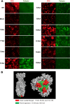
Analysis of the effects of GP64 fusion loop substitutions on membrane merger, pore formation, and pore expansion. (A) Each GP64 construct and WT GP64 were examined by dual-dye labeling and membrane fusion analysis as described in Materials and Methods. RBCs were labeled with both membrane dye (R18 [red]) and cytosolic dye (calcein AM [green]) and bound to Sf9 cells that were transiently expressing the indicated GP64 constructs. After low-pH treatment (pH 5.0 for 5 min), transfer of each dye was monitored by fluorescence microscopy. (B) A space-filling model of the GP64 postfusion trimer is shown in a side view (left), and from the end containing fusion loops (right). The positions of residues associated with inhibition of outer membrane leaflet merger (red) and expansion of the fusion pore (green) are indicated.
Fig 4
Fig 4
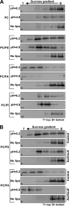
AcMNPV BV association with liposomes at pH 6.2 or 4.8. (A) Purified WT AcMNPV BV was incubated with liposomes at either pH 6.2 or pH 4.8 for 60 min. Virion interactions with liposomes were analyzed by flotation in 20 to 50% sucrose gradients. Lanes represent fractions collected from top to bottom of each gradient. The virions were TCA precipitated from each fraction and detected by Western blot analysis with an anti-VP39 antiserum. For a negative control for each experiment, BV was incubated similarly but in the absence of liposomes (No lipo). The phospholipid composition of each liposome preparation (see Materials and Methods) is indicated to the left of the blots. (B) Comparison of control and Ac23null BV interactions with liposomes. Budded virions containing either a WT complement of envelope proteins (Control) or with no Ac23 envelope protein (Ac23null) were purified and used for analysis of virion-liposome interactions. The results of flotation assays with PC/PS or PC/PA liposomes at pH 6.2 are shown. For convenience of comparisons, results shown for control virions in panel B (B1, B2, B5, and B6) are duplicates of results shown in panel A (subpanels A4, A6, A7, and A9, respectively) from the same experiment.
Fig 5
Fig 5
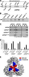
Interactions of soluble GP64 proteins with liposomes at pH 6.2 and 4.8. (A) A C-terminally truncated and six-His-tagged soluble form of GP64 was generated for analysis of GP64-membrane interactions. The GP64 protein was truncated after amino acid 482, and a six-His tag was added to the C terminus. The diagram shows the relative locations of features of the soluble GP64 construct (SP, signal peptide; FL1, fusion loop 1; FL2, fusion loop 2; AcV1, conformational epitope; AcV5, epitope). Soluble GP64 constructs that contain 2 amino acid substitutions in positions 81 and 82 (81AA), positions 153 and 154 (153AV), or both were also generated, and the positions of the substitution mutations are indicated. The position of the 462-amino-acid (aa) ectodomain is shown at the top of the panel. (B) Immunoprecipitation of soluble GP64 constructs with a conformation-specific monoclonal antibody. Purified soluble GP64 was immunoprecipitated with prefusion conformation-specific MAb AcV1 and detected on Western blots with MAb AcV5. For a negative control (N), soluble WT GP64 was incubated with protein A resin but no AcV1. The position of the GP64 band is indicated by a black arrowhead labeled 64 to the left of the gel, and the lower bands represents the heavy chain of MAb AcV1. (C) Interactions of soluble GP64 constructs with PC/PA liposomes at neutral (6.2) or low (4.8) pH. Purified soluble GP64 proteins (indicated to the left of the blots) were incubated with PC/PA liposomes at pH 6.2 or 4.8 for 60 min and analyzed by liposome flotation gradients. For each experiment, the three lanes (T, M, and B) contain three equal fractions collected from the top, middle, and bottom of the gradient. Proteins were recovered from the fractions by immunoprecipitation with an anti-6His antiserum and detected by Western blot analysis using MAb AcV5. The experiment was performed in the absence of liposomes as a negative control (No lipo). (D) Semiquantitative analysis of the effects of pH and fusion loop substitutions on GP64-liposome interactions. For each soluble GP64 protein, the flotation assay was repeated 3 times, and Western blot results were scanned and quantified by ImageJ and then used to generate averages and standard deviations (error bars). Percent flotation represents the percentage of GP64 detected in the top and middle fractions versus the total detected in all fractions. (E) Space-filling model of the GP64 postfusion trimer structure viewed from the end of the long axis of the molecule with the fusion loops in the foreground. The positions of the fusion loop residues that were substituted in the analysis of liposome interactions (above) are shown in red and blue.

Similar articles

Cited by

References

    1. Airenne KJ, Makkonen KE, Mahonen AJ, Yla-Herttuala S. 2011. Baculoviruses mediate efficient gene expression in a wide range of vertebrate cells. Methods Mol. Biol. 737:279–301 - PubMed
    1. Backovic M, Jardetzky TS. 2011. Class III viral membrane fusion proteins. Adv. Exp. Med. Biol. 714:91–101 - PubMed
    1. Backovic M, Jardetzky TS. 2009. Class III viral membrane fusion proteins. Curr. Opin. Struct. Biol. 19:189–196 - PMC - PubMed
    1. Blissard GW, Rohrmann GF. 1991. Baculovirus gp64 gene expression: analysis of sequences modulating early transcription and transactivation by IE1. J. Virol. 65:5820–5827 - PMC - PubMed
    1. Blissard GW, Wenz JR. 1992. Baculovirus GP64 envelope glycoprotein is sufficient to mediate pH-dependent membrane fusion. J. Virol. 66:6829–6835 - PMC - PubMed

Publication types

MeSH terms

LinkOut - more resources