. 2012 Sep;86(17):9416-31.
doi: 10.1128/JVI.07032-11. Epub 2012 Jun 27.

Intracytoplasmic trapping of influenza virus by a lipophilic derivative of aglycoristocetin

Affiliations

Intracytoplasmic trapping of influenza virus by a lipophilic derivative of aglycoristocetin

Evelien Vanderlinden et al. J Virol. 2012 Sep.

Abstract

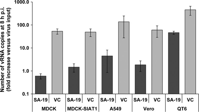
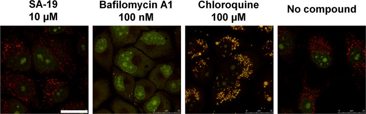
We report on a new anti-influenza virus agent, SA-19, a lipophilic glycopeptide derivative consisting of aglycoristocetin coupled to a phenylbenzyl-substituted cyclobutenedione. In Madin-Darby canine kidney cells infected with influenza A/H1N1, A/H3N2, or B virus, SA-19 displayed a 50% antivirally effective concentration of 0.60 μM and a selectivity index (ratio of cytotoxic versus antiviral concentration) of 112. SA-19 was 11-fold more potent than unsubstituted aglycoristocetin and was active in human and nonhuman cell lines. Virus yield at 72 h p.i. was reduced by 3.6 logs at 0.8 μM SA-19. In contrast to amantadine and oseltamivir, SA-19 did not select for resistance upon prolonged virus exposure. SA-19 was shown to inhibit an early postbinding step in virus replication. The compound had no effect on hemagglutinin (HA)-mediated membrane fusion in an HA-polykaryon assay and did not inhibit the low-pH-induced refolding of the HA in a tryptic digestion assay. However, a marked inhibitory effect on the transduction exerted by retroviral pseudoparticles carrying an HA or vesicular stomatitis virus glycoprotein (VSV-G) fusion protein was noted, suggesting that SA-19 targets a cellular factor with a role in influenza virus and VSV entry. Using confocal microscopy with antinucleoprotein staining, SA-19 was proven to completely prevent the influenza virus nuclear entry. This virus arrest was characterized by the formation of cytoplasmic aggregates. SA-19 appeared to disturb the endocytic uptake and trap the influenza virus in vesicles distinct from early, late, or recycling endosomes. The aglycoristocetin derivative SA-19 represents a new class of potent and broad-acting influenza virus inhibitors with potential clinical relevance.


Figures

Fig 1
Fig 1
Chemical structures of SA-19 and related glycopeptide derivatives. SA-19 is a derivative of aglycoristocetin in which the primary amine group is coupled to a phenylbenzylgroup via a cyclobutenedione group. The identical lipophylic substitution is present in SA-48 (the teicoplanin analogue of SA-19); ERJ-147 (which lacks the chloro substituents on rings C and E of SA-48); and SA-26 (the vancomycin analogue of SA-19). The glycopeptide ristocetin (not shown) differs from aglycoristocetin in having three sugar substituents at the positions marked with an asterisk: d-mannose on ring A, l-ristosamine next to ring C, and the tetrasaccharide d-arabinose–d-mannose–d-glucose–l-rhamnose on ring D.
Fig 2
Fig 2
SA-19 inhibits influenza virus replication in different cell lines. Cells were infected with A/X-31 virus, at an MOI of 0.0004 PFU per cell, in the presence of 10 μM SA-19. At 8 h p.i., total cellular RNA was extracted and the number of vRNA copies was determined by real-time RT-PCR. Virus replication is shown as the fold increase in vRNA copy number relative to the virus input at time zero. Data shown represent the means ± standard errors of the means (SEM) of the results of 3 to 5 independent experiments. VC: virus control receiving no compound.
Fig 3
Fig 3
SA-19 inhibits virus entry yet has no effect on virus binding or on low-pH-induced HA refolding. (A) Inhibitory effect of SA-19 on vRNA synthesis as a function of the time of compound addition. MDCK cells were infected with A/X-31 influenza virus at an MOI of 0.0004 PFU per cell, and compounds were added at −30 min, 0 h, 30 min, 1 h, 3 h, 5 h, or 8 h p.i. At 10 h p.i., the cells were subjected to RNA extraction for subsequent vRNA analysis by real-time RT-PCR. The vRNA synthesis is presented as the fold increase in the number of vRNA copies at 10 h p.i. relative to the amount of virus particles added at time zero. Data shown represent the means ± SEM (numbers of experiments are shown in parentheses). (B) Effect of SA-19 and related derivatives on influenza virus binding to MDCK cells. After 2 h of incubation with compounds at 35°C, MDCK cells were cooled and allowed to bind the A/X-31 virus (MOI: 0.04 PFU per cell) for 1 h on ice in the further presence of compound. In order to quantify the virus bound to the cells, cellular RNA extracts were prepared and two-step real-time RT-PCR was performed. Virus binding is shown as the vRNA copy number, relative to that in the virus control receiving no compound. Compound concentrations were 10 μM for SA-19, SA-26, and aglycoristocetin and 200 μM for NMSO3. Data shown represent the means ± SEM of 3–6 independent determinations. (C) SA-19 does not inhibit the conformational change of HA at low pH, as demonstrated by trypsin digestion assay. A/X-31 virus was incubated at 37°C for 15 min in the presence of various concentrations of SA-19 or 4c, and the pH was lowered to 5.0. After neutralization, the mixtures were treated with trypsin. The lysates were subjected to Western blot analysis under reducing conditions and using an anti-HA1 antibody. The low-pH-induced conformational change renders the HA sensitive to trypsin digestion, causing disappearance of the HA1 band. (D) SA-19 has no effect on low-pH-induced polykaryon formation in HA-transfected HeLa cells. HeLa cells expressing A/X-31 HA were treated with trypsin to cleave HA0, washed, and incubated during 15 min in the presence of compound. Then, the pH was lowered to 5.0 and the cells were incubated for 15 min at 37°C in the presence of compound. Following syncytium formation for 3 h at 37°C in the presence of compound, the cells were fixed, stained with Giemsa, and examined by microscopy (original magnification, ×200). Representative fields are shown.
Fig 4
Fig 4
Localization of influenza virus NP in infected MDCK cells treated with (panels A to D) SA-19 or reference compounds or (panels F to I) analogues of SA-19. MDCK cells were infected with A/X-31 influenza virus (MOI: 4 PFU per cell) in the presence of compound and incubated for 1 h at 35°C. After fixation and permeabilization, the cells were stained with anti-NP antibody and Alexa Fluor 488-labeled secondary antibody (green). The nuclei were visualized with DAPI (blue). Bar, 25 μm. See Fig. 1 for the chemical structures of (aglyco)ristocetin, SA-19, SA-48, and SA-26.
Fig 5
Fig 5
Virus trapping by SA-19 is reversed by washing. MDCK cells were infected with A/X-31 influenza virus (at 4 PFU per cell) in the presence of 10 μM SA-19. After 1 h of incubation at 35°C, the cells were washed three times (if indicated) and incubated for an additional hour at 35°C. Then, the cells were fixed, permeabilized, and stained. Influenza virus was detected with anti-NP antibody and Alexa Fluor 488-labeled secondary antibody (green). The nuclei were visualized with DAPI (blue). Bar, 25 μm.
Fig 6
Fig 6
Dual staining for influenza virus NP and the endosomal markers EEA1 and LBPA. MDCK cells were infected with A/X-31 virus (at 4 PFU per cell) in the presence of 10 μM SA-19 or 100 μM 4c and incubated at 35°C for 1 h. After fixation and permeabilization, the cells were double labeled. Influenza virus was visualized with anti-NP antibody and Alexa Fluor 488-labeled secondary antibody (green). Analysis was performed with anti-EEA1 and LBPA primary antibodies as early and late endosome markers and Alexa Fluor 568-labeled secondary antibodies (red). The nuclei were visualized with DAPI (blue). Bar, 25 μm.
Fig 7
Fig 7
Colocalization between influenza virus NP and Rab or LAMP-1 markers of endosomal-lysosomal compartments. MDCK cells were transfected with expression plasmids for GFP-coupled Rab4, Rab5, Rab7, or Rab11 or for YFP-coupled LAMP-1 (green), incubated for 23 h at 35°C, and subsequently infected with A/X-31 virus (at 16 PFU per cell) in the presence or absence of SA-19. After 1 h of incubation at 35°C, the cells were fixed, permeabilized, and stained with anti-NP and Alexa Fluor 633-labeled secondary antibody (red). The nuclei were visualized with DAPI (blue). Bar, 10 μm.
Fig 8
Fig 8
SA-19 has no effect on acidification of the endosomes. After treatment of MDCK cells with compound during 1 h at 35°C, acridine orange was added and the cells were subsequently analyzed by confocal microscopy. Acridine orange is an acidotropic dye. Its fluorescence is green at low concentrations, which changes to orange-red at high concentrations (as obtained in the lysosomes in which acridine orange is protonated and sequestered). Acridine orange also intercalates in DNA, resulting in green fluorescence in the nucleus. Upon treatment with the V-ATPase inhibitor bafilomycin A1, lysosomal acidification is completely inhibited, and, hence, the dye's accumulation in the lysosomes is prevented. Chloroquine induces lysosomal swelling and a decrease in lysosomal pH, giving a shift to yellow fluorescence. In the presence of SA-19, the acridine orange staining is identical to that observed in untreated cells. Bar, 25 μm.
Fig 9
Fig 9
Inhibition of transduction by GFP-expressing retroviral pseudoparticles carrying influenza virus HA and NA (upper panel) or VSV-G (lower panel). The test compounds (SA-19 at 1, 5, or 10 μM or chloroquine at 50 μM) were added to MDCK cells together with the pseudoparticles. After 72 h, the cells were subjected to flow cytometry for quantification of transduction efficiency. The bars represent the percentages of GFP-positive cells (relative to the untreated control) (data represent averages ± SEM of the results of three tests).

References

    1. Balzarini J, et al. 2006. Inhibition of feline (FIPV) and human (SARS) coronavirus by semisynthetic derivatives of glycopeptide antibiotics. Antiviral Res. 72:20–33 doi:10.1016/j.antiviral.2006.03.005 - DOI - PMC - PubMed
    1. Balzarini J, et al. 2003. Antiretroviral activity of semisynthetic derivatives of glycopeptide antibiotics. J. Med. Chem. 46:2755–2764 - PubMed
    1. Bardsley B, Williams DH, Baglin TP. 1998. Cleavage of rhamnose from ristocetin A removes its ability to induce platelet aggregation. Blood Coagul. Fibrinolysis 9:241–244 - PubMed
    1. Bauer K, Richter M, Wutzler P, Schmidtke M. 2009. Different neuraminidase inhibitor susceptibilities of human H1N1, H1N2, and H3N2 influenza A viruses isolated in Germany from 2001 to 2005/2006. Antiviral Res. 82:34–41 - PubMed
    1. Belser JA, et al. 2007. DAS181, a novel sialidase fusion protein, protects mice from lethal avian influenza H5N1 virus infection. J. Infect. Dis. 196:1493–1499 - PubMed

Publication types

MeSH terms

LinkOut - more resources