. 2012 Aug 17;287(34):28943-55.
doi: 10.1074/jbc.M111.306423. Epub 2012 Jun 27.

X-linked sideroblastic anemia due to carboxyl-terminal ALAS2 mutations that cause loss of binding to the β-subunit of succinyl-CoA synthetase (SUCLA2)

Affiliations

X-linked sideroblastic anemia due to carboxyl-terminal ALAS2 mutations that cause loss of binding to the β-subunit of succinyl-CoA synthetase (SUCLA2)

David F Bishop et al. J Biol Chem. .

Abstract

Mutations in the erythroid-specific aminolevulinic acid synthase gene (ALAS2) cause X-linked sideroblastic anemia (XLSA) by reducing mitochondrial enzymatic activity. Surprisingly, a patient with the classic XLSA phenotype had a novel exon 11 mutation encoding a recombinant enzyme (p.Met567Val) with normal activity, kinetics, and stability. Similarly, both an expressed adjacent XLSA mutation, p.Ser568Gly, and a mutation (p.Phe557Ter) lacking the 31 carboxyl-terminal residues also had normal or enhanced activity, kinetics, and stability. Because ALAS2 binds to the β subunit of succinyl-CoA synthetase (SUCLA2), the mutant proteins were tested for their ability to bind to this protein. Wild type ALAS2 bound strongly to a SUCLA2 affinity column, but the adjacent XLSA mutant enzymes and the truncated mutant did not bind. In contrast, vitamin B6-responsive XLSA mutations p.Arg452Cys and p.Arg452His, with normal in vitro enzyme activity and stability, did not interfere with binding to SUCLA2 but instead had loss of positive cooperativity for succinyl-CoA binding, an increased K(m) for succinyl-CoA, and reduced vitamin B6 affinity. Consistent with the association of SUCLA2 binding with in vivo ALAS2 activity, the p.Met567GlufsX2 mutant protein that causes X-linked protoporphyria bound strongly to SUCLA2, highlighting the probable role of an ALAS2-succinyl-CoA synthetase complex in the regulation of erythroid heme biosynthesis.


Figures

FIGURE 1.
FIGURE 1.
Genotype analysis of the proband and family members. DNA isolated from a normal individual, the proband, and his mother, sister, and two daughters was PCR-amplified, digested with NlaIII, and analyzed by agarose gel electrophoresis. Lanes 1 and 9, molecular weight standards; lane 8, a no-template PCR control; lane 2, DNA from an unrelated normal control; lanes 3–7, digested DNAs from the proband's sister (II, 1), mother (I, 1), male proband (II, 2), and two daughters (III, 1&2), respectively. The lane 2 control shows the expected 120- and 166-bp NlaIII fragments, with the additional 33-bp fragment having migrated below the picture frame. Lane 5 shows the 120-bp fragment and uncut 166 + 33 = 199 bp band for the hemizygote, whose mutation ablates the restriction site, whereas the other lanes contain all three bands, showing that all females in the pedigree are heterozygous carriers.
FIGURE 2.
FIGURE 2.
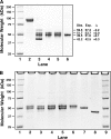
SDS-PAGE analysis of ALAS2 purification products. Protein size analysis of each step of the purification of recombinant human ALAS2 was done by denaturing polyacrylamide gel electrophoresis. A, lanes 2–5, wild type ALAS2. Lane 1, size standards; lane 2, affinity-purified MBP-ALAS2 fusion protein; lane 3, post-Factor Xa cleavage of fusion protein; lane 4, post-FPLC Superose gel filtration chromatography of the cleaved fusion protein; lane 5, same purification stage, sample stored at −80 °C for 3.75 years; lane 6, post-FPLC fraction of p.Phe557Ter enzyme (electrophoresed in the same gel, but intervening lanes excised). Obs., observed size estimated from the size markers; Exp., expected size calculated from peptide mass as discussed under “Results”; Δ, Exp. minus Obs. B, typical SDS-PAGE profiles of wild type and mutant ALAS2 proteins purified to the post-FPLC stage. Lanes 1 and 9, size standards; lane 2, wild type; lane 3, p.Arg452Cys; lane 4, p.Arg452His; lane 5, p.Met567Val; lane 6, p.Ser568Gly; lane 7, p.Phe557Ter; lane 8, MBP.
FIGURE 3.
FIGURE 3.
Lineweaver-Burk plots of wild type and Arg-452 ALAS2 mutants. Lineweaver-Burk plots were constructed for the 5-min end point reaction rates of wild type and mutant ALAS2 with varying concentrations of succinyl-CoA at 37 °C with glycine at 100 mm and the rest of the reaction conditions as described under “Experimental Procedures.” The reactions were initiated by the addition of the succinyl-CoA to reaction tubes preincubated at 37 °C. A, wild type enzyme. B, wild type enzyme data plotted with the Hill transformation of the Lineweaver-Burk plot using 1/Sn. Hill n was estimated iteratively using the Hill n derived from the slope of a plot of log(Vmax/(1 − Vmax)) versus log S and recalculation of Vmax from Lineweaver-Burk plots using 1/Sn until Hill n was constant. C, the Hill-transformed Lineweaver-Burk plot of reaction rate data for the p.Arg452Cys mutant enzyme. D, same as in C for the p.Arg452His mutant enzyme.
FIGURE 4.
FIGURE 4.
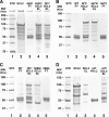
SUCLA2 affinity chromatography of ALAS2. Amylose resin columns were loaded with affinity-purified recombinant human MBP-SUCLA2 fusion protein (SCL2) and washed, and then aliquots of wild type and mutant post-FPLC purified ALAS2 enzyme preparations were applied, washed, and then eluted with maltose as described under “Experimental Procedures.” A, SDS-PAGE of SCL2 and of WT and p.Arg452Cys ALAS2. Lane 1, molecular weight standards; lane 2, SCL2 after purification by amylose resin affinity chromatography; lane 3, flow-through fraction containing excess p.Arg452Cys protein that did not bind to the amylose/SCL2 affinity column; lane 4, material eluted by maltose from the amylose/SCL2 column to which the p.Arg452Cys protein had been loaded and washed; lane 5, material eluted from an amylose/SCL2 column to which wild type ALAS2 had been loaded and washed. Note that both SCL2 and bound ALAS2 are eluted in the material electrophoresed in lanes 4 and 5. B, similar to A but showing the wild type flow-through fraction in lane 2 and the maltose-induced elution of the wild type enzyme along with SCL2 in lane 3. Lane 4, maltose-eluted material from an amylose/SCL2 column to which the p.Met567Val protein had been applied; lane 5, flow-through from that column during the p.Met567Val application step. Note that in contrast to the co-elution of SCL2 and ALAS2 bands in lane 3 for the wild type enzyme, only SCL2 bands were eluted in lane 4, showing that no p.Met567Val protein was bound to SCL2. C, similar to B, showing again that wild type enzyme binds and is co-eluted with SCL2, whereas the mutant p.Ser568Gly protein does not bind (lane 5), and only SCL2 bands are eluted (lane 4). The line between lanes 3 and 4 indicates an excised region of the gel. D, as in A; lane 2, amylose affinity resin-purified SCL2 protein before loading onto a second amylose column; lane 3, elution of that material by maltose. Lane 4, FPLC-purified p.Met567GlufsX1 protein resulting from the ΔAT ALAS2 mutation; lane 5, co-elution of this mutant protein with SCL2, showing that unlike the p.Met567Val mutation, this mutation binds strongly to SCL2.
FIGURE 5.
FIGURE 5.
ALAS2 affinity chromatography of SCS. Amylose resin columns were loaded with purified recombinant human wild type or mutant MBP-ALAS2 fusion proteins and washed, and then aliquots of purified human SCS enzyme preparations were applied, washed, and then eluted with maltose as described under “Experimental Procedures.” Lanes 1 and 7, molecular weight standards; lane 2, purified wild type MBP-ALAS2; lane 3, purified M567V mutant MBP-ALAS2; lane 4, material eluted by maltose from the amylose/WT MBP-ALAS2 column to which the SCS protein had been loaded and washed; lane 5, material eluted from an amylose/M567V MBP-ALAS2 column to which SCS had been loaded and washed; lane 6, purified SCS preparation that was loaded onto the columns eluted to give the material for lanes 4 and 5. Note that in contrast to lane 4, where the SCS was co-eluted with wild type ALAS2, in lane 5, essentially no SCS was co-eluted, and thus it failed to bind to the M567V mutant ALAS2.
FIGURE 6.
FIGURE 6.
Mutations in the ALAS2 carboxyl terminus resulting in XLSA and XLP. Wild type and mutant ALAS2 sequences are for carboxyl-terminal amino acids beginning with ALAS2 residue Ala-553 and the corresponding cDNA nucleotides, numbering from the start codon. The 15-amino acid region at the carboxyl terminus that lacked any reported XLSA mutations is indicated in italic type. The mutated regions are boxed. The nucleotide change (c.1701G→?) was not reported for the published p.Met567Ile mutation. The third line shows the synthetic truncation mutation that had normal or enhanced ALAS2 activity and stability, whereas the last two lines show the two published ALAS2 mutations that resulted in XLP. The region where mutations may alter binding to SUCLA2 to wild type ALAS2 was confirmed by demonstration of loss of binding for XLSA mutant proteins p.Met567Val, p.Ser568Gly, and p.Phe557Ter and considered possible for the mutations at residues 559, 560, and 562 reported to result in XLSA.

References

    1. Bishop D. F., Henderson A. S., Astrin K. H. (1990) Human δ-aminolevulinate synthase. Assignment of the housekeeping gene to 3p21 and the erythroid-specific gene to the X chromosome. Genomics 7, 207–214 - PubMed
    1. Anderson K. E., Sassa S., Bishop D. F., Desnick R. J. (2001) Disorders of heme biosynthesis. X-linked sideroblastic anemia and the porphyrias. in The Metabolic and Molecular Bases of Inherited Disease, 8th Ed. (Scriver C. R., Beaudet A. L., Sly W. S., Valle D., Childs B., Kinzler K. W., Vogelstein B., eds) pp. 2991–3062, McGraw-Hill, New York
    1. Ferreira G. C., Neame P. J., Dailey H. A. (1993) Prot. Sci. 2, 1959–1965 - PMC - PubMed
    1. Ferreira G. C., Vajapey U., Hafez O., Hunter G. A., Barber M. J. (1995) Aminolevulinate synthase. Lysine 313 is not essential for binding the pyridoxal phosphate cofactor but is essential for catalysis. Protein Sci. 4, 1001–1006 - PMC - PubMed
    1. Cotter P. D., Baumann M., Bishop D. F. (1992) Enzymatic defect in “X-linked” sideroblastic anemia. Molecular evidence for erythroid δ-aminolevulinate synthase deficiency. Proc. Natl. Acad. Sci. U.S.A. 89, 4028–4032 - PMC - PubMed

Publication types

MeSH terms

LinkOut - more resources