Case Reports
. 2012;39(2):249-54.

Hypoplastic right-heart syndrome presenting as multiple miscarriages

Affiliations
Case Reports

Hypoplastic right-heart syndrome presenting as multiple miscarriages

Chadi Dib et al. Tex Heart Inst J. 2012.

Abstract

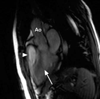
Reversible causes of miscarriage are many, but they affect only 1% of women who are trying to conceive. Herein, we describe the case of a 23-year-old woman who presented for evaluation of repeated miscarriages and was found to have hypoxemia and erythrocytosis. Further evaluation revealed hypoplastic right-heart syndrome with an intracardiac shunt. She underwent hybrid repair with pulmonary valve balloon valvuloplasty, followed by surgery to perform atrial septal defect closure and a Glenn anastomosis. The erythrocytosis and hypoxemia resolved, and she was able to conceive and deliver a healthy baby at term 2 years later. This is a unique case of a rare congenital heart defect that went unnoticed until adulthood, when attempts at pregnancy failed because of the associated hypoxemia. Timely and appropriate treatment led to a successful pregnancy after repeated miscarriages. This case exemplifies the need for a comprehensive medical evaluation of every woman with a history of multiple miscarriages to determine whether a reversible cause exists.

Keywords: Abortion, spontaneous/etiology; heart defects, congenital/complications/physiopathology/therapy/surgery; treatment outcome; ventricular function, right/physiology.


Figures

None
Fig. 1 Transthoracic echocardiograms (subcostal views) show A) a hypoplastic right heart with increased wall thickness, and B) an atrial septal defect with left-to-right shunt (arrow). LA = left atrium; LV = left ventricle; RA = right atrium; RV = right ventricle
None
Fig. 2 Transthoracic echocardiograms (parasternal views) show A) a thickened pulmonary valve (arrows) with limited systolic excursion, and B) with Doppler interrogation, moderate pulmonary stenosis. LV = left ventricle; PA = pulmonary artery; PV = pulmonary valve; RV = right ventricle
None
Fig. 3 Color-flow Doppler echocardiograms show a large atrial septal defect (arrows) with A) left-to-right shunt and B) right-to-left shunt. LA = left atrium; RA = right atrium; SVC = superior vena cava
None
Fig. 4 Transthoracic echocardiogram (basal 4-chamber view) shows a small right ventricle and tricuspid valve annulus and an atrial septal defect (asterisks). LA = left atrium; LV = left ventricle; RA = right atrium; RV = right ventricle
None
Fig. 5 Transthoracic echocardiograms (longitudinal views) show A) a thickened pulmonary valve (arrow) at the level of the pulmonary artery, with limited systolic excursion; and B) with color-flow Doppler interrogation, turbulence caused by pulmonary stenosis (arrow). LA = left atrium; LV = left ventricle; PA = pulmonary artery; PV = pulmonary valve
None
Fig. 6 Cardiovascular magnetic resonance (4-chamber view) shows a small right ventricle (RV) in comparison with the left ventricle (LV). The right atrium (RA) is of normal size; however, the interatrial septum bows toward the left atrium (LA).
None
Fig. 7 Diastole: Cardiovascular magnetic resonance (3-chamber view, equivalent to echocardiographic parasternal long-axis view) shows that the right ventricular outflow tract (arrowhead) is mildly enlarged in diastole. The left ventricular outflow tract is also shown (arrow). Ao = aorta
None
Fig. 8 Systole: Cardiovascular magnetic resonance (3-chamber view, equivalent to echocardiographic parasternal long-axis view) shows that the right ventricular outflow tract (arrowhead) is markedly dilated in systole. The left ventricular outflow tract is also shown (arrow). Ao = aorta
None
Fig. 9 Cardiovascular magnetic resonance (oblique coronal view) of the right ventricular (RV) outflow tract shows a systolic jet (arrow), indicating residual pulmonary stenosis. The main pulmonary artery is dilated.

References

    1. Tzioras S, Polyzos NP, Economides DL. How do you solve the problem of recurrent miscarriage? Reprod Biomed Online 2009;19(3):296–7. - PubMed
    1. Griesshammer M, Struve S, Harrison CM. Essential thrombocythemia/polycythemia vera and pregnancy: the need for an observational study in Europe. Semin Thromb Hemost 2006;32(4 Pt 2):422–9. - PubMed
    1. D'Uva M, Di Micco P, Strina I, Ranieri A, Alviggi C, Mollo A, et al. Etiology of hypercoagulable state in women with recurrent fetal loss without other causes of miscarriage from Southern Italy: new clinical target for antithrombotic therapy. Biologics 2008;2(4):897–902. - PMC - PubMed
    1. Brickner ME, Hillis LD, Lange RA. Congenital heart disease in adults. Second of two parts [published erratum appears in N Engl J Med 2000;342(13):988]. N Engl J Med 2000; 342(5):334–42. - PubMed
    1. Prasad K, Singh M, Radhakrishnan S. Hypoplastic right ventricle with mild pulmonary stenosis in an adult. Int J Cardiol 1992;37(2):260–2. - PubMed

Publication types

MeSH terms