

Partial hydatidiform mole progression into invasive mole with lung metastasis following in vitro fertilization
- PMID: 22740971
- PMCID: PMC3362542
- DOI: 10.3892/ol.2011.542
Partial hydatidiform mole progression into invasive mole with lung metastasis following in vitro fertilization
Abstract
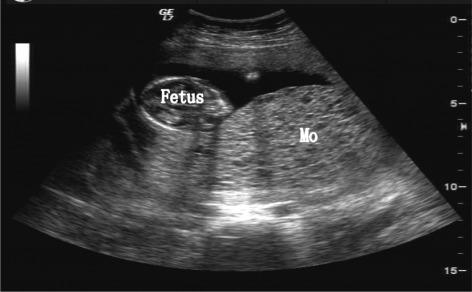
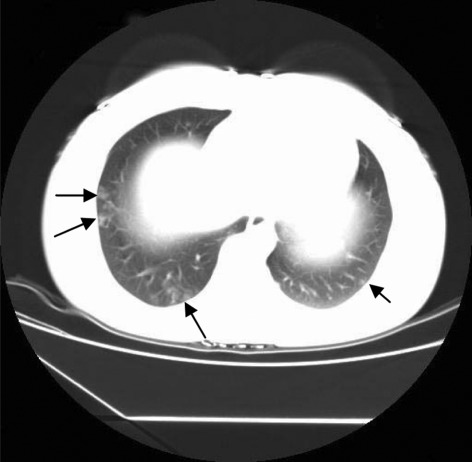
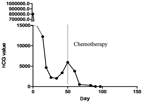
In the present study, the case of a 34-year-old Chinese female who underwent in vitro fertilization resulting in a twin pregnancy was reported. Following in vitro fertilization, the patient was found to have a partial hydatidiform mole (PHM) with a co-existing twin by transvaginal sonography (TVS). At 16 weeks, the pregnancy was terminated and a normal-looking fetus with a HM placenta was delivered, in addition to a normal fetus with a normal placenta. Following termination of the pregnancy, the PHM progressed into an invasive mole with lung metastasis, a rare event. Serum human chorionic gonadotrophin (hCG) concentrations decreased in the first week following delivery, but over the following 21 days hCG levels showed a continuous increase. Following 2 cycles of combinative chemotherapy consisting of fluorouracil (5-FU) and dactinomycin (KSM), hCG concentrations decreased to normal levels. The patient was then administered 1 cycle of repeated chemotherapy and hCG levels remained negative for the following 2 years.
Figures
Similar articles
-
Twin pregnancy and partial hydatidiform mole following in vitro fertilization and embryos transfer: a novel case of placental mosaicism.Chin Med J (Engl). 2012 Dec;125(24):4517-9. Chin Med J (Engl). 2012. PMID: 23253730
-
What is the optimal duration of human chorionic gonadotrophin surveillance following evacuation of a molar pregnancy? A retrospective analysis on over 20,000 consecutive patients.Gynecol Oncol. 2018 Feb;148(2):254-257. doi: 10.1016/j.ygyno.2017.12.008. Epub 2017 Dec 9. Gynecol Oncol. 2018. PMID: 29229282
-
Term delivery of a complete hydatidiform mole with a coexisting living fetus followed by successful treatment of maternal metastatic gestational trophoblastic disease.Taiwan J Obstet Gynecol. 2014 Sep;53(3):397-400. doi: 10.1016/j.tjog.2013.02.005. Taiwan J Obstet Gynecol. 2014. PMID: 25286799
-
Multiple metastatic gestational trophoblastic disease after a twin pregnancy with complete hydatidiform mole and coexisting fetus, following assisted reproductive technology: Case report and literature review.Taiwan J Obstet Gynecol. 2018 Aug;57(4):588-593. doi: 10.1016/j.tjog.2018.06.020. Taiwan J Obstet Gynecol. 2018. PMID: 30122584 Review.
-
Twin pregnancy with a partial hydatidiform mole and a coexistent live fetus. Diagnostic and therapeutic dilemmas. A case report and the review of literature.Ginekol Pol. 2020;91(10):589-594. doi: 10.5603/GP.a2020.0109. Ginekol Pol. 2020. PMID: 33184826 Review.
Cited by
-
Rapid progression from complete molar pregnancy to post-molar gestational trophoblastic neoplasia: a rare case report and literature review.Front Oncol. 2023 Dec 15;13:1303249. doi: 10.3389/fonc.2023.1303249. eCollection 2023. Front Oncol. 2023. PMID: 38162509 Free PMC article.
-
A Case of Rapid Transformation from Hydatidiform Mole to Invasive Mole: The Importance of β-hCG (Human Chorionic Gonadotropin) Serum Levels in Follow-Up Evaluation.Am J Case Rep. 2021 Jun 15;22:e931156. doi: 10.12659/AJCR.931156. Am J Case Rep. 2021. PMID: 34127641 Free PMC article.
-
Tegafur Substitution for 5-Fu in Combination with Actinomycin D to Treat Gestational Trophoblastic Neoplasm.PLoS One. 2015 Nov 23;10(11):e0143531. doi: 10.1371/journal.pone.0143531. eCollection 2015. PLoS One. 2015. PMID: 26599757 Free PMC article.
-
Chemotherapy-resistant invasive mole following partial hydatidiform molar pregnancy necessitating hysterectomy for hemorrhagic complications: A rare case report.Int J Surg Case Rep. 2025 Aug;133:111577. doi: 10.1016/j.ijscr.2025.111577. Epub 2025 Jun 27. Int J Surg Case Rep. 2025. PMID: 40582066 Free PMC article.
-
Medical termination of a partial hydatidiform mole and coexisting fetus during the second trimester: A case report.Oncol Lett. 2015 Dec;10(6):3625-3628. doi: 10.3892/ol.2015.3743. Epub 2015 Sep 24. Oncol Lett. 2015. PMID: 26788180 Free PMC article.
References
-
- Vassilakos P, Kajii T. Letter: Hydatidiform mole: two entities. Lancet. 1976;1:259. - PubMed
-
- Montes-de-Oca-Valero F, Macara L, Shaker A. Twin pregnancy with a complete hydatidiform mole and co-existing fetus following in-vitro fertilization: case report. Hum Reprod. 1999;14:2905–2907. - PubMed
-
- Lee SW, Kim MY, Chung JH, Yang JH, Lee YH, Chun YK. Clinical findings of multiple pregnancy with a complete hydatidiform mole and coexisting fetus. J Ultrasound Med. 2010;29:271–280. - PubMed
-
- Massardier J, Golfier F, Journet D, et al. Twin pregnancy with complete hydatidiform mole and coexistent fetus: obstetrical and oncological outcomes in a series of 14 cases. Eur J Obstet Gynecol Reprod Biol. 2009;143:84–87. - PubMed
-
- Seckl MJ, Sebire NJ, Berkowitz RS. Gestational trophoblastic disease. Lancet. 2010;376:717–729. - PubMed
LinkOut - more resources
Full Text Sources