

doi: 10.1364/BOE.3.001381.
Epub 2012 May 15.
Review of tissue simulating phantoms with controllable optical, mechanical and structural properties for use in optical coherence tomography
- PMID: 22741083
- PMCID: PMC3370977
- DOI: 10.1364/BOE.3.001381
Item in Clipboard
Review of tissue simulating phantoms with controllable optical, mechanical and structural properties for use in optical coherence tomography
Biomed Opt Express.
.
Abstract
We review the development of phantoms for optical coherence tomography (OCT) designed to replicate the optical, mechanical and structural properties of a range of tissues. Such phantoms are a key requirement for the continued development of OCT techniques and applications. We focus on phantoms based on silicone, fibrin and poly(vinyl alcohol) cryogels (PVA-C), as we believe these materials hold the most promise for durable and accurate replication of tissue properties.
Keywords: (110.4500) Optical coherence tomography; (170.3660) Light propagation in tissues; (170.7050) Turbid media.
Figures
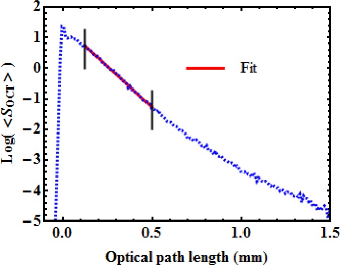
Typical average OCT signal, on a logarithmic scale, as a function of depth. The straight line illustrates a fit performed between the two vertical lines to extract the parameters A and μt.

(a) Backscattered amplitude and (b) total attenuation coefficient of silicone phantoms with different concentrations of alumina. Red line in (a) illustrates a fitted square-law dependency of the backscattered amplitude upon the concentration of alumina.

(a) SEM image of fibrin gel, and; (b) Attenuation coefficient, μt, measured for phantoms with different % w/v concentrations of Intralipid.
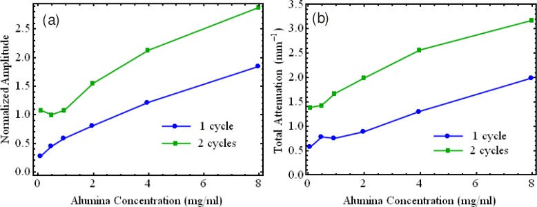
(a) Backscattered amplitude and (b) total attenuation coefficient for PVA-C with one and two FTCs for various concentrations of alumina.
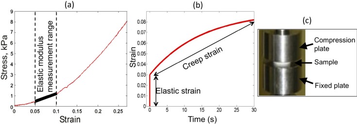
Measurements of elasticity and viscoelasticity of materials and apparatus. (a) Stress-strain curve, highlighting the region of linear elasticity from which elastic modulus is calculated. (b) Creep curve for characterization of viscoelastic materials. (c) Photograph of Instron compression tester.
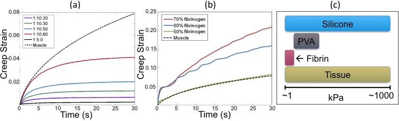
Creep strain curves for: (a) silicone fluid phantoms; and (b) fibrin phantoms. (c) Comparison of range of elastic moduli of phantom materials and soft tissue

Tensile test results for a PVA-C artery phantom, a silicone artery phantom and human media and intima. Reproduced from [48] with permission.

OCT imaging of 2D-structured silicone phantoms: (a) Skin simulating phantom; (b) Its 3-D reconstruction; (c) 200-μm channel filled with 20% Intralipid; and (d) its 3-D reconstruction. Reproduced from [39] with permission.

Cross-sectional OCT images of a 3D-structured phantom: (a) B-scan view (x-z plane); (b) y-z plane view; (c) en face view (x-y plane); (Scale bars: 100 μm) and (d) Orientation of planes with respect to the features. Solid renderings of: (e) Phantom with no scatterers in surrounding layer; and (f) Phantom with scatterers in surrounding layer. Reproduced from [77] with permission.
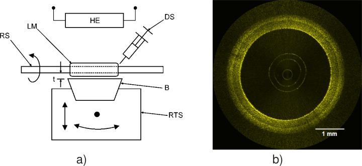
(a) Experimental setup to fabricate multilayer tubular silicone phantoms and (b) OCT image of an artery phantom. (RS rotating shaft, LM layer mixture, DS deposition syringe, B blade, RTS rotation and translation stage, HE heating element, t thickness). Reproduced from [31] with permission.

Bilayer fibrin phantom, with different Intralipid concentration in both layers.
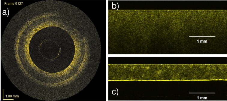
OCT images of: (a) a multilayer PVA-C artery phantom; (b) a 4 mm-thick PVA-C sample with 2 FTCs; and (c) a 0.380 mm-thick PVA-C sample with 2FTCs. Reproduced from [48] with permission.
References
-
- Nordstrom R. J., “Phantoms as standards in optical measurements,” Proc. SPIE 7906, 79060H (2011). 10.1117/12.876374 - DOI
-
- V. V. Tuchin, Tissue Optics: Light Scattering Methods and Instruments for Medical Diagnosis (SPIE, 2000).
LinkOut - more resources
Full Text Sources
Other Literature Sources
Medical
Miscellaneous