

Efficacy of I-scan endoscopy in the diagnosis of gastroesophageal reflux disease with minimal change
- PMID: 22741109
- PMCID: PMC3363044
- DOI: 10.5946/ce.2011.44.1.27
Efficacy of I-scan endoscopy in the diagnosis of gastroesophageal reflux disease with minimal change
Abstract
Background/aims: The aim of the study was to evaluate the efficacy of i-scans for the diagnosis of gastroesophageal reflux disease, especially where only minimal change is involved.
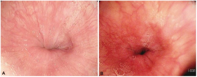
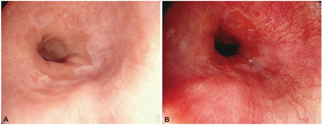
Methods: The esophageal mucosa was inspected using an i-scan following conventional white light endoscopy. The examination with iscan was performed under tone enhancement (TE) esophagus (e) mode. Patients with subtle distal esophageal mucosal changes without definite mucosal breaks, such as blurring of Z-line (B), mucosal coarseness (C), hyperemic or purplish discoloration (D), erythema (E), ectopic gastric mucosal islet (I) and mixed type were classified as minimal change.
Results: A total of 156 patients were included. Using i-scan endoscopy, the number of minimal change was found to further increase from 94 (conventional endoscopy; 19B, 9C, 29D, 13E, 5I, 19 mixed type) to 109 (i-scan; 15B, 8C, 29D, 16E, 5I, 36 mixed type). And 14 patients who had single type by conventional endoscopy were converted to mixed type after i-scan. Therefore, 29 of 156 patients were upgraded after i-scan, they were account for 19% (p<0.0001; 95% confidence interval, 0.13 to 0.25).
Conclusions: The use of i-scan endoscopy significantly improves the identification of minimal change and helps to identify more precisely the type of minimal change.
Keywords: Gastroesophageal reflux; I-scan; Minimal change.
Conflict of interest statement
The authors have no financial conflicts of interest.
Figures
References
-
- DeVault KR, Castell DO American College of Gastroenterology. Updated guidelines for the diagnosis and treatment of gastroesophageal reflux disease. Am J Gastroenterol. 2005;100:190–200. - PubMed
-
- Locke GR, 3rd, Talley NJ, Fett SL, Zinsmeister AR, Melton LJ., 3rd Prevalence and clinical spectrum of gastroesophageal reflux: a population-based study in Olmsted County, Minnesota. Gastroenterology. 1997;112:1448–1456. - PubMed
-
- Kim HY, Kim N, Kim SM, et al. Clinical spectrum and risk factors of erosive and non-erosive GERD in health check-up subjects. Korean J Med. 2006;71:491–500.
-
- Youn YH, Kang YW, Ahn SH, Park SK. Prevalence alteration of reflux esophagitis in recent years. Korean J Gastrointest Endosc. 2001;23:144–148.
-
- Jeon SG, Sohn CI, Kim JE, et al. Prevalence of gastroesophageal reflux in routine check-up subjects. Korean J Med. 2000;58:145–151.
LinkOut - more resources
Full Text Sources
Other Literature Sources
Medical
