

Tumor-derived hepatocyte growth factor is associated with poor prognosis of patients with glioma and influences the chemosensitivity of glioma cell line to cisplatin in vitro
- PMID: 22741575
- PMCID: PMC3447698
- DOI: 10.1186/1477-7819-10-128
Tumor-derived hepatocyte growth factor is associated with poor prognosis of patients with glioma and influences the chemosensitivity of glioma cell line to cisplatin in vitro
Abstract
Background: We examined the association of tumor-derived hepatocyte growth factor (HGF) with the clinicopathological features of gliomas and investigated the effect of HGF inhibition on the biological behavior of tumor cells in vitro in order to determine whether HGF is a valuable prognostic predictor for glioma patients.
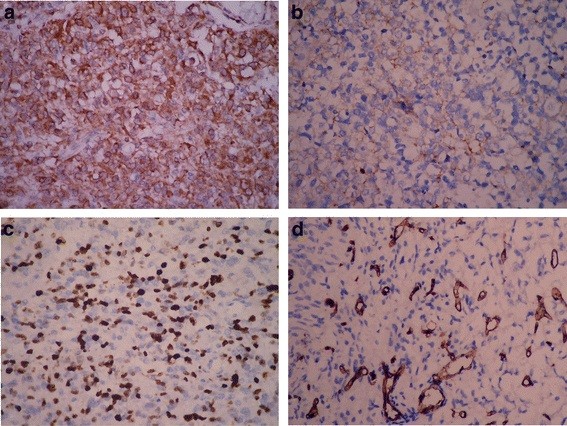
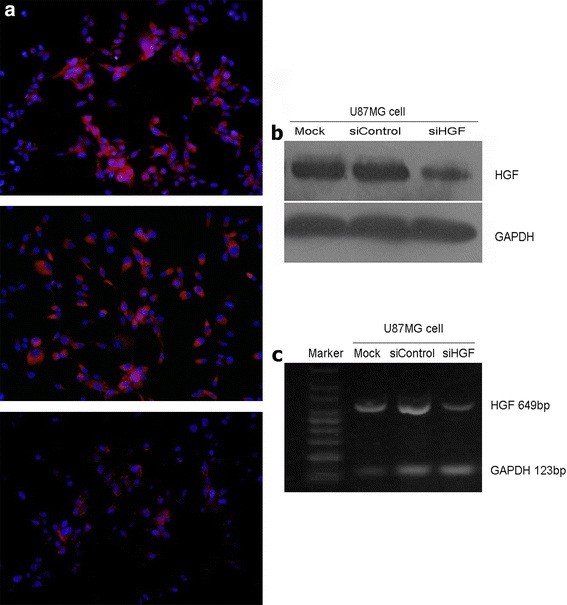
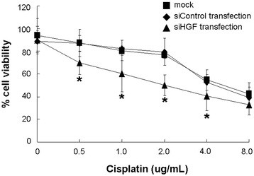
Methods: Seventy-six cases of glioma were collected. The tumor-derived HGF expression, cell proliferation index (PI) and intratumoral microvessels were evaluated by immunohistochemistry. Correlation between immunostaining and clinicopathological parameters, as well as the follow-up data of patients, was analyzed statistically. U87MG glioma cells were transfected with short interference (si)-RNA for HGF, and the cell viability, migratory ability and chemosensitivity to cisplatin were evaluated in vitro.
Results: Both high HGF expression in tumor cells (59.2%, 45/76) and high PI were significantly associated with high-grade glioma and increased microvessels in tumors (P < 0.05). However, only histological grading (P = 0.004) and high-expression of HGF (P = 0.008) emerged as independent prognostic factors for the overall survival of glioma patients. The tumor-derived HGF mRNA and protein expressions were significantly decreased in vitro after transfection of HGF siRNA. HGF siRNA inhibited the cell growth and reduced cell migratory ability. Moreover, HGF siRNA transfection enhanced the chemosensitivity of U87MG glioma cells to cisplatin.
Conclusion: This study indicated that there was significant correlation among tumor cell-derived HGF, cell proliferation and microvessel proliferation in gliomas. HGF might influence tumor progression by modulating the cell growth, migration and chemoresistance to drugs. Increased expression of HGF may be a valuable predictor for prognostic evaluation of glioma patients.
Figures


Similar articles
-
Impact of MACC1 on human malignant glioma progression and patients' unfavorable prognosis.Neuro Oncol. 2013 Dec;15(12):1696-709. doi: 10.1093/neuonc/not136. Epub 2013 Nov 11. Neuro Oncol. 2013. PMID: 24220141 Free PMC article.
-
MiR-136 targets E2F1 to reverse cisplatin chemosensitivity in glioma cells.J Neurooncol. 2014 Oct;120(1):43-53. doi: 10.1007/s11060-014-1535-x. Epub 2014 Aug 20. J Neurooncol. 2014. PMID: 25139024
-
REV3L confers chemoresistance to cisplatin in human gliomas: the potential of its RNAi for synergistic therapy.Neuro Oncol. 2009 Dec;11(6):790-802. doi: 10.1215/15228517-2009-015. Neuro Oncol. 2009. PMID: 19289490 Free PMC article.
-
Exosomes in the Chemoresistance of Glioma: Key Point in Chemoresistance.J Cell Mol Med. 2025 Feb;29(4):e70401. doi: 10.1111/jcmm.70401. J Cell Mol Med. 2025. PMID: 39950738 Free PMC article. Review.
-
Cell-Based Glioma Models for Anticancer Drug Screening: From Conventional Adherent Cell Cultures to Tumor-Specific Three-Dimensional Constructs.Cells. 2024 Dec 17;13(24):2085. doi: 10.3390/cells13242085. Cells. 2024. PMID: 39768176 Free PMC article. Review.
Cited by
-
Molecular Determinants for Photodynamic Therapy Resistance and Improved Photosensitizer Delivery in Glioma.Int J Mol Sci. 2024 Aug 9;25(16):8708. doi: 10.3390/ijms25168708. Int J Mol Sci. 2024. PMID: 39201395 Free PMC article. Review.
-
Hepatocyte growth factor is a prognostic marker in patients with colorectal cancer: a meta-analysis.Oncotarget. 2017 Apr 4;8(14):23459-23469. doi: 10.18632/oncotarget.15589. Oncotarget. 2017. PMID: 28423584 Free PMC article.
-
miR-378a-3p regulates glioma cell chemosensitivity to cisplatin through IGF1R.Open Life Sci. 2021 Nov 2;16(1):1175-1181. doi: 10.1515/biol-2021-0117. eCollection 2021. Open Life Sci. 2021. PMID: 34761108 Free PMC article.
-
Contributions of immune cell populations in the maintenance, progression, and therapeutic modalities of glioma.AIMS Allergy Immunol. 2018;2(1):24-44. doi: 10.3934/allergy.2018.1.24. Epub 2018 Mar 21. AIMS Allergy Immunol. 2018. PMID: 32914058 Free PMC article.
-
TERT rs2853676 polymorphisms correlate with glioma prognosis in Chinese population.Oncotarget. 2016 Nov 8;7(45):73781-73791. doi: 10.18632/oncotarget.12064. Oncotarget. 2016. PMID: 27655710 Free PMC article.
References
-
- Zagzag D, Esencay M, Mendez O, Yee H, Smirnova I, Huang Y, Chiriboga L, Lukyanov E, Liu M, Newcomb EW. Hypoxia- and vascular endothelial growth factor-induced stromal cell-derived factor-1alpha/CXCR4 expression in glioblastomas: one plausible explanation of Scherer’s structures. Am J Pathol. 2008;173:545–560. doi: 10.2353/ajpath.2008.071197. - DOI - PMC - PubMed
Publication types
MeSH terms
Substances
LinkOut - more resources
Full Text Sources
Other Literature Sources
Medical
Research Materials
Miscellaneous
