. 2012 Jun 28:4:41.
doi: 10.1186/1758-3284-4-41.

Effect of human beta-defensin-3 on head and neck cancer cell migration using micro-fabricated cell islands

Affiliations

Effect of human beta-defensin-3 on head and neck cancer cell migration using micro-fabricated cell islands

Kevin Wang et al. Head Neck Oncol. .

Expression of concern in

  • Comment: Head and Neck Oncology.
    BioMed Central. BioMed Central. BMC Med. 2014 Feb 5;12:24. doi: 10.1186/1741-7015-12-24. BMC Med. 2014. PMID: 24499430 Free PMC article. Review.

Abstract

Background: To examine the effect of the natural antimicrobial peptide human β-defensin-3 (hBD-3), on the migration of a head and neck cancer cell line in vitro using microfabrication and soft-lithographic techniques.

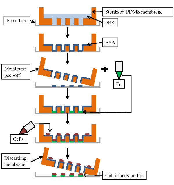
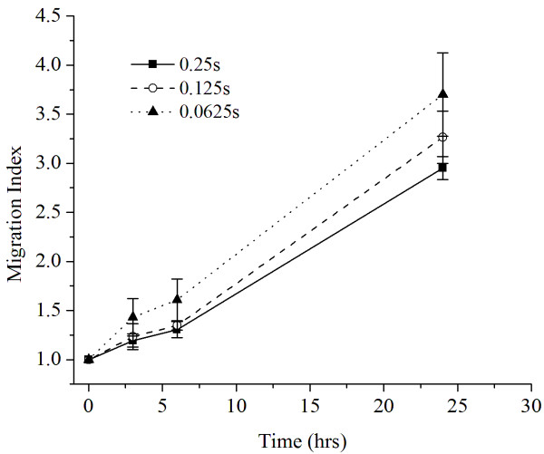
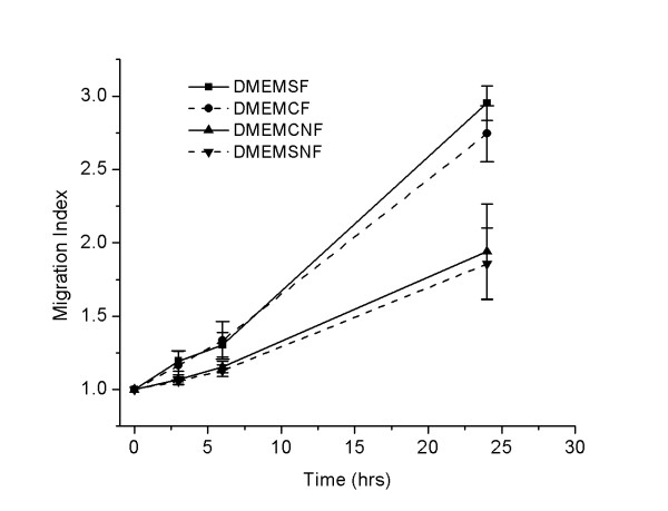
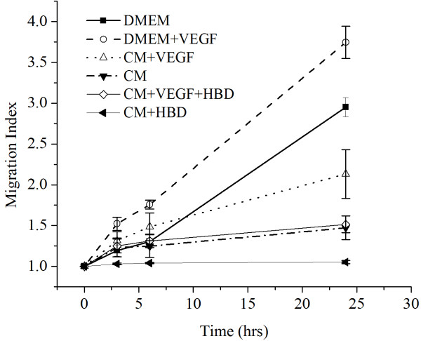
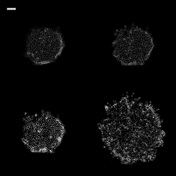
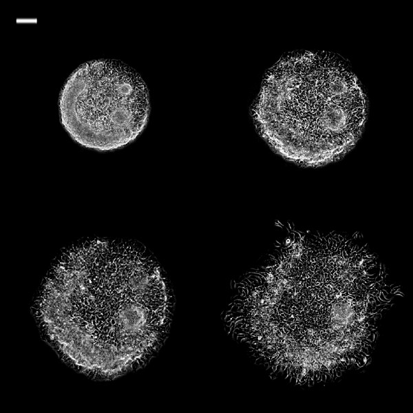
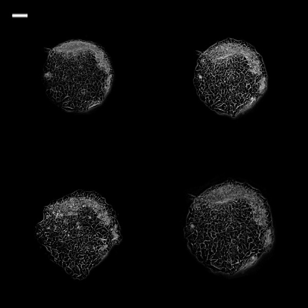
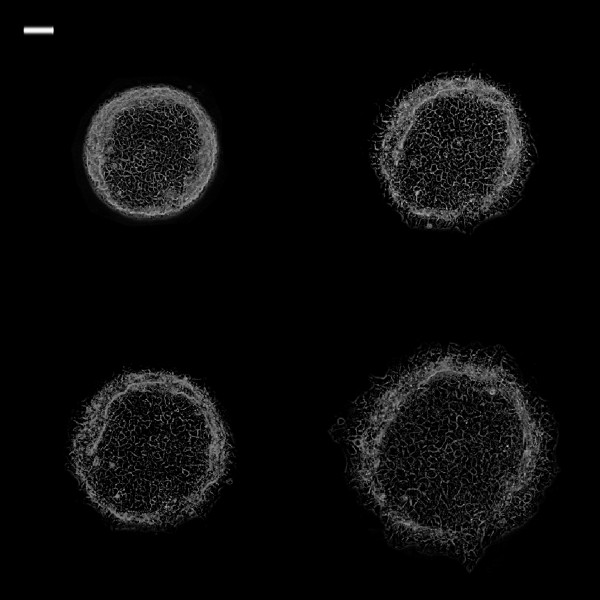
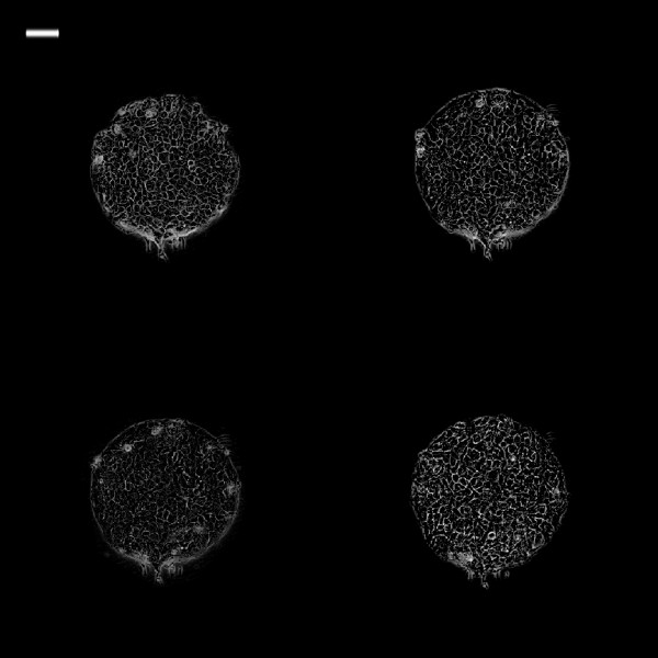
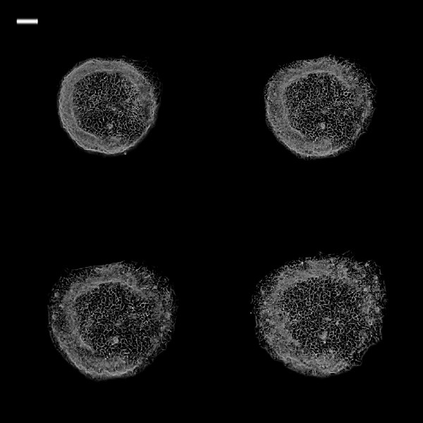
Methods: TR146 cancer cells were seeded in Petri dishes with microfabricated wells for cell migration assays. Total 54 cell islands were used of various shape and size and experimental media type. Cell migration assays were analyzed in six group media: Dulbecco's modified medium (DMEM); DMEM with vascular endothelial growth factor (VEGF); Conditioned media of human embryonic kidney cells (HEK 239) expressing hBD-3 via transfected cloned pcDNA3 as CM/hBD-3; CM/hBD-3+ VEGF; conditioned medium from non-transfected HEK 239 (not expressing hBD-3) as control (CM); and the last group was CM + VEGF. Cell islands were circular or square and varied in size (0.25 mm(2), 0.125 mm(2), and 0.0625 mm(2)). Cell islands were imaged at t=0 h, 3 h, 6 h, and 24 h.

Results: The results show cancer cell islands that originally were smaller had higher migration indices. There was no difference of MIs between circular and square cell islands. MIs at the end point were significantly different among the groups except between CM and CM-hBD-3+ VEGF.

Conclusions: VEGF enhanced cancer cell migration. The combination of DMEM and VEGF showed a synergistic effect on this phenomenon of cancer cell migration. Conditioned medium with hBD-3 suppressed cancer cell migration. hBD-3 suppressed VEGF enhancement of TR146 cancer cell migration.


Figures

Figure 1
Figure 1
Steps for cell island fabrication using PDMS membranes.
Figure 2
Figure 2
Migration indices of square cell islands (0.0625, 0.125, and 0.25 mm 2 ) in DMEM medium as function of time.
Figure 3
Figure 3
Cell migration with and without fibronectin using square and circle cell islands at different time intervals. DMEMSF = square cell islands in DMEM, treated with fibronectin; DMEMCF = circular cell islands in DMEM, treated with fibronectin; DMEMSNF = square cell islands in DMEM without fibronectin treatment; DMEMCNF = circular cell islands in DMEM without fibronectin treatment. Cell island size was 0.25 mm2.
Figure 4
Figure 4
Migration indices of the 0.25 mm 2 cell islands in the different experimental media.
Figure 5
Figure 5
Phase contrast images of 0.25 mm2 TR146 cell islands in DMEM at different time intervals. White bar on the upper left is 100 μm. (Top left: 0 h; top right: 3 h; bottom left: 6 h; bottom right: 24 h).
Figure 6
Figure 6
Phase contrast images of 0.25 mm2 TR146 cell islands in DMEM + VEGF at different time intervals. Phase contrast images of 0.25 mm2 TR146 cell islands in DMEM + VEGF at different time intervals. White bar on the upper left is 100 μm. (Top left: 0 h; top right: 3 h; bottom left: 6 h; bottom right: 24 h)(Top left: 0 h; top right: 3 h; bottom left: 6 h; bottom right: 24 h).
Figure 7
Figure 7
Phase contrast images of 0.25 mm2 TR146 cell islands in CM at different time intervals. White bar on the upper left is 100 μm. (Top left: 0 h; top right: 3 h; bottom left: 6 h; bottom right: 24 h).
Figure 8
Figure 8
Phase contrast images of 0.25 mm2 TR146 cell islands in CM + VEGF at different time intervals. White bar on the upper left is 100 μm. (Top left: 0 h; top right: 3 h; bottom left: 6 h; bottom right: 24 h).
Figure 9
Figure 9
Phase contrast images of 0.25 mm2 TR146 cell islands in CM + hBD at different time intervals. White bar on the upper left is 100 μm. (Top left: 0 h; top right: 3 h; bottom left: 6 h; bottom right: 24 h).
Figure 10
Figure 10
Phase contrast images of 0.25 mm2 TR146 cell islands in CM + VEGF + hBD at different time intervals. White bar on the upper left is 100 μm. (Top left: 0 h; top right: 3 h; bottom left: 6 h; bottom right: 24 h).

References

    1. Cancer Facts and Figures 2011. Atlanta: American Cancer Society. October; 2011.
    1. Brodt P. Cell adhesion and invasion in cancer metastasis. New York: Chapman & Hal; 1996.
    1. Jones D, Nakashima T, Sanchez O. et al.Regulation of cancer cell migration and bone metastasis by RANKL. Nature. 2006;440:692–696. doi: 10.1038/nature04524. - DOI - PubMed
    1. Diamond G, Ryan LK. Beta-defensins: what are they REALLY doing in the oral cavity? Oral Dis. 2011;17:628–635. doi: 10.1111/j.1601-0825.2011.01799.x. - DOI - PMC - PubMed
    1. Diamond G, Kimball J, Krisanaprakornki S, Dale B. Detection of β-defensins secreted by human oral epithelial cells. J Immune Meth. 2001;256:65–76. doi: 10.1016/S0022-1759(01)00442-2. - DOI - PubMed

Publication types

MeSH terms

Substances