

doi: 10.1186/1865-1380-5-30.
Carbon monoxide poisoning: Novel magnetic resonance imaging pattern in the acute setting
Affiliations
- PMID: 22742510
- PMCID: PMC3517430
- DOI: 10.1186/1865-1380-5-30
Item in Clipboard
Carbon monoxide poisoning: Novel magnetic resonance imaging pattern in the acute setting
Int J Emerg Med.
.
Abstract
The presentation of carbon monoxide (CO) poisoning is non-specific and highly variable. The diagnosis is made when a compatible history and examination occur in a patient with elevated carboxyhaemoglobin levels. The severity of intoxication is difficult to assess accurately based on laboratory markers alone. Magnetic resonance imaging (MRI) has been shown to have superior sensitivity to computed tomography for the detection of abnormalities post CO poisoning. We report a novel imaging pattern on MRI undertaken in the acute setting in a patient with CO intoxication. We also discuss the management and follow up of patients with CO poisoning.
Figures
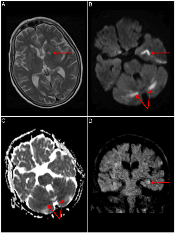
a T2-weighted MRI of brain with abnormal signal in the left medial temporal lobe (arrow). b Diffusion-weighted imaging (DWI) showing symmetrical bilateral restricted diffusion in cerebellar white matter (arrow) and in left medial temporal lobe (arrow). c Apparent diffusion co-efficient (ADC) map: corresponding areas are dark on ADC map, implying acute ischaemia. d Coronal reconstruction: Subtle signal abnormality in left hippocampus (arrow) possibly post-ictal in aetiology.
References
-
- Fan HC, Wang AC, Lo CP. et al.Damage to cerebellar white matter due to carbon monoxide poisoning: a case report. Am J Emerg Med. 2009;27:757. - PubMed
-
- Lapresle J, Fardeau M. The CNS and carbon monoxide poisoning. Anatomical study of brain lesions following intoxication with CO. Prog Brain Res. 1967;24:31–74. - PubMed
LinkOut - more resources
Full Text Sources
